Oral Pyogenic Granuloma: Report of Two Cases
Cariappa Marg, Cross Road No. 9, Sainik Farms, M.B. Road, New Delhi, India
Abstract
Diverse group of pathologic process can produce the enlargement of soft tissues in the oral cavity and often present a diagnostic challenge. This soft tissue enlargement may represent a variation of normal anatomic structure, inflammatory reaction, cyst, neoplasm and developmental anomalies. Pyogenic granulomas (PG) are common benign lesions of the skin and mucosa. They are neither infective, purulent nor granulomatous as the name might suggest-rather a reactive enlargement that is an inflammatory response to local irritation such as calculus, fractured tooth, minor trauma, rough dental restorations and foreign materials.
Clinically, oral Pyogenic Granuloma is smooth or lobulated exophytic lesion manifesting as small, red, erythematous papules on pedunculated or sometimes sessile base.
The purpose of this article is to describe different cases of oral pyogenic granuloma.
Keywords- Pyogenic Granuloma, local irritation, exophytic lesion
INTRODUCTION
Pyogenic granuloma (also known as a “Granuloma gravidarum” and “Pregnancy tumor” ) is a fast growing reactive proliferation of endothelial cells commonly on the gingiva and usually in response to chronic irritation, physical trauma or hormonal factors.
Pyogenic granuloma (PG) was first originally described in 1897 by two French surgeons, Poncet and Dor, who named this lesion ostyomycosis hominis. Hullihen’s description in 1844 was most likely the first PG reported in English literature, but the term “pyogenic granuloma” or “granuloma pyogenicum” was introduced by Hartzell in 1904.1
It usually present as smooth or lobulated red to purple mass that may be either pedunculated or sessile. Granuloma varies in size from a few millimetres to several centimetres. They are soft and not tender on palpation. Early lesions bleed easily due to extreme vascularity, even a minor trauma to the lesion may cause considerable bleeding. On maturation, vascularity decreases and the clinical appearance is more collagenous and pink. If left alone, a number of PG undergo fibrous maturation and resemble and/or become fibroma. In a number of cases, mastication on the lesion causes bleeding and pain and requires surgical intervention before parturition.2,3,4
The term pyogenic granuloma is a misnomer in that it is not pus producing and it does represent granulomatous inflammation. In fact, no relationship exists between bacteria and emergence of this reactive proliferation.2
As it is a benign lesion; therefore surgical excision is the treatment of choice. To avoid possibility of recurrence the lesion must be excised down to the under lying periosteum and predisposing irritant must be removed.
CASE REPORTS
Case One
A 30 year old male patient reported with a chief complaint of growth in mouth involving lower left anterior region of jaw and dirty teeth. The patient had noticed a small painless growth about 1 year back however he did not seek medical attention at that time. There was gradual increase in size, which leads to discomfort while eating and speech also. Patient has a habit of smoking and chewing 5 pouches of gutka per day.
Intraoral examination reveals a solitary diffuse growth, red in color measuring around 2 X 1.5 cm, arising from half of the central incisor of right quadrant to left lateral incisor involving attached gingiva (Figure 1). The growth had a pudunculated base. Mobility of grade I was seen in lower left central and lateral incisior.
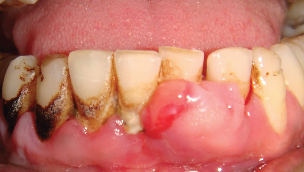
Fig 1: Intraoral photograph showing swelling in the lower left anterior region
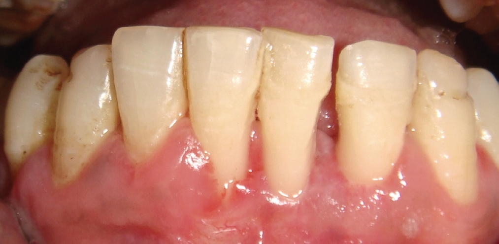
Fig 2: After healing post operative view

Fig 3: Excised tissue
The hemogram of the patient was within normal limits and the lesion was excised deep to periosteum, raising the full thickness flap, proper debridement of the area and biopsy specimen was sent for microscopic examination and was recalled to check the healing. (Figure 2 & 3)
Histopathological examination of the growth revealed stratified squamous keratinized epithelium covering cellular connective tissue. The epithelium shows area of ulceration below which can be seen inflammation in the connective tissue. The connective tissue shows proliferating fibroblasts and collagen fibres interposed in which can be seen lot of epithelial lined spaces within the connective tissue can be seen patchy distribution of lymphocytes and plasma cells.(Figure 4)

Fig 4: Photomicrograph after H&E staining
Case Two
A 24 year old lady reported with a chief complaint of growth in mouth involving lower right anterior region of jaw. The patient had noticed a small painless growth about 2 years back. There was gradual increase in size, which leads to discomfort while eating and speech also.
On general examination, patient was healthy built, no relevant medical history of intake of drugs, nutritional deficiency or any family history. There was no history of any hormonal supplements.
Extraoral examination did not reveal any facial asymmetry. Lymph nodes were not palpable. Intraoral examination reveals a diffuse growth of swelling, measuring around 2 X 2 cm, seen in interdental region in relation to lower right lateral incisor and canine involving attached gingiva extending upto mucogingival junction.
Lesion had divided into two parts, lower part was hemorrhagic (reddish in color) and upper part is fibrotic (pink in color). The growth has pudunculated base. Oral hygiene of patient was fair.(Figure 5)
Radiographically, there was no bone loss, the hemogram of the patient was within normal limits and the lesion was excised deep to periosteum, raising the full thickness flap, proper debridement of the area and biopsy specimen was sent for microscopic examination.
After complete stoppage of bleeding periodontal pack was given and patient was revisited after 1week and then after 15 days for re-evaluvation.
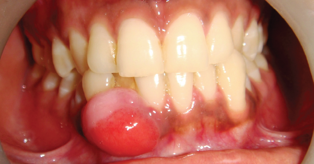
Fig 5: Intraoral photograph showing swelling in the lower right anterior region
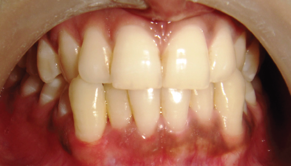
Fig 6: After healing
Complete healing of excised area was observed after 2 months follow up.(Figure 6)
Histopathologial findings of photomicrograph shows Hematoxillin-Eosin stained section showing the presence of tags of stratified squamous epithelium overlying the connective tissue stroma. Epithelium is non-keratinized and present as tags with ulcerated areas. The bulk of section reveals the connective tissue which is highly fibro-cellular comprising large number of plump active fibroblasts intermingled throughout the delicate myxomatous stroma. Areas of highly active proliferation and mature fibroblasts with collagen fibre bundles arranged in streaming pattern are seen. Small ovoid areas of dystrophic calcification characterized by concentric rings of basophilic mineralized material were scattered throughout the connective tissue stroma. Abundance of thick and thin endothelium lined small and large capillaries along with inflammatory cells composed chiefly of lymphocytes and plasma cells were seen throughout the connective tissue stroma.
Overall histopathological feature suggestive of PYOGENIC GRANULOMA.
DISCUSSION
In the oral cavity pyogenic granulomas show a striking predilection for the gingiva, mainly, buccal gingiva with interdental papillae being the most common site in 70-75 % of the cases. They are more common in the anterior region than any other area in the mouth. Gingival irritation and inflammation that result from poor oral hygiene, dental plaque and calculus or over-hanging restorations may be the precipitating factors in many cases.5 It is possible that micro-ulceration from these irritants in an already inflamed gingiva allows the ingress into the gingival connective tissue of low virulent oral micro-flora. This evokes an exaggerated vascular hyperplastic response in the connective tissue resulting in the formation of pyogenic granuloma.6
Majority of PGs are found on the marginal gingiva with only 15% of the tumors on the alveolar part. Depending upon its rate of proliferation and vascularity, there are two histological variants of pyogenic granuloma called lobular capillary hemangioma (LCH type) and non-lobular capillary hemangioma (non-LCH).7
Pyogenic granuloma was first thought to be a mycotic infection contracted from horses. Subsequently, it was claimed without scientific evidence that pyogenic granuloma results from a purulent change within benign oral tumors. Recently, the angiogenesis associated factors Tie-2, angiopoietin-1, angiopoietin- 2, ephrin B2 and B4 have been detected in pyogenic granuloma by immunochemistry. It is now generally accepted that the lesion is an exaggerated localized connective tissue reaction to minor injury or irritation.6
Females are far more susceptible than males because of the hormonal changes that occur in women during puberty, pregnancy, and menopause. The pyogenic granuloma has been called a "pregnancy tumor" and does occur in 1% of pregnant women.4
Treatment of PGs consists of conservative surgical excision which is usually curative. For gingival lesions, excising the lesion down to the periosteum and scaling adjacent teeth to remove any calculus and plaque that may be a source of continuing irritation is recommended. Other conventional surgical modalities are cryosurgery in the form of either liquid nitrogen spray or a cryoprobe. Nd: YAG, CO2 and flash lamp pulsed dye lasers have also been used for the treatment of oral pyogenic granuloma.6,8
No complications are anticipated with removal of this lesion other than the chance of a cosmetic gingival defect. The prognosis is excellent, and the lesion usually does not recur unless inadequately removed.
Focus patient education on better oral hygiene, and consider recommending pulsating mechanical toothbrushes with timers. These toothbrushes encourage better hygiene.
CONCLUSION
Pyogenic granuloma is a non-neoplastic growth in the oral cavity. Proper management including diagnosis, treatment and further prevention is very important. Regular, careful maintenance of oral hygiene, removal of dental plaque and use of soft toothbrush are important to avoid the occurrence of pyogenic granuloma.
REFERENCES
- Shah C, Buhiraj S. Oral Pyogenic Granuloma- A Case Report. J Ind Dent Assc. 2011;5(10):1074-1075.
- Neville BW et al. Oral and Maxillofacial pathology 3rd ed, WB Saunders, Philadelphia 2009, p. 517-518.
- Jafarzadeh H et al. Oral Pyogenic Granuloma. A Rewiew. Journal of Oral Science 2006;48(4):167-175.
- Venugopal S et al. Pyogenic Granuloma- A Case Report. Journal of Denal Sciences and Research 1(1); 80-85.
- Amirchaghmaghi M et al. Extragingival Pyogenic Granuloma. A Case Report. Cases Journal 2008;1:371.
- Rizwanulla T et al. Pyogenic Granuloma. A Case Report. Health Renaissance 2010;8(3):196-198.
- Greenburg MS, Glick M. Burkit’s Oral Medicine: Diagnosis and Treatment. 10th ed, BC Decker, Hamilton, p. 141-142.
- Sandhu M et al. Management of Pyogenic Granuloma. A Case Report. Journal of Innovative Dentistry 2011;1(3): 12-15.
Disclaimer from Guident
The views and opinions expressed in articles published in Guident are solely those of the authors and do not necessarily reflect the views of the editors, the editorial board or the publisher, Ivory India. Guident does not endorse any product, technique or treatment described. Clinical content is intended for qualified dental and medical professionals and is not a substitute for individual clinical judgement. Guident, its editors and the publisher accept no liability for any consequence arising from the use of information published in the journal.
How to cite this article
Dr. Nipun Dhalla, Dr. Anil Dhalla, Dr. Niharika Dhalla, Dr. Dhalla Dental Health Care Centre 197- E. Oral Pyogenic Granuloma: Report of Two Cases. Guident. 2013 Oct;6(12). Available from: https://guident.in/articles/periodontics/oral-pyogenic-granuloma-report-of-two-cases.html