Varied Treatment Modalities for Management of Horizontal Root Fracture
Department of Conservative Dentistry & Endodontics Subharti Dental College Meerut, India
Abstract
Management of mid root fractures presents a formidable challenge for clinicians because of the difficulty of achieving a stable reunion of fracture fragments. Root fractures in permanent teeth are uncommon injuries among dental traumas accounting for 0.5-0.7% of the cases. The most common type of root fracture is in the middle third, followed by apical and coronal part. This case report discusses two different treatment modalities for intra- radicular stabilization of horizontal root fractures.
Keywords: horizontal root fracture, intra-radicular splinting, canal reinforcement.
INTRODUCTION
Traumatic injuries to a tooth can vary inseverity from a simple enamel infraction to a complete ex-articulation of tooth. Among these injuries, tooth fracture (crown fractures, crown-root fractures and root fractures) are considered to be the third most common cause of tooth loss1. Andreasen states that the root fractures in permanent teeth are relatively infrequent occurrences, implicated in .5 % to 7% of all dental traumas2and from 2% to 4% in primary teeth for all traumatic dental injury3.
Root fractures are defined as fractures involving the dentine, cementum and pulp and commonly occur between the age group of 11 to 20 years. These fractures are generally oblique to transverse and may be single or multiple, complete or incomplete4.
Horizontal root fractures occur most commonly in the middle-third of the root and rarely in the apical third . They occur most commonly in the anterior region of maxilla i.e. maxillary central incisor region in fully erupted teeth with complete root formation owing to the frontal impact5.
The success of the treatment of a root fracture dependsupon the degree of fragment displacement and fracture localization. This case report discusses two different treatment modalities for intra-radicular stabilization of horizontal root fractures
CASE REPORT 1
A 32 year old male was referred to the department of conservative dentistry and endodontics, with the chief complaint of pain in upper anterior region . He gave history of trauma one month back.
Careful intra-oral examination showed intact 21however 11 was severely tender on percussion and exhibited slight mobility. Pulp vitality test showed negative response in relation to 11. An IOPA radiographrevealed the presence of horizontal root fracture and fractured segments appeared to be closely approximated. Bone loss was not visible on the radiograph (Fig 1).
Root canal treatment was planned for 11 followed by intra-radicular stabilization by fibre post and Ribbond.
Endodontic treatment of 11 was initiated under local anaesthesia. Access cavity preparation was done by Endo access bur (Dentsply Maillefer) and the patency of root canal was assessed by passing ISO size #15K- filein the root canal. Radiographic determination of working length was done (Fig. 2).and the canal was biomechanically

preparedby protaper file ( Dentsply Maillefer) till F2 size. An intracanal medicament of Ca(OH)2was placed and the patient was recalled after one week.
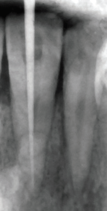
Fig 3: Master apical cone
On recall appointment, as the patient was asymptomatic, master cone selection was done (Fig.3) and sectional obturation with F2 protaper was carried out in the apical 4mm of canal using AH plus sealer(Dentsply Maillefer).
Post space preparation was done till No. 3 peeso reamer . A No. 2 light transmitting fibre post ( Glassix plus, Nordin) was selected and RIBBOND( Ribbond Inc. seattle Washington USA)3mm wide and double the length of radicular pulp space was cut with special scissors and tried in the canal(Fig. 4,5).The post and Ribbond were removed and
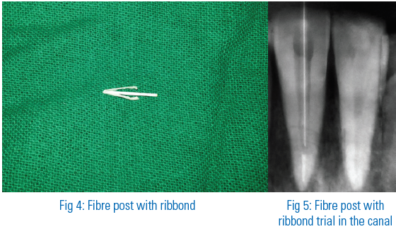
thereafter etchant was applied on the canal wall and access cavity (Scotchbond Etching Gel, 3M ESPE). After rinsing and drying with absorbent paper points, bonding agent ( 3M ESPE Adper Single Bond Plus Adhesive) was applied and light cured for 20 seconds. The canal walls were coated with dual cure resin ( RelyX 3M ESPE), and the fibre post with RIBBOND placed in a V-shape was inserted in the post space and light cured for 40 seconds(Fig. 6,7). Subsequently, the access cavity was restored with composite.
At a four-month recall visit, the left centralincisor was asymptomatic, exhibited no mobility and responded normallyto percussion and palpation.
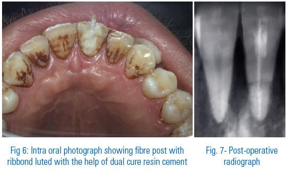
CASE REPORT 2
A 22 year old male was referred to the department of conservative dentistry and fendodontics, with the chief complaint of pain in upper anterior tooth region. He had suffered a trauma in the upper anterior teeth due to fall from the bike three weeks back.
On clinical examination,tooth demonstrated slight mobility, and responded non-vital to electric pulp test and thermal test. Radiographic examination revealed a horizontal root fracture in the middle third of left central incisor(Fig. 8) The space between the fractured segments appeared minimal and there was no bone loss.
Root canal treatment was planned for 11 followed by intra-radicular splinting. Endodontic treatment of right central incisors was initiated under local anaesthesia. Access cavity preparation was done and ISO size #15 K-file was inserted into the canal to assess the patency of the canal and record the working length radiographically (Fig. 9). The root canal was prepared with ISO size # 60K-file size. The canal was dried with sterile paper points and calcium hydroxide was placed in the root canal for intra canal medication. On recall after one week there was still mild bleeding from the fracture site which excluded the option of use of any adhesive materials as proper isolation could not be achieved hence intra- radicular splinting by use of ISO size # 80 K-file and MTA was planned..
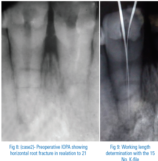
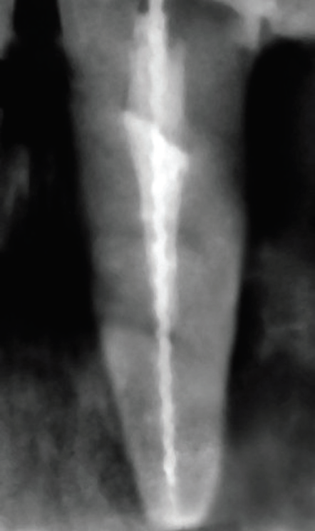
Fig 10: Post-operative radiograph showing intra-radicular splinting with 80 No. K-file size with MTA
An ISO size #80 k- file size was selected which passed beyond fracture line .A groove was prepared on the shaft of file to serve as a weak point. MTA ( ProRoot, Dentsply, Tulsa Dental. Tulsa USA) was mixed in a 3:1 proportion and placed into the canals with the help of MAP(Micro-Apical Placement System,DENTSPLY Maillefer) and condensed by plugger. The file was then inserted into the canal and by circumferential motion advanced into the canal until it reached beyond the fracture line and got wedged into the canal . By further winding the file got separated at the previously made groove. A moist cotton pellet was placed in the access to allow the MTA to set and cavity was sealed with temporary restoration. At the next appointment, the cotton pellet was removed, set of MTA was evaluated and the access cavity was subsequently restored with composite resin(Fig.10).The tooth exhibited no mobility and was asymptomatic at 3 months recall.
DISCUSSION
Following a root fracture, if there is no mobility or displacement of the coronal segment, the patient may have no apparent complaint and may not require dental treatment. However, symptomatic teeth with mobility do require intervention6. The treatment principles for horizontally fractured teeth mainly involve maintaining pulp vitality by immobilizing the coronal segment. According to Hjorting-Hansen, there are four healing patterns that can affect the prognosis and tissue response to dental trauma i.e. Healing with calcified tissue, Healing with interproximal connective tissue, Healing with interproximal bone and connective tissue7.
In the present cases, endodontic treatmentwas carried out as the tooth responded non-vital to thermal and electric pulp test. Although a negative response tothe electric pulp test at an early examination is notindicative for endodontic treatment, this was justified in both the cases as necrotic pulp tissue wasnoticed in the canal after access cavity preparation. Radically, both fragmentswere treated endodontically because of the risk ofapical fragment to have been infected due to thenecrosis.
Two different methods for intraradicular splinting, stabilization and canal reinforcement have been used in the above cases. Fibre posts have been used in such clinical situations however fibre-reinforced composite(FRC) materials when used, as posts are more advantageous than the conventional post systems. RIBBOND which is made up of polyethylene fibres, is a spectrum of 215 fibres with a very high molecular weight. These fibres have a very high coefficient of elasticity(117GPA), so it has an excellent resistance to distortion. They also have very high resistance to traction(3GPA) as a result of their stitch configuration and a good adaptability8,9. However, curing Ribbond inside the canal is questionable as the depth of penetration from light curing unit is only 5mm.
Hence, use of light transmitted fiber post ensured easeof carryingRibbond in the canal, ensuring maximum adaptation to the canal wall and additionally transmitted light via light transmitting post which ensures complete curing of the RIBBOND inside the root canal.
Use of adhesive techniques rely on complete isolation of the canal however, in case of horizontal root fracture often this is not achievable due to bleeding from the fracture line hence MTA along with a ISO# 80 K-filewas used in case 2 for intra radicular stabilization. MTA has many favourable properties including biocompatibility, bactericidal effect, radioopacity and the most important in this case i.e. ability to set in the presence of blood and promote hard tissue healing along the fracture line10.
CONCLUSION
Growth of adhesive dentistry leading to the introduction of highly bondable, high strength RIBBOND which serves as an excellent splint with the availability of biocompatible and osteogenic materials like MTA have built in roads to successfully manage cases of horizontal root fractures thus retaining the natural teeth.
REFERENCES
- Orhan K, Orhan AI, Tulga F. Managementof untreated traumatized permanentincisors with crown and root fractures: a case report. Quintessence Int.2009;40:647–654.
- Ernani da Costa Abad, Luis Claudio Campos, Mauricio Santa Ceciliaand Amauri Favieri.Outcome of treatment of horizontal root fracture using thermafil and surgical: acase report.Brazilian Journal of Dental Traumatology 2009;1(1):7-12.
- Geetha I B, Dr. Shuaib Razvi and Mohamed Asifulla.Non - invasive and invasive management of horizontal root fracture : a report of two cases.Journal of Health Sciences and Research 2011;2:17-19.
- Schweiz Monatsschr Zahnmed.Permanent teeth with horizontal root fractures after dental trauma. Research and Science 2010;120:200-206.
- Joo-Hee Shin and Ryan Jin-Young.Management of horizontal root fractures by fabrication of canine protected occlusion using composite resin.Restor Dent Endod 2012 Aug;37(3):180-184.
- Melahat Görduysus,Alper Akçay, Arlin Kiremitçi.Horizontal root fracture in maxillary first premolar: a case report. Clinical dentistry and research 2011;35(1):53-56.
- Andreasen FM and Andreasen JO. Resorption and mineralization processes following root fracture of permanent incisors. Endod Dent Traumatol 1988; 4:202.
- M. C. Vitale, C. Caprioglio, A. Matrignone, U. Matrignone, and A. R. Botticelli.Combined technique with polyethylene fibers and composite resins in restoration of traumatized anterior teeth. Dental Traumatology 2004; 20:172–177.
- Vallittu PK. Ultra high modulus polyethylene ribbon as reinforcement for denture poly methyl methacrylate, a short communication. Dent Mater 1997; 13:381 -382.
- Sibel Kocak, Serkan Cinar, M. Murat Kocak, GuvenKayaoglu.Intraradicular splinting with endodontic instrumentof horizontal root fracture: acase report. Dental traumatology2008;24:578-580.
Disclaimer from Guident
The views and opinions expressed in articles published in Guident are solely those of the authors and do not necessarily reflect the views of the editors, the editorial board or the publisher, Ivory India. Guident does not endorse any product, technique or treatment described. Clinical content is intended for qualified dental and medical professionals and is not a substitute for individual clinical judgement. Guident, its editors and the publisher accept no liability for any consequence arising from the use of information published in the journal.
How to cite this article
Dr. Shikha Jaiswal, Dr. Sachin Gupta, Dr. Vaikunth Aggarwal. Varied Treatment Modalities for Management of Horizontal Root Fracture. Guident. 2013 Oct;6(12). Available from: https://guident.in/articles/endodontics/varied-treatment-modalities-for-management-of-horizontal-root-fracture.html