Buccal Fat Pad (Bichat,S Lobule): Simple, Reliable and Versatile Flap
Abstract
Taurodontism is a type of variation in tooth morphology that occurs predominantly in molars and it refers to change in tooth shape caused by the failure of Hertwig’s epithelial sheath diaphragm to invaginate at the proper horizontal level. This anomaly is characterised by presence of enlarged pulp chamber, apical displaced pulpal floor, and absence of constriction at the level of the cemento-enamel junction. Endodontic management in taurodont teeth has been described as complex and difficult. The present paper describes the successful completion of endodontic treatment in taurodontic teeth with appropriate use of instruments and techniques.
The pedicled buccal fat pad is a most reliable and versatile flap for the reconstruction of small to medium size of defects like OAF, palatal defects, buccal mucosal defects. It is durable, easy to harvest, and should be considered in settings where access to free flaps is limited and in cases other options have failed. The indications, anatomy and techniques of successful harvest are discussed here in this article.
INTRODUCTION
Oral cavity is known to affect by various pathologic conditions leading to formation of defects of various size compromising function. The aim is not only to fill the defect but at the same time it should restore the function. Variuos factors are responsible for the selection of flap such as size, region, type, quality and quantity of tissue available. The buccal fat pad flap is an pedicle flap and may be used to fill small-to-medium sized soft tissue and bony defects in the palate, superior and inferior alveoli and buccal mucosa. It is often encountered as it bulges into the surgical field during surgery in the pterygomandibular region.[1] The use of the buccal fat pad as a grafting source in the closure of intra-oral defects has gained popularity because of the ease of access and rich blood supply. The purpose of this article is to share the experience of buccal fat pad.
Anatomy [2, 5]
The buccal fat pad is an anatomically rounded and biconvex structure.
- The buccal fat pad is an encapsulated, rounded, biconvex, specialised fatty tissue which is distinct from subcutaneous fat which acts as gliding pads when masticatory muscles contract, and cushions important structures from the forces generated by muscle contraction.
- The buccal fat pad is attached by 6 ligaments to the maxilla, posterior zygoma, and inner and outer rims of the infra-orbital fissure, temporalis tendon and buccinator membrane.
- It has Four processes (buccal, pterygoid, superficial and deep temporal) extend from the body to surrounding tissue spaces
- The parotid duct passes along the lateral surface of the buccal fat pad or penetrates the body of the fat pad before it opens on the buccinator muscle.
- The body is divided into 3 lobes – anterior, intermediate and posterior, in accordance with the structure of the lobar envelopes, the ligaments and the source of the nutritional vessels.
- The anterior lobe is located below the zygoma, extending to the front of the buccinator, maxilla and the deep space of the quadrate muscle of the upper lip and zygomaticus major muscle. The canine muscle originates from the infraorbital foramen and passes through the medial part of the anterior lobe. The parotid duct passes through the posterior part, and the anterior facial vein passes through the antero-inferior margin. The anterior lobe also envelops the infra-orbital vessels and nerve, and together enters the infra-orbital canal. The branches of the facial nerve lie on the outer surface of the capsule.
- The intermediate lobe lies in and around the posterior lobe, lateral maxilla and anterior lobe. It is a membrane-like structure with thin fatty tissue in adults, but is a large mass in children.
- The posterior lobe is situated in the masticatory and neighbouring spaces. It extends up to the infra-orbital fissure and surrounds the temporalis muscle, and extends down to the superior rim of the mandibular body, and back to the anterior rim of the temporalis tendon and ramus. In doing so, it forms the buccal, pterygopalatine and temporal processes.
FEEDING VESSELS
It is an axial flap. The facial artery, transverse facial vessel and the internal maxillary artery as well as their anastomosing branches enter the fat tissue and form a lobar subcapsular vascular plexus by anastomosing with each other. When precisely dissected and mobilised, the buccal fat pad provides a 7x4x3 cm pedicled graft.1
Indications
Reconstruction of small to medium (<5 cm) acquired or congenital soft-tissue and bone defects in the oral cavity,
- Oro-nasal and oro-antral communications following dental extraction;
- Surgical defects following tumour excision and excision of leukoplakia and submucous fibrosis.
- Primary and secondary palatal cleft.
- Other medium defects of oral cavity.
- In Tempromandibular joint reconstruction
Coverage of maxillary and mandibular bone grafts.
Alternative or backup for other failed local flaps.
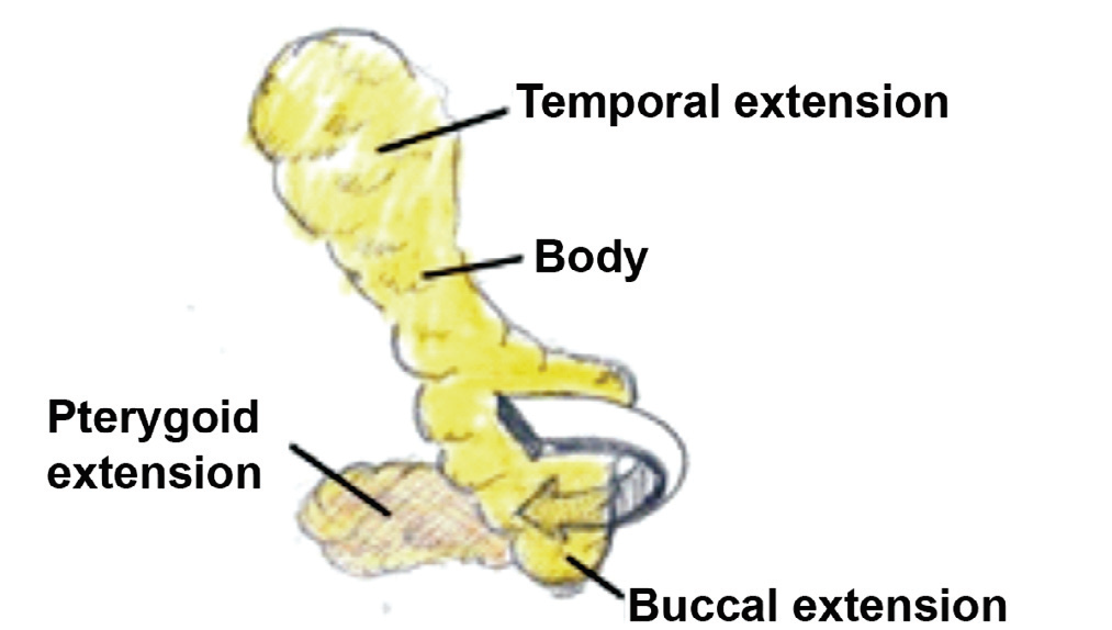
Technique of harvesting buccal fat pad. [3]
- Approach no 1:-An incision through vestibular mucosa in the molar region membrane 1 cm below opening of parotid duct. (Matarasso’s method). (Fig.3)
- Approach no 2:- A vertical mucosal incision slightly lateral to the anterior margin of the ascending ramus . (Stuzin’s method) (Fig. 4)
- Approach no 3:-Elevate mucoperiosteal flap in the molar region on the lateral aspect of the maxillary alveolar process and [ 24 ] incision of the periosteum at the level of buccal sulcus.
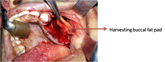
Fig – 1
Advantages
- Easy to harvesting.
- Simple.
- Versatile.
- Hardly any complications.
- Can be done under local anaesthesia.
- Fat pad located in the same surgical field as the defect
- Minimal donor site morbidity.5
Complications
Complications are very rare (3 - 7%), and comprise postoperative infection, partial necrosis, bleeding, scarring and granulation, and sulcus obliteration
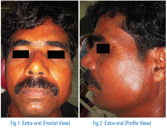
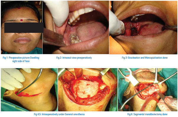
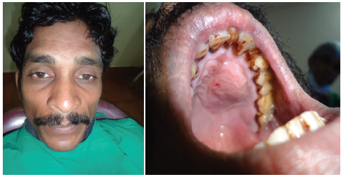
OUR CASES
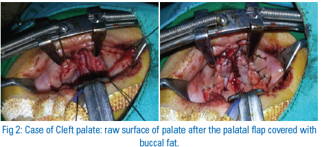
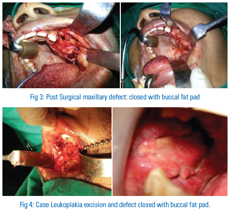
DISCUSSION
The repair of oral defects is always a challenge for a surgeon because of anatomical constraints and the specialized nature of intra oral tissues. Various surgical techniques have been suggested for the closure of oral defects such as primary closure, buccal mucosal graft, split thickness skin graft, allogenic graft, Regional flaps, distant flaps have also been successfully used for intra-oral reconstruction but, they are generally preferred for defects of much larger dimensions. The use of buccal fat pad has gained popularity in the reconstruction of various defects because of the following reason,
- Easy to harvest.
- Local flap.
- Rich blood supply.
- Little postoperative morbidity,
- Good patient acceptance
Initially buccal fat pad was considered a surgical nuisance because of its accidental encounter during various operative procedures, but in 1977 Egyedi[4] reported the use of fat pad in reconstruction of maxillary defect. After it become popular among the surgeon for the reconstruction of minor to average oral defect. Size limitations of BFP must be understood in order to provide successful outcome. Defects of larger sizes and distant, the possibility of partial dehiscence of the flap is high due to the impaired vascularity because of stretching of flap[5].
It is important to preserve the thin capsule around the fat pad to avoid the damage to the blood vessel supplying it. The nature of healing of buccal fat pad[6]
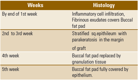
Reported complications associated with the buccal fat pad flap are haematoma, partial necrosis, scarring, infection and facial nerve injury[1, 7,8, 9]. The use of buccal fat pad in patients with previously radiated, malar hyperplasia, thin cheek, facial microsomia or other local defects.
SUMMARY
The buccal fat pad is a quick, simple, reliable flap for repair of small to medium sized oral defects. It has an excellent blood supply and causes minimal donor site morbidity. Furthermore it is closely located to the defect. In the light of these features, it is thought that the buccal fat pad will be used more often for the repair of various defects in the future.
REFERENCES
- Rapidis AD, Alexandridis CA, Eleftheriadis E, et al. The use of the buccal fat pad for reconstruction of oral defects: Review of the literature and report of 15 cases. J Oral Maxillofac Surg 2000;58(2):158-163.
- Martin-Granizo R, Naval L, Costas A, Goizueta C, Rodriguez F, Monje F, et al. Use of buccal fat pad to repair intraoral defects: review of 30 cases. Br J Oral Maxillofac Surg 1997;35:81–4
- Liversedge RL, Wong K. Use of the buccal fat pad in maxillary and sinus grafting of the severely atrophic maxilla preparatory to implant reconstruction of the partially or completely edentulous patient: Technical note. Int J Oral Maxillofac Implants 2002;17:424-428.
- Egyedi P. Utilization of the buccal fat pad for closure of oro-antral and/or oro-nasal communications. J Craniomaxillofac Surg 1977; 5:241.
- Zhang H, Yan Y, Qi K, Wang J, Liu Z. Anatomical structures of the buccal fat pad and its clinical adaptation. Plast Reconstr Surg 2002; 109(7):2509-2518.
- Samman N, Cheung LK,Tideman H.The buccal fat pad in oral reconstruction. Int J Oral Maxillofac Surg 1993; 22:2–6.
- Dean A, Alamillos F, Garcia-Lopez A, Sanchez J, Penalba M. The buccal fat pad in oral reconstruction. Head Neck 2001;23(5):383-388.
- El-Hakim IE, El-Fakharany AM. The use of pedicled buccal fat pad and palatal rotating flaps in closure of oroantral communication and palatal defects. J Laryngol Otol 1999; 113:834–8.
- Seward GR, Harris M, McGowan DA. Killey and Kay’s Outline of oral surgery Part 1. 2nd ed pp 241- 247. Bristol: IOP Publishing Ltd, 1987.
Disclaimer from Guident
The views and opinions expressed in articles published in Guident are solely those of the authors and do not necessarily reflect the views of the editors, the editorial board or the publisher, Ivory India. Guident does not endorse any product, technique or treatment described. Clinical content is intended for qualified dental and medical professionals and is not a substitute for individual clinical judgement. Guident, its editors and the publisher accept no liability for any consequence arising from the use of information published in the journal.
How to cite this article
Dr. K.N.V. Sudhakar Consultant, Dr. Rajesh Sabnis, Dr. Abdul Hameed. Buccal Fat Pad (Bichat,S Lobule): Simple, Reliable and Versatile Flap. Guident. 2013 Oct;6(12). Available from: https://guident.in/articles/oral-surgery/buccal-fat-pad-bichats-lobule-simple-reliable-and-versatile-flap.html