Unicystic Ameloblastoma of the Mandible
Department of Oral and Maxillofacial Surgery K.D. Dental College & Hospital, Mathura, India
ABSTRACT
Ameloblastoma is a true neoplasm of odontogenic epithelial origin. It is the second most common odontogenic neoplasm. Its incidence combined with its clinical behavior, makes ameloblastoma the most significant odontogenic neoplasm. Unicystic ameloblastoma refers to those cystic lesions that show clinical, radiographic, or gross features of a mandibular cyst, but on histologic examination show a typical ameloblastomatous epithelium lining part of the cyst cavity.1 It accounts for 5-15% of all intraosseous ameloblastomas. This article report a case of 30-year-old female with unicystic ameloblastoma associated with right impacted mandibular molar.
Keywords: Ameloblastoma, unicystic,Mandibular reconstruction
INTRODUCTION
Ameloblastoma develops from epithelial cellular elements and dental tissues in their various phases of development. It is a slow-growing, persistent, and locally aggressive neoplasm of epithelial origin. It is the second most common odontogenic neoplasm. The tumor is often asymptomatic presenting as slowly enlarging facial swelling. It is often associated with an unerupted third molar2. It may be detected during the course of routine radiography. The vast majority of ameloblastomas arise in the mandible, and the majority of these are found in the angle and ramus region. There are three forms of ameloblastomas, namely multicystic, peripheral, and unicystic tumors3. Multicystic ameloblastoma is the most common variety and represents 86% of cases. Peripheral tumors are odontogenic tumors, with the histological characteristics of intraosseous ameloblastoma that occur solely in the soft tissues covering the tooth-bearing areas of the jaws. Unicystic tumors include those that have been variously referred to as mural ameloblastomas, luminal ameloblastomas, and ameloblastomas arising in dentigerous cysts4. Various treatment methods for the lesion in relation to many factors, such as the tumour size and location, have been suggested. These include enucleation, marginal resection and aggressive resection. When a diagnosis of ameloblastoma is obtained, the treatment must be aggressive and radical. The goal of treatment of ameloblastoma is to achieve complete excision and appropriate reconstruction. We present a case of a large unicystic mandibular ameloblastoma in a 30 year old female.
CASE REPORT
A 30 year old female came with a chief complaint of a slowly growing swelling on the right side of the face since one year (Figure1). Initially, the swelling was small in size but has gradually increased over a period of time to attain the present size. It was not associated with pain or difficulty in opening the mouth, chewing or articulating (Figure2). No abnormality was detected upon systemic examination. On inspection, a diffuse swelling measuring approximately 7x4 cms in size is present on the right side of face extending anteroposteriorly 2 cms from the symphisis mentii to the ramus of mandible and superoinferiorly from alatragal line to
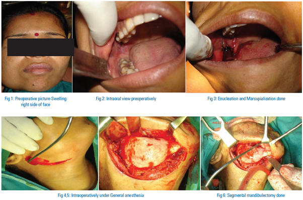

the inferior border of mandible. The surrounding oral mucosa was normal. On palpation, the inspection findings were confirmed. The swelling was non tender and hard in consistency. The lymph nodes were non palpable. An orthopantomogram (OPG) was done, which showed large cystic lesion in the right side of mandible. The patient was taken up for enucleation and marsupialisation (Figure3) and after that for surgery under general anaesthesia (Figure 4, 5). A segmental mandibulectomy was done (Figure 6) and reconstruction plate was put (Figure7). The resected specimen (Figure 11, 12) was sent for histopathological examination and it revealed features consistent with that of the unilocular ameloblastoma.
DISCUSSION
Unicystic ameloblastoma, a variant of ameloblastoma was first described by Robinson and Martinez5 .Unilocular ameloblastoma is a rare type of ameloblastoma, accounting for about 6% of ameloblastomas. It usually occurs in a younger age group, with about 50% of the cases occurring in the second decade of life. More than 90% are located in the mandible6-8. Between 50 and 80% of cases are associated with tooth impaction, the mandibular third molar being most often involved. Patients most commonly present with swelling and facial asymmetry, pain being an occasional presenting symptom. Mucosal ulceration is rare, but may be caused by continued growth of the tumor. Small lesions are sometimes discovered more on routine radiographic screening examinations or as a result of local effects (like tooth mobility, occlusal alterations and failure of eruption of teeth) produced by the tumor9. Histologically, the minimum criterion for diagnosing a lesion as Unilocular ameloblastoma is the demonstration of a single cystic sac lined by odontogenic (ameloblastomatous) epithelium often seen only in focal areas. Unilocular ameloblastoma should be differentiated from odontogenic cysts because the former has a higher rate of recurrence than the latter10 According to Reichart relative frequency has been reported as between 5% and 22% of all subtypes of ameloblastomas. No data is available concerning prevalence and incidence of unicystic ameloblastoma. There have been many debates regarding whether unicystic ameloblastoma develops or arises in an existing cyst11. Leider et al, proposed three pathogenic mechanisms for the evolution of Unicystic ameloblastoma: reduced enamel epithelium, from dentigerous cyst and due to cystic degeneration of solid ameloblastoma. 12 .It often involves an impacted tooth and the focal area of the cystic tumor lining is often composed of a nonspecific, thin epithelium that mimics the dentigerous cyst lining.13 In the present case the association with an impacted tooth and presence of non specific thin epithelium lining in focal areas of cystic tumor supporting the second hypothesis i.e. arising from preexisting dentigerous cyst. Based on the character and extent of tumor cell proliferation within the cyst wall, several histologic subtypes of unicystic ameloblastoma are recognized, which include simple cystic type, intraluminal and mural type. Gardner in 1984 has pointed out that there is a difference in biological behaviour between these lesions14. Till now no recurrence has been seen and patient is still regularly being followed up. Perhaps the most important consideration regarding unicystic ameloblastoma is that of biologic behaviour. It has been widely stated that these lesions are less aggressive than their solid or multicystic counterparts and should be treated by enucleation or curettage. However, Gardner has pointed out that there is a difference in biological behaviour between those lesions that are simply cystic or show intraluminal proliferation and those in which the epithelium penetrated and breaches the fibrous wall, therefore having the capacity to invade the cancellous bone. It has been suggested that recurrence following conservative surgery is more likely to occur in the third group therefore these lesions should be treated in the same manner as solid ameloblastomas.15 Whatever surgical approach the surgeon decides to take, long-term follow-up is mandatory, as recurrence of unicystic ameloblastoma may be long delayed .16
CONCLUSION
Unicystic ameloblastoma is a tumor with a strong propensity for recurrence, especially when the ameloblastic focus penetrates the adjacent tissue from the wall of the cyst. The ability to predict this potential occurrence prior to surgery would greatly enhance therapeutic strategies for reducing the incidence. Hence, the Pathologist should examine the tissue sections carefully in an attempt to determine whether ameloblastoma has penetrated the wall of the cyst or not so that the complications can be minimized.
REFERENCES
- Kalaskar R, Unawane AS, Kalaskar AR, and Pandilwar P. Conservative management of unicystic ameloblastoma in a young child: Report of two cases. Contemp Clin Dent. 2011 Oct-Dec; 2(4): 359–363.
- Gerzenshtein J, Zhang F, Caplan J, Anand V, Lineaweaver W. Immediate mandibular reconstruction with microsurgical fibula flap transfer following wide resection for ameloblastoma. J Craniofac Surg. 2006;17(1):178–182.
- Philipsen HP, Reichart PA. Odontogenic tumors and allied lesions. Quintessence Pub. Co. Ltd; 2004. Classification of odontogenic tumors and allied lesions; pp. 21–3.
- Chana, Jagdeep S, Yang-Ming Chang, Wei, Fu-Chan, Shen, Yu-Fen, Chan Chiu-Po, Lin Hsiu-Na, Tsai Chi-Ying, Jeng Seng-Feng. Segmental mandibulectomy and immediate free fibula osteoseptocutaneous flap reconstruction with endosteal implants: An ideal treatment method for mandibular ameloblastoma. Plast Reconstr Surg. 2004;113(1):80–87.
- Robinson L, Martinez MG. Unicystic ameloblastoma: A prognostically distinct entity. Cancer 1977;40:2278-85
- Philipsen HP, Reichart PA. Odontogenic tumors and allied lesions. London: Quintessence Pub. Co. Ltd; 2004. Unicystic ameloblastoma; pp. 77–86.
- Pizer ME, Page DG, Svirsky JA. Thirteen-year follow-up of large recurrent unicystic ameloblastoma of the mandible in a 15-year-old boy. J Oral Maxillofac Surg. 2002;60:211–5.
- Navarro CM, Principi SM, Massucato EM, Sposto MR. Maxillary unicystic ameloblastoma. Dentomaxillofac Radiol. 2004;33:60–2.
- Roos RE, Raubenheimer EJ, van Heerden WF. Clinico-pathological study of 30 unicystic ameloblastomas. J Dent Assoc S Afr. 1994;49:559–62.
- Konouchi H, Asaumi J, Yanagi Y, Hisatomi M, Kawai N, Matsuzaki H, Kishi K. Usefulness of contrast enhanced-MRI in the diagnosis of unicystic ameloblastoma. Oral Oncol. 2006;42:481–6.
- Ackermann GL, Altini M, Shear M. The unicystic ameloblastoma: A clinicopathological study of 57 cases. J Oral Pathol 1988;17:541-6
- Leider AS, Eversole LR, Barkin ME. Cystic ameloblstoma. Oral Surg Oral Med Oral Pathol 1985;60:624-30.
- Li TJ, Wu YT, Yu SF, Yu GY. Unicystic ameloblastoma: A clinicopathologic study of 33 Chinese patients. Am J Surg Pathol 2000;24:1385-92
- Gardner DG. A pathologist's approach to the treatment of ameloblastoma. J Oral Maxillofac Surg 1984;42:161-6.
- Thankappan S, Thomas V, Kandamparambil S, Nair S. Unicystic ameloblastoma: 3 case reports and review of literature. J Indian Acad Oral Med Radiol 2008;20:65-70.
- Li TJ, Kitano M, Arimura K, Sugihara K. Recurrence of unicystic ameloblatoma: A case report and review of the literature. Arch Pathol Lab Med 1998;122:371-4.
Disclaimer from Guident
The views and opinions expressed in articles published in Guident are solely those of the authors and do not necessarily reflect the views of the editors, the editorial board or the publisher, Ivory India. Guident does not endorse any product, technique or treatment described. Clinical content is intended for qualified dental and medical professionals and is not a substitute for individual clinical judgement. Guident, its editors and the publisher accept no liability for any consequence arising from the use of information published in the journal.
How to cite this article
Dr. Arti. Unicystic Ameloblastoma of the Mandible. Guident. 2013 Sep;6(11). Available from: https://guident.in/articles/oral-surgery/unicystic-ameloblastoma-of-the-mandible.html