Taurodontism - Engima to Endodontist
s superspeciality dental and orthodontic Center, Delhi Gate, Agra, India
Abstract
Taurodontism is a type of variation in tooth morphology that occurs predominantly in molars and it refers to change in tooth shape caused by the failure of Hertwig’s epithelial sheath diaphragm to invaginate at the proper horizontal level. This anomaly is characterised by presence of enlarged pulp chamber, apical displaced pulpal floor, and absence of constriction at the level of the cemento-enamel junction. Endodontic management in taurodont teeth has been described as complex and difficult. The present paper describes the successful completion of endodontic treatment in taurodontic teeth with appropriate use of instruments and techniques.
INTRODUCTION
Taurodontism is considered as a variation in tooth morphology that occurs more often in molars and occasionally in premolars. The term “taurodontism” was first used by Sir Arthur Keith in 1913 to describe the teeth of prehistoric people, the Neanderthals and Heidelberg. He coined this term from the Latin Word “tauro” (for bull) and Greek term “dont” (for tooth) in which the tall root trunk encloses a high pulp chamber and short roots. This anomaly is characterized by enlarged pulp chamber with more apical positioning of floor of pulp chamber and furcation of root1. Witkop2 defined Taurodontism as “teeth with large pulp chambers in which the bifurcation or trifurcation are displaced apically, so that the chamber has greater apico-occlusal height than in normal teeth and it lacks the constriction at the level of cemento-enamel junction (CEJ). The distance from the trifurcation or bifurcation of the root to the CEJ is greater than the occluso-cervical distance”.
The exact etiology of taurodontism is unclear. However, according to Hamner3 et al, taurodontism is caused by the failure of Hertwig’s epithelial sheath diaphragm to invaginate at the proper horizontal level.
Taurodontism has been graded according to its severity: least pronounced (hypotaurodontism), moderate (mesotaurodontism), and most severe (hypertaurodontism)4 .
Previously, taurodontism was related to syndromes such as Down’s and Klinefelter’s5. Today, it is considered as an anatomic variance that could occur in a normal population6. The prevalence of taurodontism is reported to range from 2.5% to 11.3% of the human population8. This range is accounted for by variations in race and differences in diagnostic criteria9. The present article describes the diagnosis and management of hypertaurodontism by endodontic treatment in a left mandibular second molar.
CASE REPORT
A 25-year-old male patient came with the chief complaint of pain in lower left back teeth region. Clinical examination revealed a deep carious lesion with pulpal involvement with respect to lower left second molar. The patient’s medical history was non contributory. At the time of examination, the tooth was asymptomatic. Intraoral examination revealed a normal shaped crown. The tooth was sensitive to percussion. On periapical radiography, occlusal carious lesion was found on the left mandibular second molar, with endodontic involvement. A vitality test of the tooth was negative, and a diagnosis of pulp necrosis with symptomatic apical periodontitis was made. Radiographic examination of the affected tooth revealed an abnormal tooth anatomy and resorbed root on the distal aspect and a large periapical radiolucency.
The radiographic findings were:
• The pulp chamber extended beyond the cervical area reaching the furcation.
• Three short roots were seen at the furcation area in the apical third.
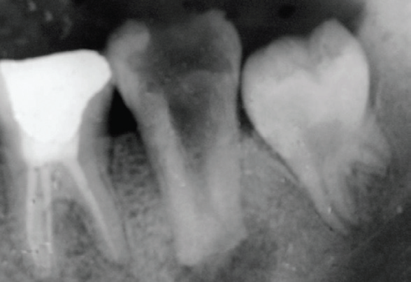
Fig 1: Preoperative IOPA showing taurodontism in lower left second molar
From these radiographic findings, the tooth was diagnosed to be a hypertaurodont (Figure 1).
ENDODONTIC MANAGEMENT OF THE MANDIBULAR LEFT SECOND MOLAR
Endodontic access to the cavity was prepared on the occlusal surface. A huge pulp chamber was found, but the root furcation was difficult to identify. Under local anesthesia and proper isolation, access opening was done with cutting concentrated more towards centre due to the large central orifice of pulp chamber. Canals were negotiated with ISO no 10 K- file, ISO 15 No. K-file was used to determine the working length in the division of the large central orifice into two at apical third {Figure 2}. Tooth was disoccluded and the two small canals were prepared till size Hand Protaper till F3 while the coronal and middle third i.e the central large canal was filed sequentially till size 80 with intermittent copious irrigation of 1.25% sodium hypochlorite with ultrasonic tips. Calcium hydroxide mixed with 2% Chlorhexidine solution was used as an interappointment dressing. The patient was recalled after 7 days. The patient was
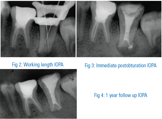
asymptomatic. A final irrigation with 17% EDTA was performed. Bifurcation was sealed with F3 cones (Dentsply) and AH plus sealer (Dentsply). The gutta percha was sheared off with a warm vertical plugger. The wide main canal was then back-filled with obtura and AH plus sealer. Final radiographic analysis showed a well condensed obturation with slight amount of sealer extrusion on the distal aspect {Figure 3}. Post endodontic restoration was done. Patient was recalled at 1 month and after every 6 months. One year radiographic analysis showed complete healing of the periapical lesion {Figure 4}.
DISCUSSION
Taurodontism refers to a condition in which the pulp chamber is widened apico-occlusally and thus, the furcations are positioned more apically than normal4. Taurodontism is frequently associated with other anomalies and syndromes5. The patient discussed above, systemic disturbances or malformations could not be identified and hence considered to be of non syndromic taurodontism. The teeth most frequently affected are the molars, although it can be occasionally seen in premolar and incisors and are mostly diagnosed by radiographic study. Endodontic treatment in taurodont teeth has been described as complex and difficult. Durr10 et al suggested that the morphology could hamper the location of the orifices, thus creating difficulty in instrumentation and obturation. The number of root canals varied in each case. Since the pulpal chamber was huge, mechanical debridement and shaping was achieved with circumferential filing using K- files of standard ISO size and taper. Sodium hypochlorite irrigation was limited to the initial use as the apical foramina were wide open and as a precautionary measure to avoid a hypochlorite accident. A modified filling technique, which consists of combined lateral compaction in the apical region with vertical compaction of the elongated pulp chamber, has been proposed11. In this case apical plug with lateral condensation followed by backfilling with Obtura (Spartan) was used, as the main pulp chamber was huge and large wide apical foramina were present. This technique allows good apical control and adapts well to the canal configuration. Some amount of sealer extruded from the resorbed root which gradually resorbed with due course of time.
The present case describes successful completion of endodontic treatment of a taurodont mandibular second molar, which was difficult to perform with conventional techniques.
Success was mostly attributed to the use of ultrasonic irrigation which led to effective debridement of the root canal as well as a modified obturation technique.
SUMMARY
Although taurodontism is a rare occurrence its discovery can allow insight into other associated syndromes and anomalies and proper understanding of the root canal morphology allows us to carry out successful endodontic treatment prolonging the longevity of the tooth in function.
REFERENCES
- Keith A. Problems relating to the teeth of the earlier forms of prehistoric man. J Royal Soc Med 1913;6:103-24.
- Witkop CJ. Clinical aspects of dental anomalies Int Dent J 1976;26:378-90.
- Hamner J E, Witkop CJ, Metro PS. Taurodontism. Report of a case. Oral Surg Oral Med Oral Pathol 1964;18:409-18.
- Shaw J C M. Taurodont teeth in South African Races. J Anat 1928;62:476-99.
- Regezi JA, Scuibba JJ. Oral pathology: clinical pathologic correlations. WB Saunders Company, 1999:458.
- Shifman A, Chanannel I. Prevalence of taurodontism found in radiographic dental examination of 1200 young adult Israeli patients. Community Dent Oral Epidemiol 1978;6:200–3.
- Guttal KS, Naikmasur VG, Bhargava P, Bathi RJ. Frequency of Developmental Dental Anomalies in the Indian Population. Eur J Dent 2010;4:263-9.
- Joseph M. Endodontic treatment in three taurodontic teeth associated with 48, XXXY Klinefilter syndrome: a review and case report. Oral Surg Oral Med Oral Pathol Oral Radiol Endod 2008;105:670-7.
- Durr DP, Campos CA, Ayers CS. Clinical significance of taurodontism. J Am Dent Assoc 1980;100:378–81.
- Tsesis I, Shifman A, Kaufman AY. Taurodontism: an endodontic challenge: report of a case. J Endod 2003;29:353-5.
Disclaimer from Guident
The views and opinions expressed in articles published in Guident are solely those of the authors and do not necessarily reflect the views of the editors, the editorial board or the publisher, Ivory India. Guident does not endorse any product, technique or treatment described. Clinical content is intended for qualified dental and medical professionals and is not a substitute for individual clinical judgement. Guident, its editors and the publisher accept no liability for any consequence arising from the use of information published in the journal.
How to cite this article
Dr. Mohita Dhingra, Dr. Chand Sawhney, Dr. Santosh Kumar, Dr.Sawhney. Taurodontism - Engima to Endodontist. Guident. 2013 Oct;6(12). Available from: https://guident.in/articles/endodontics/taurodontism-engima-to-endodontist.html