Root Coverage - a Case Series in the Treatment of Isolated Gingival Recession Defects.
ABSTRACT
Gingival recession is the exposure of root surfaces due to apical migration of the gingival tissues. Though it seldom results in tooth loss, marginal tissue recession poses esthetic complaints, root hypersensitivity and root caries. This article emphasizes on some surgical procedures for the coverage of exposed root surfaces, including their indications, advantages and disadvantages. The results obtained in this study substantiate the effectiveness of root coverage procedures with predictability and long term stability of the results.
KEY WORDS:Gingival recession; Laterally displaced flap; Free gingival graft; Subepithelial connective tissue graft; Coronally advanced flap; Root coverage
INTRODUCTION
Gingival recession associated with root surface exposure is a complex phenomenon that may present numerous therapeutic challenges to the clinician. Recession may be accompanied by root caries or cervical abrasion which may cause the patients esthetic or root sensitivity concerns.1
A variety of periodontal procedures have the potential to correct gingival recession by increasing the width of attached gingiva and also attain partial or complete root coverage. Today, gingival reconstruction has become a routine part of periodontal practice. The success of these root coverage procedures depends upon several factors and ideal selection of cases.
CONDITIONS NECESSARY FOR SUCCESSFUL ROOT COVERAGE
1) Sufficient interdental papilla and interdental bone dimensions
2) Sufficient blood supply to donor tissue
3) Root surface to be covered with thick donor tissue
4) Close adaptation of donor tissue with the recipient site
5) No severe decay or abrasion of the tooth2
6) Thick gingival biotype (≥0.8mm)3
OVERVIEW OF VARIOUS TECHNIQUES FOR ROOT COVERAGE
Several techniques have been proposed for the treatment of isolated gingival recession defects. The main treatment goals are to achieve recession reduction and increasing keratinized tissue width. Some of the pedicle soft tissue grafts like lateral pedicle graft4, double papilla flap5 and oblique rotational flap6 and advanced flaps like coronally advanced flap7 and semilunar flap8have shown successful root coverage.Non submerged free soft tissue autografts either one stage (free gingival graft) or two stage (free gingival graft plus coronally advanced flap) has shown 48.1% of mean root coverage9. Connective tissue grafts predictably achieved 90% or greater root coverage most of the time. It has been clearly established as a highly effective means of achieving root coverage.10
Coronally advanced flap combined with additive treatments like non resorbable barier11, resorbable barrier12, enamel matrix derivative13, platelet rich plasma14, acellular dermal matrix15, growth factors16, tissue engineering17 and low intensity laser therapy18 have also shown predictable root coverage. In this paper we intend to discuss three predictable and simple techniques of achieving root coverage.
LATERALLY POSITIONED PEDICLE GRAFT (LPPG)
This technique was first introduced by Grupe and Warren in 1956.4This technique cannot be performed without sufficient attached gingiva lateral to the recession site. A shallow vestibule may also jeopardize the outcome. Although LPPG provides an ideal colour match, it is often inadequate for the treatment of multiple recessions.
The indications of LPPG are to cover isolated, denuded roots that have adequate donor tissue laterally and vestibular depth. The contraindications are diseased periodontium, excessive root prominences, deep caries, extensive root abrasion and significant loss of interproximal bone height.
CASE REPORT-1
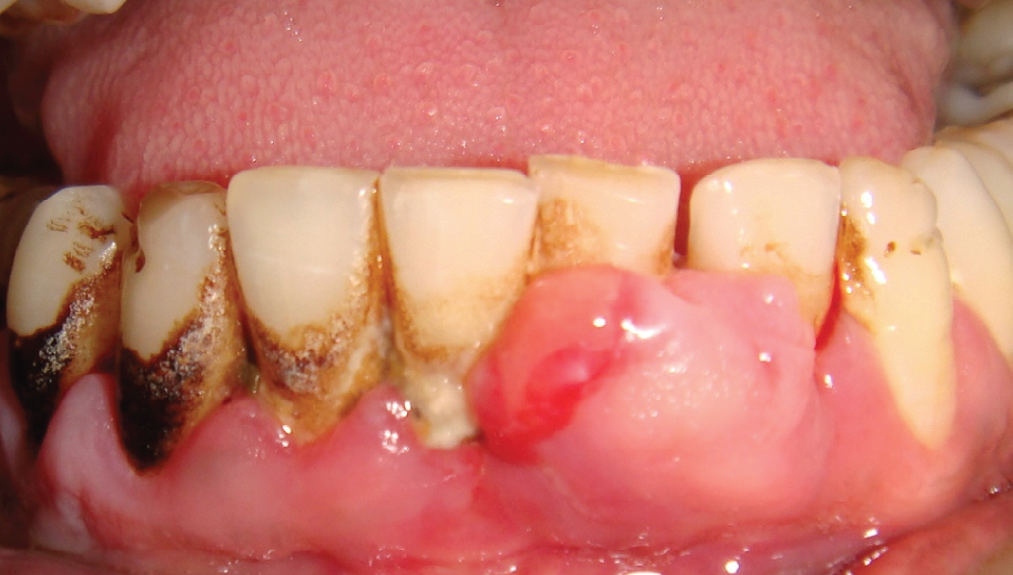
A 27 year male patient, with isolated Miller’s class II gingival recession in 33 reported to the Department of Periodontology, Krishnadevaraya college of dental sciences desiring esthetic improvement. Intra oral examination revealed the presence of 7 mm of gingival recession with mild labial root prominence. The recipient site was prepared over which the lateral donor split thickness flap was displaced and sutured. Post-operative instructions were given. Complete root coverage was seen that was maintained for 2 years.

FREE GINGIVAL GRAFT (FGG):
FGG was introduced by Bjorn in 196319 to increase the width of attached gingiva. FGG is indicated when the palatal tissue facilitates a graft of sufficient thickness (1.5-2.0 mm) and preferred in mandibular anterior teeth and premolars (because of colour mismatch). The disadvantages of FGG are necrosis of the graft with poor recipient site preparation , open palatal wound, scarring occurs with wound healing and graft stability.
CASE REPORT-2
A male patient aged 33 reported with Miller’s class III isolated gingival recession defect in 41. A split thickness flap was prepared in the recipient site and the papillae of 41 were also deepithelialized to facilitate the reception of the graft. Graft was harvested from palatal gingiva adjacent to 24 and 25. Graft was stabilized with sutures. Healing was uneventful. Successful root coverage was seen after 6 months.

CORONALLY ADVANCED FLAP (CAF) WITH SUBEPITHELIAL CONNECTIVE TISSUE GRAFT (SCTG):
Coronally advanced flap (CAF) was first described by Bernimoulin in 1975.7 Subepithelial connective tissue graft (SCTG) was first described by Edel in 1964.20 CAF with SCTG has been the most successful technique forroot coverage.21The advantages of this technique arepossibility of multiple recession defect coverage, non-involvement of adjacent teeth, high success rate and even if this procedure does not work, it does not increase the existing problem.
CASE REPORT-3
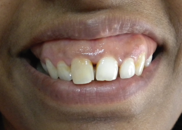
A female patient aged 28 had multiple Miller’s class II recession defects from 12 to 15. Flap was coronally advanced by a full-split thickness flap. Subepithelial connective tissue graft was harvested from palate using trap-door technique. The graft was stabilized using simple interrupted sutures and the flap was coronally advanced and sutured with sling sutures without tension. Healing was uneventful. Complete root coverage was seen after 6 months.

DISCUSSION
The variability in the percentage of sites with complete and mean root coverage is probably associated with a set of factors such as the type of defect, amount and quality of adjacent gingival tissue and the inclusion criteria. Better results in terms of root coverage can be obtained when the recession defects are < 4mm. Smoking affects the results obtained by periodontal plastic surgery procedures.22 Root conditioning has not been found to have a significant clinical benefit in root coverage procedures.23
Coronally advanced flap with subepithelial connective tissue graft has shown the highest success rate (95%) for gingival recession defects.10 Besides the primary objective of attaining root coverage, these procedures invariably increase keratinized tissue width, which in turn facilitates better plaque control.
The results obtained by these procedures as seen in the case reports are also stable for a long period of 2 years. In a long term RCT by Kuis et al (2013)24 results obtained with CAF+ CTG were stable for 60 months.Rossberg et al in 2008 in their case series followed their patients for a longer period (6 to 22 yrs, mean 11 yrs) to find stable results.25
CONCLUSION
Root coverage, on contrary to general opinion of the practitioners is not an unachievable feat. With proper selection of cases, ideal surgical procedures and motivation of patients to maintain good oral hygiene, successful root coverage is attainable with long term stability of results.
REFERENCES
- Newman MG, Takei HH, Klokkevold PR, Carranza FA. Clinical Periodontology. 10th ed. Missouri: Elsevier Saunders; 2006.p.370.
- Sato N. Periodontal Surgery. A Clinical Atlas. Illinois: Quintessence publishing Co, Inc; 2000.p. 340.
- Baldi C, Pini-Prato G, Pagliaro U, et al. Coronally advanced flap procedure for root coverage. Is flap thickness a relevant predictor to achieve root coverage? A 19-case series. J Periodontol 1999;70:1077-1084.
- Grupe HE, Warren RF Jr. Repair of gingival defects by a sliding flap operation. J Periodontol 1956; 27: 92.
- Cohen ES, Ross S. The double papilla flap in periodontal therapy. J Periodontol 1968;39:65-70.
- Pennel BM, Higgison JD, Towner TD. Oblique rotated flap. J Periodontol 1965;36:305-9.
- Bernimoulin JP, Lu¨scher B, Mu¨hlemann HR. Coronally repositioned periodontal flap. Clinical evaluation after one year. J Clin Periodontol 1975;2:1-13.
- Tarnow DP. Semilunar coronally repositioned flap. J Clin Periodontol 1986;13:182-5.
- Paolantonio M, di Murro C, Cattabriga A, Cattabriga M. Subpedicle connective tissue graft versus free gingival graft in the coverage of exposed root surfaces. A 5-year clinical study. J Clin Periodontol 1997;24:51-56.
- Greenwell H. Oral reconstructive and corrective considerations in periodontal therapy. J Periodontol 2005;76:1588-1600.
- Pini Prato G, Tinti C, Vincenzi G, Magnani C, Cortellini P, Clauser C. Guided tissue regeneration versus mucogingival surgery in the treatment of human buccal gingival recession. J Periodontol 1992: 63: 919–928.
- Roccuzzo M, Lungo M, Corrente G, Gandolfo S. Comparative study of a bioresorbable and a non-resorbable membrane in the treatment of human buccal gingival recessions. J Periodontol 1996: 67: 7–14.
- Castellanos AT, de la Rosa MR, de la Garza M, Caffesse RG. Enamel matrix derivative and coronal flaps to cover marginal tissue recessions. J Periodontol 2006: 77: 7–14.
- Keceli HG, Sengun D, Berberog˘lu A, Karabulut E. Use of platelet gel with connective tissue grafts for root coverage: a randomized-controlled trial. J Clin Periodontol 2008: 35: 255–262.
- Harris RJ. Root coverage with a connective tissue with partial thickness double pedicle graft and an acellular dermal matrix graft: a clinical and histological evaluation of a case report. J Periodontol 1998: 69: 1305–1311.
- Rubins RP, Tolmie PN, Corsig KT, Kerr EN, Kim DM. Subepithelial connective tissue graft with growth factor for the treatment of maxillary gingival recession defects.Int J Periodontics Restorative Dent. 2013 Jan-Feb;33(1):43-50.
- Wilson TG Jr, McGuire MK, Nunn ME. Evaluation of the safety and efficacy of periodontal applications of a living tissue-engineered human fibroblast-derived dermal substitute. II. Comparison to the subepithelial connective tissue graft: a randomized controlled feasibility study. J Periodontol 2005: 76: 881–889.
- Ozturan S, Durukan SA, Ozcelik O, Seydaoglu G, Haytac MC. Low-intensity laser therapy. Coronally advanced flap adjunct with low intensity laser therapy: a randomized controlled clinical pilot study.J Clin Periodontol. 2011 Nov;38(11):1055-62.
- Bjorn H. Free transplantation of gingival propria. Sven TandlakTidskr 1963;22:684.
- Edel A. Clinical evaluation of free connective tissue grafts used to increase the width of keratinized gingiva. J Clin Periodontol 1974;1:185-196.
- Chambrone L, Sukekava F, Arau´ jo MG, Pustiglioni FE, Chambrone LA, Lima LA. Root-coverage procedures for the treatment of localized recession-type defects: A Cochrane systematic review. J Periodontol 2010; 81: 452-478.
- Chambrone L, Lima LA, Pustiglioni FE, Chambrone LA. Systematic review of periodontal plastic surgery in the treatment of multiple recession-type defects. J Can Dent Assoc 2009;75:203a-203f.
- Oliveira GH, Muncinelli EA. Efficacy of Root Surface Biomodification in Root Coverage: A Systematic Review. J Can Dent Assoc 2012;78:c122.
- Kuis D, Sciran I, Lajnert V, Snjaric D, Prpic J, Ribaric SP, Bosnjak A. Coronally Advanced Flap Alone or With Connective Tissue Graft in the Treatment of Single Gingival Recession: A Long-Term Randomized Clinical Trial.J Periodontol.In press 2013.
- Rossberg M, Eickholz P, Raetzke P, Kruger PR. Long term results of root coverage with connective tissue in the envelope technique: A report of 20 cases. Int J Periodontics Restorative Dent 2008;28:19-27.
Disclaimer from Guident
The views and opinions expressed in articles published in Guident are solely those of the authors and do not necessarily reflect the views of the editors, the editorial board or the publisher, Ivory India. Guident does not endorse any product, technique or treatment described. Clinical content is intended for qualified dental and medical professionals and is not a substitute for individual clinical judgement. Guident, its editors and the publisher accept no liability for any consequence arising from the use of information published in the journal.
How to cite this article
Dr. Divakaran R, Dr. Joann Pauline George, Dr. Mohammed Azamullah. Root Coverage - a Case Series in the Treatment of Isolated Gingival Recession Defects. Guident. 2013 Nov;6(13). Available from: https://guident.in/articles/periodontics/root-coverage-a-case-series-in-the-treatment-of-isolated-gingival-recession-defects.html