Virtual Articulators: Changing Trends in Prosthodontics
INTRODUCTION
The future of dental practice is closely linked to the utilization of computer-based technology, specifically virtual reality, which allows the dental surgeon to simulate true life situations in patients. In daily practice, mechanical articulators are used to diagnose and simulate the functional effects of malocclusions and morphological alterations upon dental occlusion.However, this mechanical scenario, so very different from the real life biological setting, poses a series of problems.
In effect, the movements reproduced by the mechanical articulator follow the margins of the structures that conform the mechanical joint, which remain invariable over time, and which cannot simulate masticatory movements that are dependent upon the muscle patterns and resilience of the soft tissues and joint disc. Moreover, tooth mobility cannot be simulated by plaster models; as a result, the latter are unable to reproduce the real life dynamic conditions of occlusion.
The virtual articulator offers the possibility of significantly reducing the limitations of mechanical articulators , due to a series of advantages: full analysis can be made of static and dynamic occlusion, of the inter-maxillary relationships, and of the joint conditions, thanks to dynamic visualization in three dimensions (3D) of the mandible, the maxilla or both, and to the possibility of selecting section planes allowing detailed observation of regions of interest such as for example the temporomandibular joint. Combined with CAD/CAM technology, this tool offers great potential in planning dental implants, since it affords greater precision and a lesser duration of treatment.
ADVENT OF VIRTUAL ARTICULATORS
Szentpetery’s virtual articulator: It was introduced by Szentpetery in 1999. It is based on a mathematical simulation of the articulator movements. It is a fully adjustable 3D virtual dental articulator capable of reproducing the movement of an articulator. It offers possibilities that are not offered by some of the mechanical articulators as curved Bennet angle movements which make it more versatile than mechanical articulator.
Virtual articulator of Kordass and Gartner: It was introduced by Kordass and Gartner in 2003, based on the exact registration of mandibular movement with the help of jaw movement analyzer. This virtual articulator system requires digital 3D representation of the jaws as input data generates an animation of the jaw movement and delivers a dynamic and tailored visualization of the collision points.
Virtual articulator based on mechanical dental articulator: It was introduced by graphic design and engineering project developments, the University of the Basque Country in 2009. The project was focused on developing a different virtual articulator based on mechanical dental articulator, knowing which setting parameters can be registered and transferred to the patient. The main advantage of this approach is that the user can choose the most suitable articulator to use in the simulation.
TYPES OF VIRTUAL ARTICULATOR
- Completely Adjustable Virtual Articulator
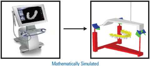
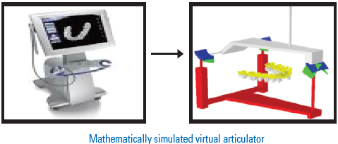
- Mathematically Simulated Virtual Articulator
Completely Adjustable Virtual Articulator:
The ultrasonic measurement system, Jaw Motion Analyzer (Zebris, Germany) is used to record and implement the movement pattern of the mandible Recording the reference plane using special digitizing sensor. Three transmitters are attached to the lower sensor. Four receivers are attached to a head gear opposite the lower sensor . Detection of all rotative and translative components . A special digitizing sensor is used to determine the reference plane, which is composed of the hinge axis infraorbital plane and special points of interest (eg, on the occlusal surface).Movement data is calculated using digitized points. Silicon-based jaw relation registrations are used to reproduce the best occlusion in the position of intercuspidation. It is important that the registration remain attached to the upper teeth during opening. The registration then should be stabilized with impression plaster on metal carrier plate. The digitizing Sensor is attached to detect three main reference points on the rear of this metal plate. These three points are used to combine movement data and the digitized dental arches.
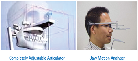
- Silicon-based jaw relation registrations are used to reproduce the best occlusion in the position of intercuspidation. It is important that the registration remain attached to the upper teeth during opening. The registration then should be stabilized with impression plaster on metal carrier plate. The digitizing Sensor is attached to detect three main reference points on the rear of this metal plate.These three points are used to combine movement data and the digitized dental arches. First, the impression of the upper teeth is digitized and then the record material and the plaster of the lower teeth are scanned.Both dental arches are correctly related to each other. The digitized impressions of the lower and upper jaw can be combined with the scanned data from casts without losing the predefined jaw relationship. By defining and calculating the same reference, both data sets, which come from three-dimensional scanner and jaw movement recordings, were matched and presented in the virtual articulator. The program detects the collision points of the teeth.

The contact points are displayed using a different colour (e.g. red). It is able to calculate and visualize static and kinematic occlusal collisions. It is further planned to integrate the system into the design and correction of occlusal surfaces in CAD-systems.
Mathematically simulated virtual articulator
It is a fully adjustable three-dimensional virtual articulator capable of reproducing the movements of an mechanical articulator.It is simple ,adjustment free articulator.It is used as average value articulator
NEED FOR A VIRTUAL ARTICULATOR
In contrast to the conventional mechanical procedure, the VR tools enable three- dimensional navigation through the occlusion based on every point of view while the mandible moves along predefined pathways (as the mechanical articulator would do) or reproduce movement patterns of mastication that never can be simulated in mechanical systems. The digitizing of tooth surfaces opens possibilities in manipulation procedures to improve the occlusion. CAD/ CAM tools for a virtual set- up of the teeth could be linked.
Currently, the virtual articulator is concerned with better visualization of details and supports the use of mechanical tools, but it will replace them in the future. Importantly, it would influence the quality of the networking communication between dental practice and laboratory, helping to produce the best- fitted occlusal restorations possible.
FUNCTIONING OF THE VIRTUAL ARTICULATORS
Ideally, the virtual articulator is equipped with a device for registering the specific mandibular movements of a given patient (such as the JMA), and can integrate the movements recorded in the animation.
If no device is available for registering the mandibular movements, specific movements must be defined by means of parameters, in a way similar to the practice used with mechanical articulators. Some parameters of interest in these cases would be the following: protrusion, retrusion, laterotrusion, and aperture / closure slope. After defining the movement parameters, collision detection is required in order to identify the movement restrictions.
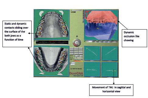
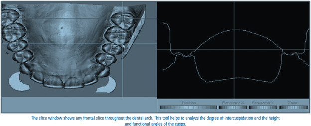
As an example, the software of the Dent-CAM virtual articulator uses three main windows that show the same movement pattern, distinguishing a series of aspects:
a. interpretation window: this shows both maxilla in dynamic occlusion and allows us to obtain unusual points of view, e.g., observation from an occlusal surface of closing of the opposing tooth during mastication;
b. occlusion window: this shows the points of contact that appear on the occlusal surfaces of the upper and lower teeth as a function of time; and,
c. section window: this offers different frontal sections along the dental arch. This tool can be used to analyze the degree of intercuspidation, as well as the height and functional angles of the cuspids.
The latest software versions incorporate an orthodontic module allowing the creation of a virtual setup. The program has also been equipped with the representation of the condylar trajectories in the sagittal and horizontal planes. This tool allows us to observe the inter-relationship between the incisal guide and the condylar guide, and the effects of joint mobility upon occlusion.
One of the most recent new developments in the virtual articulators is the 3D virtual articulation system (Zebris company, D-Isny). This system requires the following:
a. an input unit in the form of a 3D scanner;
b. the software for prosthesis modeling and collision detection, based on a virtual articulator; and,
c. the output module (a rapid prototyping system). With this system, and in addition to mandibular movement, we can analyze masticatory movement – including force at the points of contact and the frequency of contacts in relation to time.
PROGRAMMING
Pre requisite for visualization on screen is 3D scanning/digitizing of tooth surface or restorations or denture models using 3D LASER SCANNER
TWO TYPES Of programming are
- Direct digitizing
- Indirect digitizing

VIRTUAL REALITY DENTCAM
To demonstrate virtual tools in dental articulation, the VR DENT CAM was developed at UNIVERSITY OF GREIFSWALD, GERMANY. It consists of three main windows, which show the same movement of the teeth from different aspects
FUTURE MODULES OF VIRTUAl ARTICULATOR
- In the latest version of DentCAM software, a special orthodontic CAD module was added to simulate the therapeutic result by repositioning single teeth and reforming the dental arch (virtual set-up).
- Separating single teeth from the complete data set of the upper and lower jaw is a prerequisite to the virtual set-up.
- DentCAM includes a pointer, which makes it possible to find the tooth-crown margin automatically when positioned in the middle of the occlusal table of molars or premolars or incisal rim or canine top.
- Continuing tooth by tooth, the procedure results in single-tooth-based data sets that are prepared for special software operations, such as simulation of orthodontic movements.
- Based on the images from the virtual setup tool, an active protrusion splint is included in the treatment plan
- Detection of wear or bruxism, a module semiautomically analyses the teeth for the signs.

CONCLUSION
Virtual reality enables new perspective in visualizing complex relationship in the diagnosis of the occlusion and function. The new virtual articulator provides interesting modules for presenting and analyzing the dynamic contact of the occlusal surface of the maxilla and mandible and the relation to the condylar movement. To improve the functional occlusion, the occlusal profile of the teeth can be designed with increased or decreased cusps to eliminate occlusal interferences of the dynamic pattern. The data set of newly designed animproved occlusal surfaces can be transferred to a milling machine, producing real crowns and fixed restoration with that particular, optimized functional occlusion. The virtuaarticulator is a basic tool that deals primarily with the functional aspects of the occlusion; however, it also can be regarded as a core tool in many diagnostic and therapeuticprocedures and in the CAD-CAM manufacture dental restoration. The concept of Virtual Articulator will change conventional ways of production and communication in dentistry and begin to replace the mechanical tools.
REFERENCES
- The virtual articulator in dentistry :concept and development bekorda,christian gartnerDCNA46(2002)493-506
- Virtual Articulators in prosthodontics Gugwad.R.S, Basavakumar.M, Abhijeet.K, Arvind.M, Sudhindra.M, Ramesh.C :International Journal Of Dental Clinics 2011:3(4):341
- Virtual articulator for the analysis of dental occlusion: An update :Laura Maestre-Ferrín 1, Javier Romero-Millán Med Oral Patol Oral Cir Bucal. 2012 Jan 1;17 (1):e160-3.
Disclaimer from Guident
The views and opinions expressed in articles published in Guident are solely those of the authors and do not necessarily reflect the views of the editors, the editorial board or the publisher, Ivory India. Guident does not endorse any product, technique or treatment described. Clinical content is intended for qualified dental and medical professionals and is not a substitute for individual clinical judgement. Guident, its editors and the publisher accept no liability for any consequence arising from the use of information published in the journal.
How to cite this article
Dr. Manesh Lahori, Dr. Siddharth, Dr. Dipta. S. Chakraborty. Virtual Articulators: Changing Trends in Prosthodontics. Guident. 2013 Jan;6(3). Available from: https://guident.in/articles/prosthodontics/virtual-articulators-changing-trends-in-prosthodontics.html