Scleroderma -Dentist,S Perspective -an Overview
Faculty Of Dentistry, Jamia Milia Islamia New Delhi, India, Department of Oral Medicine & Radiology, Z.A Dental College & Hospitals A.M.U Aligarh, India
Abstract
Scleroderma is a generalized auto-immune collagen disorder, characterized by fibrosis that involves skin, muscles, and other internal organs like the GIT, lungs, blood vessels and Kidneys. Systemic sclerosis can affect the facial and oral structures as well and can present as diagnostic dilemma. Oro-facial and radiographic manifestations include mask like facial appearance, microstomia, restricted mouth opening, xerostomia, periodontal diseases, malocclusion, widened periodontal ligament (PDL) space, pseudo ankylosis, pulpal calcifications, and osseous resorption. Early diagnosis and individually tailored therapy help to manage this disorder which is treatable but not curable. Hereby, presenting a review article on this uncommon connective tissue disorder with emphasis on dental findings and management.
Keywords: Connective tissue disorders, Scleroderma, Restricted mouth opening, Widened PDL space.
INTRODUCTION
Scleroderma (Gr. skleros , hard, and derma, skin) is a generic term, used to describe both systemic as well as more localized cutaneous disorders. When the systemic nature of the disease became evident, the name progressive systemic sclerosis was proposed.1 Systemic sclerosis is a chronic inflammatory disease of unknown origin and autoimmune nature characterized by excessive deposition of collagen and glycosaminoglycans in the connective tissue of the dermis and internal organs. 2,3 Though the term scleroderma is indelibly etched in the literature through common usage, the disease is currently called as “ systemic sclerosis ”. Since hidebound skin is the clinical hallmark of the disease, it is also called “ hidebound disease”.It is a disease of low incidence, with an average of 4 to 19 new cases per million inhabitants and preferentially affects females.2 The age group most affected is between the third and fifth decade of life.4 Earlier genetic, environmental, autoimmune, vascular, nervous factors were all proposed to be involved in the etiology of the disease.5,6,7 But later on, only vascular and environmental factors are found to play a major role in the isolated cases of this disease. The disease can occur in three forms: (a) morphea (circumscribed scleroderma), characterized by local thickening of the skin; (b) generalized or progressive scleroderma (diffuse form), characterized by tautness of the skin with distinctive involvement of the lungs, heart, kidneys, and gastrointestinal tract, and osteolytic changes in the skeleton; and (c) acrosclerosis, a combination of scleroderma of the extremities and Raynaud’s disease.8
A variant of this disease is known as the “CREST SYNDROME” , which is an acronym for calcinosis cutis, Raynauds phenomenon, Esophageal dysmotility with dysphagia, Sclerodactly and Telangiectases.9
The disease presents a variety of clinical features. Raynauds phenomenon is usually its first manifestation, and skin tightening, eosophageal dysmotility, restrictive pulmonary disease, pulmonary hypertension, arthralgia, myopathy, myocardiopathy and progressive renal insufficiency are also observed.2,3,4 The involvement of the skin together with the quality of its mobility, particularly in the distal portions of the extremities is by far the most obvious symptom. Cutaneous manifestations include thickening of skin, starting withpitting edema and over several months pitting edema is replaced by tightening and hardening of skin.
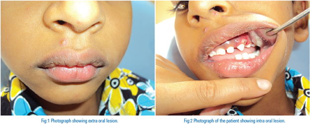
Oral and facial tissues are often affected, presenting characteristic features. Oral manifestations of progressive systemic sclerosis (PSS) may include limited ability to open the mouth; xerostomia; periodontal disease; increased periodontal ligament (PDL) width; and osseous resorption of the mandible. 10 Facial skin becomes thin and taut, which leads to a mask-like appearance.11 The most common oral radiographic manifestation of PSS, which occurs in about two-thirds of patients, is an increase in the width of the PDL around the teeth.12 Widening of the PDL affects both anterior and posterior teeth, although it is more pronounced around the posterior teeth. Despite the widening of the PDL space, the lamina dura remains normal, the teeth are often not mobile and their gingival attachments are usually intact.
Histologic features of the gingiva reveal thickening and hyalinization of the collagen fibers, atrophy of the gingiva with loss of rete pegs and sclerosis of the walls of the blood vessels. The microscopic changes in the periodontal ligament consist of a widening as well as an appearance of hyalinization with diminution in the number of connective tissue cells usually found.13
The clinical sign of stiffened skin texture is suggestive of the diagnosis and a skin biopsy may confirm the diagnosis.14 Several serological tests are also useful for confirmation of the diagnosis and for classification.15
DISCUSSION
Scleroderma is a chronic disease of unknown etiology that affects the microvasculature and loose connective tissue. It is characterized clinically by fibrous deposition and obliteration of vessels in the skin, lungs, gastrointestinal tract, kidneys, and heart.16 Scleroderma literally means “hard skin” and was first reported by William and Robert Watson in 1754. The diffuse thickening and induration of the skin in the systemic form of scleroderma, systemic sclerosis (systemic scleroderma, SSc) is accompanied by fibrosis and vascular obliteration of internal organs. Its course is often progressive and fatal. The localized form has a more restricted pattern of involvement and doesnot affect internal organs.
HISTORICAL BACKGROUND
Hippocrates (460-377 BC) was the first to describe a skin disorder compatible to scleroderma but due to a rather imprecise description this report can not be relied upon.17 The first convincing description of the disease appeared in a monography written by Carlo Curizo, in Naples in 1753. He reported on the hardness in the skin of a 17 year old woman.18 Goetz 1 proposed the name progressive systemic sclerosis in 1945, when the systemic nature of the disease was proven. The name has though been replaced by systemic sclerosis,since not all cases progress, and also to diminish the psychological impact of the diagnosis.19
PATHOGENESIS
The pathogenesis of SSc involves three findings: a. Vascular dysfunction that manifests as injury to the endothelial cells; b. Immunological activation of T cells, cytokines and inflammation; and c. Fibrosis. Inflammation manifests initially as a perivascular macrophage infiltrate.20,21,22 Injury to the endothelial cells causes prominent thickening of vessels and two of the most serious complications, pulmonary arterial hypertension and renal crises.23 Increased deposition of fibroblasts causes destruction of normal tissues which forms the basis for the tissue and organ dysfunction that occurs in the skin, lungs, kidneys, and GI tract.24 All three disease pathways are most likely linked. Research suggests thatfibroblasts and myofibroblasts that over produce collagen and other extra cellular matrix products are stimulated via the by-products of the functional vasculopathy. B cells also are involved in the pathogenesis as there are multiple scleroderma-specific antibodies; however, no correlation with specific pathological damage has been found.
CLASSIFICATION
Localised type of scleroderma may occur either as circumscribed morphea form or as linear coup de sabre form. 25 In the former type, there are white / yellowish cutaneous patches surrounded by violaceous halo of varying size and shape. In the latter form, linear ribbons that resemble marks produced by blow of a saber are seen. Lesions are mostly asymptomatic.
Systemic scleroderma is divided into two distinct subsets: limited SSc (ISSc) and diffuse SSc (dSSc).26 Sixty percent of patients with SSc are in the ISSc group, which includes individuals with the CREST syndrome, so called for its features of calcinosis cutis, Raynaud’s phenomenon, esophageal dysfunction, sclerodactyly, and telangiectasia. Patients with ISSc typically are women who are older than patients with dSSc and have a long history (10 to 15 years) of Raynaud’s phenomenon, skin involvement limited to the digits or hands, face, feet, and forearms, nail fold capillary dilatation; and early onset of facial and digital telangiectasias. Anticentromere antibodies (ACA) have been identified in 50to 96 % of patients with CREST syndrome, but in only 12 to 25 % of patients with dSSc. Systemic involvement (notably pulmonary hypertension) may not appear for years (often decades), and many patients outlive their disease and die of other causes. The typical patient with dSSc is well until the abrupt onset of swelling (non-pitting) of the hands and feet associated with Raynaud’s phenomenon and hidebound changes in the skin, often sparing only the back and buttocks. Polyarticular symmetric synovitis, tenosynovitis, and tendon friction rubs are also present. Nail fold capillary dilatation and destruction are common, and there is early onset of internal organ involvement. In some patients, skin involvement progresses rapidly initially and then subsides over years.16
CLINICAL MANIFESTATIONS
The clinical manifestations of SSc depend on the sites involved. The initial complaints are usually related either to Raynaud’s phenomenon or to chronic, usually non-pitting edema of the hands and fingers. As many as one-thirds of patients first have pain and stiffness of the fingers and knees. In some patients, the first manifestation is active, often migratory, polyarthritis. In others, severe erosive digital osteo-arthritis (related to CREST syndrome, particularly in females) occurs first. Flexion contractures and sclerodactyly are present; on x-ray, digital tuft resorption, subcutaneous calcification, joint space narrowing, and focal erosion of periarticular bone are seen. Usually, skin changes precede visceral involvement by several years, but occasionally this order is reversed.27
Raynaud’s phenomenon, defined as episodic vasoconstriction in the fingers and toes, develops in virtually every patient with SSc. In some, episodes may also affect the tip of the nose and earlobes. Attacks are trigerred by exposure to cold, a decrease in temperature, emotional stress, and vibration. Typical attacks start with pallor, followed by cyanosis of variable duration. Eventually erythema develops spontaneously or with rewarming of the digit. The progression of the three color phases reflects the underlying pathogenic mechanisms of vasoconstriction, ischaemia, and reperfusion. Some patients with Raynaud’s phenomenon may experience only pallor or cyanosis. As much as 3- 5% of the general population has Raynaud’s phenomenon, and it is more frequent in women. In the absence of associated signs and symptoms of an underlying condition, Raynaud’s phenomenon is classified as primary, and represents an exaggerated physiological response to cold. Secondary Raynaud’s phenomenon occurs as a complication of SSc and other connective tissue diseases, hematological and endocrine conditions, and occupational disorders, as well as in association with the use of beta blockers such as atenolol, anticancer drugs such as cisplastin and bleomycin and a variety of other medications. Distinguishing primary versus secondary Raynaud’s phenomenon can present a diagnostic challenge. The lack of an underlying cause for Raynaud’s phenomenon on the basis of the history and physical examination; a positive family history of Raynaud’s phenomenon; absence of digital tissue necrosis, ulceration, or gangrene; and a negative test for anti nuclear antibodies supports the diagnosis of primary Raynaud’s phenomenon. Secondary Raynaud’s phenomenon tends to develop at an older age ( ≥ 30 years), is clinically more severe ( episodes more frequent, prolonged, and painful), and is frequently associated with ischaemic lesions and infarctions in the digits. Nail fold microscopy may be useful in Raynaud’s phenomenon; patients with primary Raynauds phenomenon have normal capillaries that appear as regularly spaced parallel vascular loops, whereas in SSc and other connective tissue diseases, nailfold capillaries are distorted with widened and irregular loops, dilated lumen, and areas of vascular “dropout”.28
The disease then extends to involve the upper extremities, trunk, face, and, finally, the lower extremities, which may sometimes be spared. In the early stages, the painless, slightly pitting edema often lasts several months before tightening of the skin occurs. Isolated periorbital edema may occur in the early edematous phase in patients with few other manifestations of SSc. Often the skin feels indurated and stiff; as it progresses to the atrophic state, it becomes tense, smooth, hardened, and eventually, firmly bound to the underlying structures. The skin of the face becomes mask like and expressionless, with loss of the normal facial lines and then thinning of the lips and constriction of the opening of the mouth (microstomia). Radial furrowing around the mouth is seen. Uncommonly, the mucous membranes are involved, with painful induration of the gums and tongue. A prominent feature is tightening of the skin over the nose, giving it a small, sharp appearance. The neck sign, ridging and tightening of the skin of the neck on extending the head is common. Mat like telangiectases may develop, especially about the face and upper trunk. There may be thinning or complete loss of hair and anhidrosis in the affected areas. Generalised hyperpigmentation, similar to that of Addison’s disease but with no evidence of adrenal insufficiency or elevated plasma levels of melanocyte-stimulating hormone , can occur and may antedate the sclerosis.
Sclerodactyly causes the fingers to become tapered, with marked skin atrophy. As in systemic lupus erythematosus and dermatomyositis, periungual telengiectasia occurs. Enlarged, dilated nail fold capillaries forming “giant” or sausage-shaped loops can be seen by capillary microscopy of nail folds, and this may be useful in diagnosis.27 Another common problem is recurrent painful ulceration of the fingertips. Slow-healing ulcers over the knuckles may also occur. These may heal with stellate scarring, become chronic, or rarely,become gangrenous. Flexion contractures of the stiff fingers trouble many patients. Resorption of bone may cause dissolution of terminal phalanges, and cutaneous calcifications develop in some cases, especially around fingertips and bony prominences but also in any area involved with scleroderma. These calcifications may ulcerate, extrude calcified material, and re-epithelialize very slowly.
Gastrointestinal manifestations are common in systemic sclerosis, and the most common is esophageal dysfunction. Dysphagia,manifested by various abnormal swallowing sensation, is initially caused by impaired esophageal motility but later can result from gastroesophageal reflux disease and secondary stricture formation. Barrett’s esophagus occurs in 1/3 of patients with scleroderma; and these patients have an increased risk of complications. (stricture, adenocarcinoma).28,29
Pulmonary manifestations of systemic sclerosis include interstitial lung disease, pulmonary hypertension, pleuritis and pleural effusion, and aspiration pneumonia.30 Dyspnea and non productive cough in a patient with systemic sclerosis should raise the possibility of lung disease, and a work up for interstitial lung disease should be performed. However, chronic cough may be the only sign of pulmonary disease in systemic sclerosis.29
Clinical evidence of myocardial involvement in scleroderma is uncommon, but such involvement is more frequent in patients with diffuse scleroderma. The clinical presentation of cardiac involvement
can include pericarditis, conduction problems, and congestive heart failure. Patchy replacement of the myocardium and the conduction system by the fibrous tissue occurs in most patients.31
Scleroderma renal crisis is characterized by an abrupt rise in blood pressure over days to weeks and rapidly progressive renal failure if untreated, usually within the first 5 years of the disease.32 The spectrum of presentation varies from normal or mildly elevated blood pressure to malignant hypertension, causing elevated rennin plasma levels, elevated serum creatinine (seen in 50% of the patients), proteinuria, and microangiopathic hemolytic anemia (seen in 50% of patients).33
ORAL MANIFESTATIONS
The most frequent oral finding to precede systemic involvement appears to be trigeminal neuropathy followed by enlargement of the periodontal ligament (PDL) space. 10,34 The trigeminal neuropathy is characterized by slow and gradual facial muscle inactivity followed by pain, sometimes severe, and parasthesia. The neuropathy is associated with deposition of collagen in the perineurium and or reduced vascularity to the trigeminal nerve itself.35,36 Another oral finding of significance is the increased risk of oral cancer that has been reported for some individuals with scleroderma. In particular, a recent study found an increased risk of squamous cell carcinoma of the tongue. 37 Hence, providing regular and thorough oral cancer examinations is extremely important in patients with SSc. Of particular importance to oral healthcare professionals is the skin involvement affecting the hands and oral facial structures. One of the most prevalent oral findings is the excess collagen deposition affecting the peri-oral structures.38 Many of the debilitating resultant functional problems relating to scleroderma result from microstomia. Due to the loss of elasticity and subsequent tightening of the lips and checks, both self-care and professional care can bechallenging. Due to the fibrosis and atrophy of major and minor salivary glands, patients with SSc experience a high incidence of xero- stomia. The loss of salivary function is significant and similar to that afflicting patients after radiation therapy to the head and neck.39 Xerostomia adds other complications to the patients, diminishing quality of life by increasing the risk of caries, periodontal disease and fungal infections. Nutritional status, dietary habits, swallowing, taste, speech and tolerance to dental appliances may be adverselyaffected. A combination of oesophageal neural damage, vascular changes and fibrosis causes gastrointestinal reflux (GERD) and affects approximately 90% of SSc patients.22 Due to the acid from reflux, perimylolysis tooth erosion may occur resulting in hypersensitivity.10,36 The above mentioned microstomia and xerostomia often resultin an elevated plaque index. This elevated plaque index of course resulting in gingival inflammation, gingivitis. Accordingly gingival and even mobility indexes has shown significantly higher in patients with SSc. It is believed that the decreased vascularity and tissue ischemia may cause the increased periodontal disease and following tooth mobility. Patients often show signs of gingival recession and stripping of the buccal attached gingival tissues.40,41
RADIOGRAPHIC FEATURES
Extreme widening of periodontal ligament space i.e, two to four times the normal thickness is a characterstic feature of scleroderma.40 Stafne and Austin 42 first described this finding and noted an incidence of about 7%. They mentioned that the posterior teeth are involved more frequently than the anterior teeth. Other authors have confirmed both this incidencand more involvement of the posterior teeth. Auluck suggested that PDL widening is caused by involvement of the masticatory muscle, which becomes more bulky and leads to an increase in masticatory occlusal forces that may result in primary trauma from occlusion. Hence, widening of the PDL in patients with PSS occurred without noticeable deposits of plaqueor calculus.43 However, Mehra found that atrophy of the muscles decreases the forces of mastication and thus the chances of trauma from occlusion. In his opinion, it seems more likely that increased collagen synthesis, which leads to an increase in PDL bulk, explains the aetiology of PDL widening in these patients.44 Regarding the latter hypothesis, however, it is difficult to explain why PDL widening does not affect all patients with PSS and why it occurs more often in the posterior teeth in some patients. A number of authors have reported the presence of PDL widening in only some of the teeth in certain scleroderma patients.40
A radiographic feature in some cases of PSS includes typically bilateral, sharply demarcated and relatively symmetric mandibular erosions at regions of muscle attachment, such as the angles, coronoid process, digastric region or condyles. This resorption may be progressive in the course of the disease. 12 Mandibular resoption in scleroderma was first reported by Taveras in 1959.45 Auluck pointed out that these erosions are caused by exerting pressure on the bone via the atrophic muscles at their attachment site. Atrophy of the muscles is attributable to increased fibrosis of the muscles and a decrease in the vascularity of the muscles, secondary to the fibrosis of muscular walls of the arteries.43 Other authors have mentioned that mandibular resorption can also be caused by ischaemia of the bone secondary to vasculitis associated with PSS. In addition, the connective tissue abnormality associated with the disease can cause entrapment of the blood vessels.46 Ramon et al47 suggested that the condyle, coronoid and mandibular angles, areas that show resorption, are supplied by small arterial branches of the internal maxillary artery rather than the main inferior alveolar artery. PSS may be more likely to affect these smaller arterial branches rather than larger arteries, thus leading to bone resorption secondary to diminished vascularity in those areas.47 Tightness of the skin, which may lead to pressure resorption of the bone, can be another factor in mandibular resorption.46 Overall, it seems that bone resorption in systemic sclerosis has a multifactorial aetiology. However, root resorption may also be a feature in some scleroderma patients. This unusual observed phenomenon, i.e., resorption of the teeth root apices, can be accounted for by: (a) decrease of the oxygen tension in the environment; (b) the immunologic reactions which may take place in the course of the disease.48
DIFFERENTIAL DIAGNOSIS
Widening of the PDL may also occur in malignancies such as osteosarcoma and trauma from occlusion. In malignancy, the lamina dura does not remain intact, and PDL widening takes place in the teeth located around the lesion. Conversely, the lamina dura remains intact in PSS, and PDL widening occurs in more than one quadrant and usually in the posterior teeth.12 In trauma from occlusion, PDL widening occurs but in association with angular bone defects and mobility of teeth, conditions that are not seen in patients with systemic sclerosis.44 However, some authors have suggested that PDL widening can be localized or generalized in the repair phase of trauma from occlusion and only in advanced stages will there be bone loss or tooth resorption. Therefore, it may be the case that tooth mobility and bone loss do not always occur.
TREATMENT
The treatment of scleroderma patients depend on the extent and severity of skin and organ involvement. D-penicillamine, a drug effective for both rheumatoid arthritis and wilson’s disease, has shown promise in the management of the condition by decreasing both skin thickening and organ involvement. This drug has two mechanisms of action: interferes withcross linking of collagen and immunosuppression. Nifedipine is a calcium channel blocker that has been shown to be effective in managing Raynaud’s phenomenonand myocardial perfusion. Extra-corporeal chemotherapy has also shown promise in reversing cutaneous sclerosis in patients in early stages of scleroderma.31
DENTAL CONSIDERATIONS
The most common problem in the dental treatment of scleroderma patients is the physical limitation caused by the narrowing of the oral aperture and rigidity of the tongue. Procedures such as molar endodontics, prosthetics, and restorative procedures in the posterior portions of the mouth become difficult, and the dental treatment plan may sometimes need to be altered because of the physical problem of access. The oral opening may be increased an average of 5mm by stretching exercises. One particularly effective technique is the use of an increasing number of tongue blades between the posterior teeth to stretch the facial tissues. In addition, mechanical devices that assist the patient in performing the stretching exercises are available. If these approaches are insufficient, a bilateral commissurotomy may be necessary.
When treating a patient with diffuse scleroderma, the extent of the heart, lung, or kidney involvement should be considered, and appropriate alterations should be made before, during, and after treatment.
Patients with extensive resorption of the angle of the mandible are at risk for developing pathologic fractures from minor trauma, including dental extractions. Patients with Sjogren’s syndrome should have daily fluoride treatments and make frequent visits to the oral hygienist.31
REFERENCES
- Goetz R. Pathology of progressive systemic sclerosis with special reference to changes in the viscera. Clin Proc 1945;4:337-392
- Kayser C, Andrade LEC. Esclerose Sistêmica. In: Sato E. Guias de Medicina Ambulatorial e Hospitalar – Reumatologia. São Paulo: Manole; 2004. p. 111-20.
- Mayes MD, Lacey Jr JV, Beebe-Dimmer J, Gillespie B, Cooper B, Timothy JL et al. Prevalence, incidence, survival and disease characteristics of systemic sclerosis in a large US population. Arthritis and Rheum. 2003; 48(8): 2246-55.
- Marques Neto JF, Sampaio-Barros PD. Reumatologia: diagnostic e tratamento. Rio de Janeiro: Medsi; 2000. p. 465-80.
- Korn JH. Immunologic aspects of Scleroderma. Curr Opin Rheumatol 1990; 2; 922-928
- Welsh KL, Briggs DC. Genetic and environmental factors in scleroderma. Curr Opin Rheumatol 1990; 2; 920-1
- Wilson RH, Mc Cornick WE, Tatum CF, Creech JL. Occupational acroosteolysis. Report of 31 cases. J. Am Med Assn 1967; 201; 577- 81
- Alexandridis C, White SC. Periodontal ligament changes in patients with progressive systemic sclerosis. Oral Surg Oral Med Oral Pathol 1984; 58: 113–118.
- Veloyos EE, Masi AT, Stevens MB, Shulman LE. The CREST syndrome: Comparison with systemic sclerosis. (scleroderma) Arch Intern Med 1979; 139; 1240-5
- Wood RE, Lee P. Analysis of the oral manifestations of systemic sclerosis (scleroderma). Oral Surg Oral Med Oral Pathol 1988; 65: 172–178.
- Benetti R, Zupi A, Toffanin A. Prosthetic rehabilitation for a patient with microstomia: A clinical report. J Prosthet Dent 2004; 92: 322– 327.
- White SC, Pharoah MJ, editors. Oral radiology—principles and interpretation, 6th edition. Saint Louis: Mosby, 2009, p 467
- Shafer WG, Hine MK, Levy BM. A textbook of oral pathology. 4th ed. Philadelphia:WB Saunders Co, 1974; 789-90.
- Neville BW, Damm DD, Allen CM, Bouquot JE. oral and maxillofacial pathology (3rd edn). St Louis: Elsevier/Saunders, 2009, p 799.
- Chung L. Lin J, Furst E, Florentino D. Systemic and localized scleroderma. Clin Dermatol 2006; 24: 374–392.
- Hawk A,English JC. Localised and systemic scleroderma. Seminar Cutan Med Surger 2001: 20: 27
- Rodnan GP. Benedek TG. Medsger TA. Cammarata PJ: The association of progressive systemic sclerosis with coal miners pneumoconiosis and other forms of silicosis. Ann Intern Med 1967;66:323-33
Disclaimer from Guident
The views and opinions expressed in articles published in Guident are solely those of the authors and do not necessarily reflect the views of the editors, the editorial board or the publisher, Ivory India. Guident does not endorse any product, technique or treatment described. Clinical content is intended for qualified dental and medical professionals and is not a substitute for individual clinical judgement. Guident, its editors and the publisher accept no liability for any consequence arising from the use of information published in the journal.
How to cite this article
Dr. Shamimul Hasan, Dr. Sarah Asif, Dr. Nabeel Ishrat Khan. Scleroderma -Dentist,S Perspective -an Overview. Guident. 2013 Jan;6(3). Available from: https://guident.in/articles/oral-medicine/scleroderma-dentists-perspective-an-overview.html