Fibrous Dysplasia - a Rare Case Report with Review of Literature
Department of Oral Medicine and Radiology Sri Venkateswara Dental College and Hospital, Thalambur, Chennai, India
ABSTRACT
Fibrous dysplasia, a benign fibro osseous condition involving one or more bones of the cranial and extra cranial skeleton, consists of non-encapsulated lesion which shows replacement of a normal bone by cellular fibrous tissue containing islands of metastatic bone (Edwards 1984). It has been classified by W.H.O. as developmental in origin. Fibrous dysplasia of bone is one of the most perplexing diseases of the osseous tissue. It is a lesion of unknown etiology, uncertain pathogenesis and diverse histopathology. A case report of fibrous dysplasia is presented here and discussed in detail.
KEY WORDS: Benign fibro osseous lesion, nonencapsulated, cellular fibrous tissue, granular or ground glass pattern.
INTRODUCTION
Fibrous dysplasia (FD) is a benign disease of bone that was originally described by Lichtenstein more than sixty years ago(1,2). Although its actual incidence is not known, it accounts for between 2.5 and 10 percent of all bone tumors, thus a relatively common disease. FD is a developmental dysplastic disorder of bone in which the normal bone matrix is replaced by fibroblastic proliferation. Lesions contain irregular trabeculae of partially calcified osteoid.
Its etiology has been linked with a mutation in the Gs ? gene located at chromosome 20q13.2–13.3(4,5). The mutation was first identified in patients with McCune-Albright syndrome but later observed in monostotic and polyostotic FD cases. Clinical presentation varies depending on mutation timing and distribution(6).
CASE REPORT
A 13-year-old male patient reported with swelling on the left side of the face (Fig 1) and upper posterior teeth region for 1 year. The swelling gradually increased without pain or paresthesia.

FIG 1: EXTRAORAL VIEW SHOWING FACIAL SWELLING.
Intraoral examination showed swelling in relation to 24–27 region.
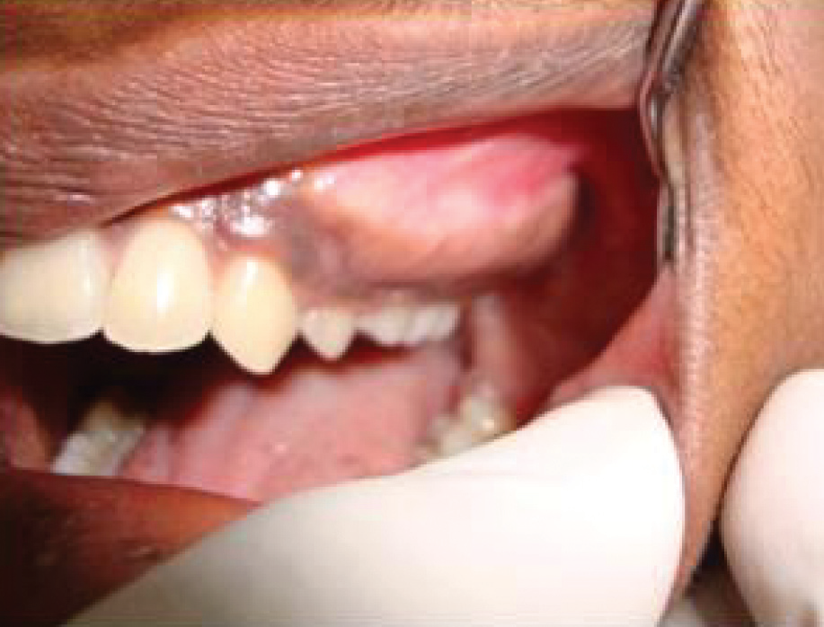
FIG 2: INTRAORAL VIEW SHOWING SWELLING IN LEFT MAXILLA.
Radiographic examination revealed mixed radiolucent-radiopaque lesions with ground glass appearance.
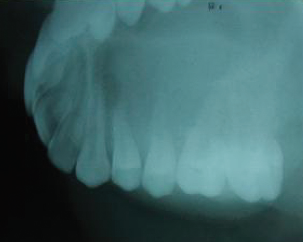
FIG 3: OCCLUSAL RADIOGRAPH SHOWING GRANULAR PATTERN.

FIG 4: OPG SHOWING DIFFUSE RADIO-OPACITY.
Histopathological findings confirmed fibrous dysplasia.

FIG 5: HISTOPATHOLOGICAL FEATURES.
Patient was advised regular follow-up every 6 months.
DISCUSSION
Craniofacial FD presents typically around age 10 and progresses during adolescence(7). Clinical presentation varies from mild swelling to severe deformity(9).
Complications include optic nerve compression, visual loss, and cystic degeneration(10–14). Management depends on severity and progression.
DIAGNOSIS / EVALUATION
Diagnosis requires correlation of clinical, radiographic, and histological findings(7). CT imaging is superior for extent evaluation(19), while MRI may sometimes mislead(24).
Biochemical markers like serum alkaline phosphatase help in monitoring disease progression.
TREATMENT
Treatment options include conservative contouring or radical excision depending on severity(7). Reconstruction may involve bone grafts or microvascular flaps.
Medical treatment includes steroids and bisphosphonates like pamidronate(47).
CONCLUSION
Fibro-osseous lesions require careful diagnosis using clinical, radiographic, and histological correlation. Biopsy remains essential for definitive diagnosis. Proper differentiation from similar conditions is crucial for effective management.
Disclaimer from Guident
The views and opinions expressed in articles published in Guident are solely those of the authors and do not necessarily reflect the views of the editors, the editorial board or the publisher, Ivory India. Guident does not endorse any product, technique or treatment described. Clinical content is intended for qualified dental and medical professionals and is not a substitute for individual clinical judgement. Guident, its editors and the publisher accept no liability for any consequence arising from the use of information published in the journal.
How to cite this article
Dr. P. E. Chandra Mouli, Dr. S.Parthiban. Fibrous Dysplasia - a Rare Case Report with Review of Literature. Guident. 2013 Mar;6(5). Available from: https://guident.in/articles/oral-medicine/fibrous-dysplasia-a-rare-case-report-with-review-of-literature.html