Micro-Endodontic Management of Middle Mesial Canal in Mandibular Molar
Department of Conservative Dentistry and Endodontics Centre for Dental Education and Research, All India Institute of Medical Sciences, New Delhi., India
Abstract
The root canal treatment of mandibular molar can be technically challenging as the management of complex anatomy requires a lot of understanding, patience and perseverance. The present case report describes the successful orthograde endodontic management of carious exposed mandibular first molar with three mesial canals. This paper also highlights the importance of detecting aberrant root canal with microultrasonics which would otherwise be left with unaided eyes thus jeopardizing the successful outcome.
Keywords : Middle mesial canal, Microultrasonics, Magnification
INTRODUCTION
The contemporary approach to manage aberrant root canal complexities often leads to surgical intervention or orthograde retreatment on follow ups. The enhanced magnification with loupes or use of microultrasonics invariably leads to better resolution of periradicular lesions with complete three dimensional reconstruction of the PDL entity in the apical region. 21 st century technologies intended for endodontic treatment has been focused largely on improving the quality of treatment by improving clinicians’ ability to detect root canals[1, 2] particularly in teeth in which accessory canals are present. Several investigations have supported the advantage of the microscope over the use of no magniï¬cation.[3, 4] One of the teeth with a complex root canal system is the mandibular molar. The mesial root in mandibular molars is commonly considered to have two canals[5, 6] with an isthmus in between.[7, 8] Within this system, the presence of an accessory mesial canal has been identiï¬edwith a prevalence ranging from 0% to 17% [9, 10, 11,12] Apart from the prevalence, the ability to negotiate the detected accessory mesial canals, including the troughing depth required to enable negotiation, has not been well characterized, hence an astute clinician should have thorough knowledge of aberrant anatomy, advanced diagnostic and treatment modalities with the recent modifications in dental materials with regards to biocompatibility because every root canal poses a new challenge to endodontists. Numerous studies describe the morphology of mandibular molars. Pineda and Kuttler, and Vertucci[13] have all reported on the presence of three or four canals in mandibular molars. In 1974, Vertucci and William, as well as Barker et al.[14, 15] described the presence of an independent middle mesial canal. The incidence of third mesial canal varies from 1 to 15 % in mandibular molars. There have been multiple case reports of
aberrant canal morphology of the mandibular first molar,[16] hence in each case care should be given for search of additional canal in the mesial root of the mandibular first molar But presence of such complexity in mandibular second molar should not be overlooked. This present case reports describes the presence of third mesial canal in mandibular first and second molars and its successful negotiation with microultrasonics for a successful outcome of root canal therapy.
CASE REPORT
A 24-year-old male reported to the department of Conservative Dentistry and Endodontics, with a chief complaint of pain in the lower left posterior teeth since 2 months. The patient’s medical history was non-contributory. Extraoral examination did not reveal any significant changes. Clinical examination revealed the decayed mesial surface in the left mandibular first molar (tooth #36) with no sinus or fistula. There was tenderness to palpation and vertical percussion, but the tooth mobility was within normal physiological limits. Thermal testing elicited a delayed and prolonged response in tooth #36. The pre-operative radiograph of tooth #36 taken from 20 degrees mesial angulation showed the presence of two roots with widening of the periodontal ligament space in relation to the mesial and distal root apex Fig 1. By clinical and radiographic findings, a diagnosis of irreversible pulpitis with acute apical periodontitis with tooth #36 was made. Tooth #36 was anesthetized by using 1.8 mL (30 mg) of 2% lidocaine containing 1:200,000 epinephrine (Xylocaine; AstraZeneca Pharma Ind Ltd., Bangalore, India) under rubber dam isolation. With the use of microscope (Carl Zeiss Surgical GmbH, Oberkochen, Germany) endodontic access opening was established in tooth #36. The pulp chamber floor was then explored with DG 16 explorer and Mesiobuccal, Mesiolingual, distobuccal and distolingual canals were then located. After access refinent

Fig 1: Preoperative Intraoral periapical radiograph
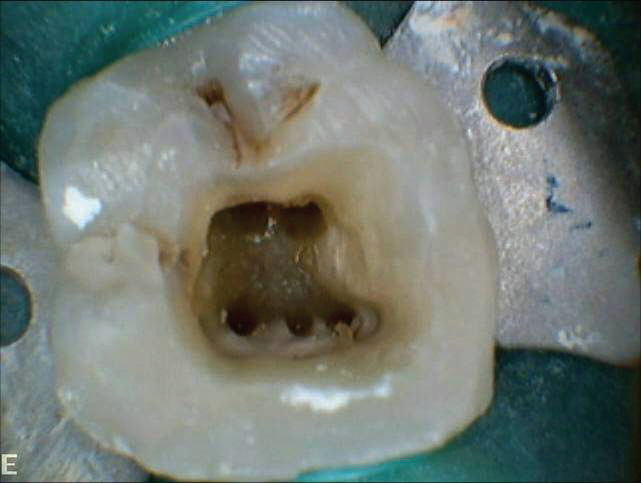
Fig 2: Intraoral photograph showing 5 canals
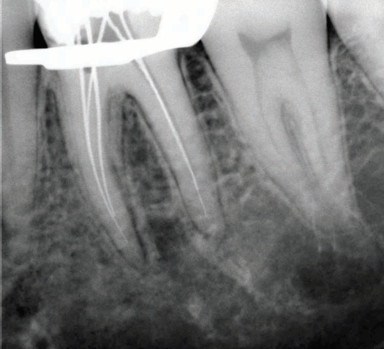
Fig 3: Working length determination
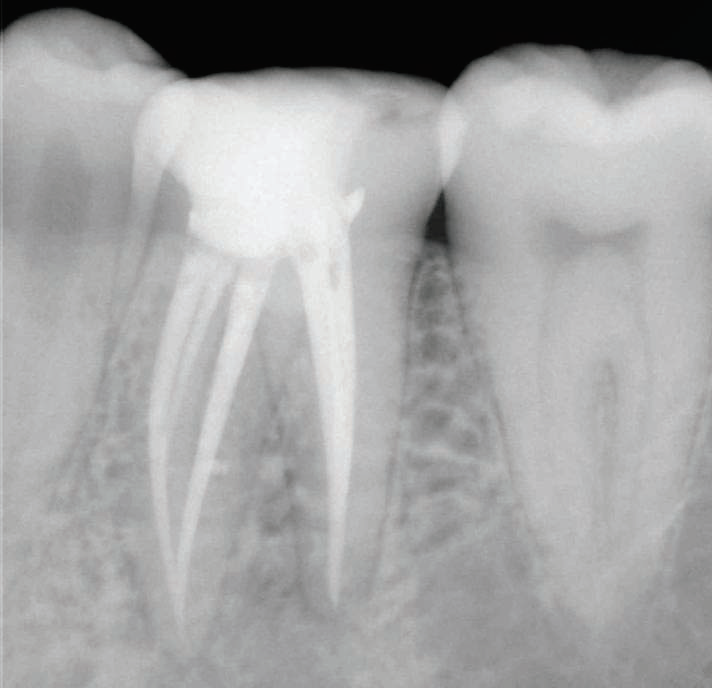
Fig 4: Post operative Intraoral periapical radiograph 7 month
with ultrasonic tips 1 and 3 (Pro Ultra Endo Tips, Dentsply Maillefer, New York, America) a hemorrhagic point was noted approximately in between the orifices of mesial canals and a middle mesial canal was also located in tooth #36 Fig 2.Coronal enlargement was done with a nickel–titanium (NiTi) ProTaper SX rotary file (Dentsply Maillefer, Ballaigues, Switzerland) to improve the straight-line access. Working length was determined with the help of an apex locator (Root ZX; Morita, Tokyo, Japan) and later confirmed by using a radiograph Fig 3. Cleaning and shaping was performed under rubber dam isolation by using ProTaper NiTi rotary instruments (Dentsply Maillefer) with a standardized technique. Irrigation was performed using normal saline, 5.25% sodium hypochlorite solution and 17% ethylenediaminetetraacetic acid. Final rinse was carried out with 2% chlorhexidine solution after saline irrigation. After completion of cleaning and shaping, the root canals were dried with absorbent points (Dentsply Maillefer). Calcium hydroxide (Calcicur; VOCO, Cuxhaven, Germany) was placed as an intracanal medicament with a lentulo spiral (Dentsply Maillefer) for 1 week and the access cavity was sealed with Cavit (3M ESPE Dental Products, St Paul, MN, USA). The patient was asymptomatic on the next visit; therefore, tooth #36 was obturated using protaper gutta percha and AH 26 root canal sealer. The tooth was permanently restored using composite resin (Z100; 3M Dental Products). The patient was clinically asymptomatic on follow-up visits. Radiographically, there
was healing of the periapical lesion after 7 months Fig 4
DISCUSSION
One of the biggest challenges a clinician faces while performing endodontic treatment in mandibular molars is the complexity of the root canal systems. Numerous studies have described the morphology of mandibular molars[17]. The morphology of the mesial root canals in mandibular molars is complex and difficult to find, with a high frequency of inter-canal communications and or isthmuses[18, 19]. The presence of a third canal (middle mesial) in the mesial root of the mandibular molars has an incidence of 0.95%- 15%[20, 21]. In most of the clinical cases reported this canal joined the mesiobuccal or mesiolingual canal in the apical third[22, 23] However a few mandibular first molars that had three independent canals in their mesial root have also been reported[24]
Hence, identification and treatment of entire root canals of mandibular molars should be the ideal goal of endodontic treatment. But management of such complex entity is impossible to achieve in the very ideal manner. But with endodontic advancement through standardized techniques and armamentarium, reaching this goal is possible. When explored with the aid of the operating microscope, an accessory mesial canal was detected in 17% of ï¬rst molars and fewer than 5% of second molars compared with 0%
when no magniï¬cation was used.[9] Mandibular first molars are known for the presence of three, four or five canals but presence of extra canals is often overlooked while managing mandibular second molars.
The present case reports described a successful orthograde endodontic management of middle mesial canal with microultrasonics. Treating additional aberrant canals can be challenging, but the inability to find root canals may cause failures.
Manning[25] looked at mandibular second molars and found that 22% had one root, 76% had 2 roots, and 2% had 3 roots. In the 2-root group, most of the teeth had one or 2 canals in the mesial root that joined before the apex (Weine Type II) and 1 canal in the distal root. About 25% of the mesial roots had 2 separate canals from pulp chamber to apex (Weine Type III).
Although the location of the accessory mesial canal oriï¬ce has been reported closer to the mesiolingual (ML) canal, its pathway converges with either the ML canal or the MB canal.
The mandibular second molars often don’t have 2 roots but a single fused root. The canal configurations of these teeth vary from one canal to 4 canals and commonly contain multiple isthmuses and anastomosis with 8% incidence of C shaped canals. The presence of a third canal in the mesial root of mandibular molars has been reported to have an incidence rate of 1 to 15%. This additional canal may be independent with a separate foramen, or the additional canal may have a separate foramen and join apically with either the mesiobuccal or mesiolingual canal. Although the high-prevalence accessory
mesiobuccal canals in maxillary molars have been well characterized the lower-prevalence accessory mesial canals in mandibular molars are not well recognized by clinicians. The accessory mesial canals invariably originate within the subpulpal groove or isthmus connecting the two main canals7 making their detection very challenging. Many clinicians tend to perceive a given tooth will contain a predetermined number of root canals however, a careful evaluation of the literature shows deviations from the normal is not uncommon hence, a thorough knowledge of root canal morphology and unusual anatomy of the tooth is critical for the treating dentist for a successful outcome of root canal therapy.
CONCLUSION
Unforeseen complexities can be managed with the thorough knowledge and armamentarium. Diagnostic tools such as multiple radiographs, careful examination of the pulpal floor with a sharp explorer, and better visualization using an operating microscope and use of ultrasonics are all important aids in the detection of aberrant root canal in mandibular molars for its successful management.
Disclaimer from Guident
The views and opinions expressed in articles published in Guident are solely those of the authors and do not necessarily reflect the views of the editors, the editorial board or the publisher, Ivory India. Guident does not endorse any product, technique or treatment described. Clinical content is intended for qualified dental and medical professionals and is not a substitute for individual clinical judgement. Guident, its editors and the publisher accept no liability for any consequence arising from the use of information published in the journal.
How to cite this article
Dr. Navin Mishra, Dr. Isha Narang, Dr. Vijay Kumar. Micro-Endodontic Management of Middle Mesial Canal in Mandibular Molar. Guident. 2013 Mar;6(5). Available from: https://guident.in/articles/endodontics/micro-endodontic-management-of-middle-mesial-canal-in-mandibular-molar.html