Selection and Arrangement of Teeth for Esthetic Complete Denture Prosthesis - an ART and Science
ABSTRACT
Face is the most exposed part of the body, and the mouth being a prominent feature, teeth are getting a greater share of attention. Beautiful and pleasing smile enhances the acceptance of an individual in our society by improving the first impression in interpersonal relationships. Poor and ugly looking smile is considered as a physical handicap. The smile is one of the most important facial expressions and is essential in expressing friendliness, agreement and appreciation. Esthetics have become increasingly important in the practice of modern prosthetic rehabilitation and are synonymous with a natural, harmonious appearance. This article highlights on the different factors influencing selection and arrangement of teeth for complete denture prosthesis to enhance esthetics.
Keywords : Esthetics, Denture Esthetics, Selection of teeth
INTRODUCTION
Denture Esthetics is the effect produced by a dental prosthesis that affects the beauty and attractiveness of the person1. Every edentulous patient desires to avoid ugly and gloomy appearance caused due to the loss of teeth and also the “denture look” associated with the poorly fabricated denture prosthesis. To avoid such circumstances in dental practice it is necessary to have an understanding of the facial musculature, normal facial appearance, and the physiological limits, or parameters, within which esthetic compromises are made once a patient become edentulous. The prosthodontists should have a visual concept of the cause- and- effect relationship. It is at this stage that guidelines relating to anterior tooth positioning may be used and these guidelines are centred on the fact that the six maxillary anterior teeth should: Appropriately support the upper lip, occupy that area of the upper anterior arch bordered by the corners of the mouth, allow for individualisation like rotation, imbrication or spacing whenever indicated.
There are certain mechanical limitations in the placement of anterior teeth that must be taken into account. According to Fish statement, “In the upper jaw there is no exception to the rule of replacing the natural teeth by setting the artificial ones in exactly the same relation to the body of the maxillae unless the patient’s appearance will be modified by some slight modification”. Fish also states, “The proper position for the teeth is not necessarily on the ridge, inside the ridge, or outside the ridge, but at a point where the tongue and cheek pressure balances”. The process of selection of teeth can be divided into four separate stages2:
- Selection of maxillary anterior teeth
- Selection of mandibular anterior teeth
- Selection of posterior teeth types and moulds
- Selection of shade of the anterior and posterior teeth.
SELECTION CRITERIA FOR ANTERIOR TEETH
Different patients require different shapes, size and colour of artificial teeth. Therefore one must have the knowledge of various criteria in order to select the right mould for the patient from the available mould chart.
Selection criteria includes: selection of tooth shape, selection of tooth size, selection of tooth colour and selection of denture material3.
Pre extraction and post extraction records are very important guidelines for the selection of anterior teeth shape to enhance esthetics.
SELECTION OF TOOTH SHAPE
Dentogenic and dynesthetics concept was proposed by Frush and Fisher (1955-1959) and authored a series of articles that presented a concept of esthetics based on gender, personality and age. According to them, to achieve a more natural- appearing denture, three things are necessary: the right teeth, teeth placed at proper position, and should be held in place by natural appearing denture base4.
Sex: Masculine: Teeth selected are strong, large and squarish; Feminine: Teeth selected are delicate, rounded, smaller.
Personality: Spectrum of personality types ranges from vigorous to medium to delicate.
Vigorous: Masculine imparts roughness; delicate imparts feminine softness whereas medium in between these two.
Age: The dignity of advancing age must be copied in the denture in terms of Translucency, shade and wear.
SELECTION OF TOOTH SIZE
Size has three dimensions: occlusogingival height, mesiodistal width, faciolingual thickness.
Preextraction and postextraction records are very important guidelines for the selection of size of the anterior teeth. The following methods could be used to select the size of the teeth:
1) Pre extraction records- Pre extraction record is a very important tool for the selection of teeth for an esthetic denture. Records such as Diagnostic casts, Pre-extraction photographs, Pre-extraction radiographs, and teeth of close relatives or could be preserved extracted teeth can be taken as an adjunct for these records. The photographs should realistically show facial views of the patient’s smiling. Such views should enable the clinician to see and to measure carefully the ratio of the patient’s horizontal intercanine distance, and relate that to the interpupillary distance in the photograph. In the clinic, the clinician may then measure the patient’s interpupillary distance and it should be possible to establish the horizontal width of the upper six anterior teeth as given by the formula below.

Fig 1
If photographs are not available then anthropometric measurements could be used to measure the width of the teeth to be selected for the denture5.
Width of the upper central incisor= circumference of the head/13
(Or)Width of the maxillary central incisor= Bizygomatic width/16
(Or)Width of the maxillary central incisor= Length of the face/20
Total width of the maxillary anteriors= Bizygomatic width/3.36
Total width of the mandibular anteriors= 4/5th of the maxillary anteriors.
Guidelines for the selection and position of maxillary anterior teeth6,7,8,9,10,11.
According to the Dentogenic concept, restoration of the lost tooth structure should be done based on sex, personality and age of the patient.
Female body form are round, smooth and soft in general. But could be vigorous and sturdy type as well. Male physical forms are cuboidal, hard, muscular and vigorous in general but few could be soft and gentle as well.
We have to keep these ideas in mind to do the proper tooth mould selection. They should express the anatomic features of an individual or can be shaped to a delicate type by grinding procedure.
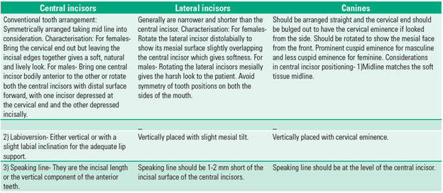
Fig 2
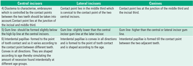
Fig 3
2) POST EXTRACTION RECORDS (ANATOMICAL LANDMARKS)
In most cases, however, no adequate photographs or other preextraction records are available and the clinician has to decide how best to select the teeth that will satisfy esthetic and functional parameters. It is at this stage that guidelines relating to anterior tooth positioning may be used and these guidelines are centred on the fact that the six upper anterior teeth should: appropriately support the upper lip, occupy that area of the upper anterior arch bordered by the corners of the mouth, allow for individualisation where indicated like rotation, imbrications or spacing.
To achieve this, the clinician should select the teeth on the basis of measurements and decisions made with the maxillary occlusal rim in place and functional and esthetic parameters are to be assessed. With the maxillary rim in place and the lip appropriately supported and the incisal point determined, the patient should be asked to smile. By marking the outline of the high smile line on the maxillary rim, the clinician should take the decision on selection of tooth moulds . Another critical point is to determine the position of the canine teeth. Earlier reference has been made to the use of preextraction records. Where these are not present,the position of the corners of the mouth, at rest should be used.
Another method advocated is to ask the patient to smile and to extend a line from the inner canthus of the eye via the lateral border of the alar cartilage and extend that onto the maxillary rim. This may be done with a thread. This corresponds in a high proportion of cases, to the position of the tip of the upper canine teeth.
If a flexible ruler was laid from one canine point to another on the upper rim, the length of the anterior teeth could be read off and this reading is the second critical dimension required to prescribe tooth moulds.
Before evaluating mould charts, however, it is of critical importance that the clinician determines how the patient desires the tooth arrangement to look. If the patient wishes spacing, then clearly that would require teeth of a smaller width to be used. The opposite holds good where imbrication or crowding is desired. The importance of the two measurements is apparent when one examines most tooth mould charts.
There are three values allocated per mould:
1) The combined widths of all six anterior teeth, that is from distal of canine to distal of the contralateral canine (in mm). This is approximately the circumference of the upper rim from one canine point to the other. 2) The height of the central incisors from the incisal edge to the highest point on the labial face of the tooth corresponding to the highest point of the crown (in mm). 3) The width of the central incisors.
Taking these three measurements into considerations, which may be compared with the record rim, we should be able to select from those moulds that lie within the selected intercanine distance. Similarly, an awareness of dental ageing changes is required when the height of the central incisors is being considered. The distance measured on the record rim is from the incisal tip to the high smile line.
In order to customise the anterior teeth to reflect the age of the patient, the clinician should select longer central incisors than would be expected, to permit incisal grinding to simulate attrition. On the other hand, some patients may not show much of their teeth when they smile. This may be the attained habit, for socio psychological reasons, a consequence of tooth wear and a long upper lip. This may be clear from a good photograph of the patient in which he or she is smiling. It may also be seen at the time of preparation of the maxillary rim.
We should consult with the patient regarding the patient’s wishes and expectations on tooth selection to avoid or to minimise any problem regarding the acceptance of the prosthesis at a later date.
Other factors could also be taken into consideration to finalise anterior tooth selection. According to Williams (1907), suggested that the frontal appearance of the face from the normal hairline to the chin could be used as a guideline to the select the shape of the central incisor (facial form- square, tapering and ovoid- the anterior teeth are also similarly grouped). The proximal form of the teeth- straight or convex which follows the lateral profile of the patient.Incisal form could be convex or flat12.
SELECTION OF LOWER ANTERIOR TEETH
As has already been discussed that pre-extraction records may be used to ensure appropriate tooth selection and the anterior form of the trial dentures. When these are not available, mould charts could be used to equate the lower anterior teeth to the selected upper anterior teeth13.
Identify the position of the lower canines taking the angle of the mouth as a landmark and then measure the canine-canine distance. As tooth moulds for lower anterior teeth have the equivalent three measurements to upper anterior teeth, we should choose for the mould that is appropriate for each patient, taking age, facial form and patient perceptions into account.
SELECTION OF POSTERIOR TEETH TYPES AND MOULDS
This part is least considered by the general dentist. The choice of posteriors is often made by technicians who never have seen the patient. The complete dentures are supposed primarily to restore function and secondarily to restore facial appearance. As a prosthodontist it is very important to discuss briefly, types of occlusion in complete denture fabrication that has to be taken into account for different clinical situations and relevant type of posterior teeth selection.
According to Lang14 posterior tooth moulds are of four types:
1. Anatomic 2. Non-anatomic 3. Zero-degree teeth 4. Cuspless teeth. According to the Glossary of Prosthodontic Terms, the following definitions apply to each type:
1. Anatomic: teeth that have cuspal inclinations greater than 0° and tend to replicate occlusal anatomy. Such teeth may have cuspal angles set to 20°, 30°, 33° or 45°.
2. Non-anatomic: teeth designed in such a way the occlusal surfaces are not the copy of the natural tooth form but rather are given the form that can fulfil the requirements of mastication.
3. Zero-degree teeth: posterior teeth that have 0° cuspal angles.
4. Cuspless teeth: teeth designed without cuspal prominence on the occlusal surface that is inverted cusp teeth.
Three factors have to be considered for complete denture prosthesis fabrication, namely occlusal factors, stability factors and esthetic factors as shown in the table down below:
OCCLUSAL FACTORS
If the patient only performs vertical mandibular movements then it is possible that cuspless teeth is acceptable. However the patient performs lateral excursive mandibular movements, then teeth with cusps will be required for balanced articulation and thus stable dentures. Examination of current dentures may help in the diagnosis . For example, if the dentures have occlusal surfaces that are evenly worn, this is usually suggestive of vertical chopping mandibular movements, whereas much greater wear of the maxillary buccal cusps especially, is suggestive of lateral excursive mandibular movements.
List of factors influencing selection of posterior tooth forms2:
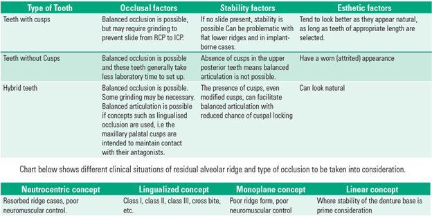
Fig 4
STABILITY FACTORS
In addition to the stability related to the muscle balance and occlusal balance in all border positions, cusps that tend to lock or cause tripping can aggravate the stability of dentures. This is particularly pronounced in flat, atrophic mandibular ridges. It is mandatory to prescribe cuspless teeth in such cases. Another factor to consider is the width of the posterior teeth. If the posterior teeth are too broad, the presence of which could lead to a major cause of instability. Thought should also be given to the number of posterior teeth. There are few clinical situations where there is sufficient mesiodistal length to incorporate two molars and two premolars without compromising stability. In some clinical situations like shortened dental arch it is advisable to drop off one molar.
ESTHETIC FACTORS
These factors are the ones that can only be determined by the patient. The patient should be informed of the options and allowed to decide on the appearance of anterior teeth as well as teeth in the esthetic zone.
SELECTION OF COLOUR AND SHADE OF TEETH
Great care has to be taken by the prosthodontist over the selection of teeth of appropriate colours and shades. We should take into account four qualities when selecting denture teeth.
1. Hue: This is a specific colour resulting from light of a particular wavelength acting on the retina. The hue is an indication of a specific colour, eg blue, green, reddish yellow. Some authorities suggest that the hue of teeth should harmonise with the hue of the patient’s face/ natural hair.
2. Saturation (chroma): This represents the amount of colour per unit area, eg a tooth may appear greyer than another tooth. The hue of both teeth could be equal or one tooth could contain a higher saturation of the grey than the other.
3. Brilliance (value): This equates to the lightness or darkness of a tooth. Variations in brilliance are affected by dilution of the colour (ie the hue) by black or white. It is the ratio of white or black on teeth to the natural hue which determines the lightness or darkness of teeth.
4. Translucency: This property enables light to pass through a body without giving any distinguishing image. The careful selection of colours and shades of teeth is an art which would enhance the look of an esthetic denture. The shade of teeth on any previous dentures is often the shade of choice for the patients. The selection of tooth shade may be based on the facial complexion. The least conspicuous shade is often the best choice. If at all available we should make use of pre-extraction records for shade selection. Its always better to have a careful consultation with patients desire regarding shades of teeth, taking into account age since teeth tend to become darker with age, patient preference and, skin colour. When all of these details have been recorded on the laboratory prescription card, the occlusal rims may be dispatched to the laboratory for final dentures to be made.
ARRANGEMENT OF MAXILLARY ANTERIOR TEETH
When arranging maxillary anterior teeth, position of the central incisors are of utmost importance. As they are the dominating actors on the stage of expression, the smile and also determine the plane of occlusion for function. The desired lip support and position is verified as was determined at the time of occlusal rim adjustment during jaw relation. Incisal edges should be straight and labial surface slightly proclined as was attained by the natural teeth. Using the occlusal rim as the guide on to which the smile lip line was drawn, teeth are arranged in a way so that neck of the teeth lies at the high lip line to minimise the show of base material when the lip is raised to its maximum extent. Both the Central incisor are arranged symmetrically according to patient’s age and gender. Middle aged men and women will have 1 mm of tooth visible below the lip line at rest( younger individuals will have 2-3 mm of visibility at rest). Once the wax has chilled, the tooth arranged is tried in the patients mouth for lip support, tooth visibility, mid line and maximum tooth visibility at maximum lip elevation (smile) are verified. In anterior hyperfunction syndrome, where anterior ridge resorption is extensive, the anteriors are arranged anterior to the ridge to produce more of lip support. This can be done by communicating with the technicians or the operator should arrange the two central incisors, and send it to the lab for other teeth setting to establish the horizontal plane of occlusion after mounting the cast on to the articulator. Laterals should be arranged having the incisal edges about 1 to 2 mm short of the centrals and inclined slightly more labially as compared to the centrals. Long axis should slope mesially,this makes cervical portion appear depressed. Maxillary canines are placed vertically or slightly mesially inclined. Cervical portion is made more prominent. The canine is located at the transition point of the arch and therefore rotated to follow this curvature. The canine is in contact to the occlusal plane. When viewed from the front, the mesial surface of the canine is more visible than the distal surface12.
ARRANGEMENT OF MANDIBULAR ANTERIOR TEETH
Mandibular centrals and laterals are arranged side by side so as to have the same occlusal plane, parallal to the long axis of the residual alveolar ridge and perpendicular to the horizontal plane.
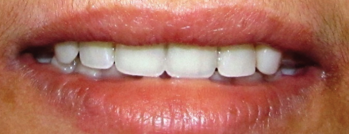
Dentogenic female arrangement of teeth and alignment of maxillary teeth following the curvature of lower lip.

All incisal edges are arranged straight and even giving an illusion of ‘false teeth’ and hence a denture look to the patient.
Canines are mesially tilted and should follow, canine to canine relation that is mesial incisal slope of the maxillary canine should be in a line with that of the distal incisal slope of the mandibular canine. Once the anteriors are arranged and verified in the patients mouth, follows the posterior teeth arrangement.
ARRANGEMENT OF THE POSTERIOR TEETH
Posterior teeth should be arranged within the neutral zone in which forces between the tongue and cheeks or lip are equal. If placed outside this zone, either buccaly or lingually can cause the cheek or tongue to displace the denture. They are arranged taking type and concepts of occlusion into consideration.
DENTURE BASE
One most important aspect of denture fabrication that we generally tend to forget is the denture base itself on to which all the artificial teeth are supported. To give natural appearance to the denture base the acrylic resin between the teeth should be made slightly convex or concave rather than just flattening it. We should lower down the papillae and stippling should be done on the buccal and labial surface of the denture base.
DISCUSSION
Face is the most expressive part of human body as it determines an individual’s social acceptance. The loss of teeth which affects facial appearance, leads to psychological trauma. It is therefore essential that esthetically pleasing and functionally comfortable dentures are provided. The selection and arrangement of anterior teeth for edentulous patients in natural and esthetically pleasing form has remained a challenging experience. Pre-extraction record is a reliable guide for selection of teeth in the absence of which arch form, face form and nasal width serve as useful guides. This is based on the premise that the size and arrangement of anterior teeth harmonises with anatomical landmarks. Determination ofthe width of the maxillary anterior teeth for an edentulous patient becomes difficult in the absence of pre-extraction records. Various techniques have been employed for selection of anterior teeth. These methods reveal a dependence on the physical characteristics of the dento-facial form. Facial landmarks such as bizygomatic width, circumference of head, facial height and interalar width have been taken into consideration while arriving at the mesiodistal dimensions of upper anterior teeth.
If proper guidelines are not followed for the fabrication of complete denture, would result in a ‘false teeth’ which would thereby result in a ‘Denture look’ for the patient. Some patient thinks that this false appearing teeth in a denture is an unavoidable consequence after losing their teeth. This could be avoided if the concept of ‘Dentogenics’ is applied for the fabrication of denture. Dentogenics is the art, practice, and technique of creating the illusion of natural teeth in artificial denture and is based on the elementary factors suggested by the sex, personality, and age of the patient. Every patient can’t afford the expense of full mouth implant reconstruction so as to avoid complete dentures. So employing dentogenic concept for the fabrication of complete denture, it is possible to restore the dignity and individuality and thereby developing self confidence in the patient.
CONCLUSION
Esthetic dentistry has become an important part of restorative dental practice .The appearance of teeth is very important to patients of all ages and is often associated with a perception of health and fitness. The change in dentistry from need based dentistry to elective dentistry for esthetic has made a significant impact. So with thorough understanding of esthetic principles we can provide pleasing smile to the patient thereby enhancing their confidence in themselves and trust on us. Excellent esthetics can be achieved by following simple guidelines such as using tooth moulds specifically carved and prepared for males and females separately, and also arranging the teeth according to the sex, personality and age. Care should be taken for the fabrication of the visible denture base simulating more natural contours.
Visual stimuli are transmitted from the eyes to the brain. The statement that “beauty is in the eye of the beholder” is complex which we are challenged to solve. While there is no shortcut to success, practical application of the above principles can achieve a successful prosthodontic result, satisfying both patient and the professional in respect to esthetics as well as function with the combination of art and science.
REFERENCES
- Tjan, A. H. L, Miller, E. D. and J. G. P. Some esthetic factors in a smile. J ProsthetDent 51(1984):24-28.
- J. F. McCord, A.A. Grant: Registration: Stage III- Selection of teeth; British Dental journal,2000; 188 (12):660-666
- Sheldon Winkler: Essentials of complete denture prosthodontics:Anterior tooth selection and guidelines for complete dentureesthetics;2nd edition, pg 202-210
- Vig, R. G. and Brundo, G. C. The kinetics of anterior tooth display. JProsthet Dent 39(1978):502-504.
- Scandrett, F. R., Kerber, P. E. and Umrigar, Z. R. A clinical evaluation oftechniques to determine the combined width of the maxillary anterior teeth and the maxillary central incisor. J Prosthet Dent 48(1982):15-22.
- Frush, J. P. and Fisher, R. D. Introduction to dentinogenic restorations.J Prosthet Dent 5(1955)5:586-595.
- Frush, J. P. and Fisher, R. D. How dentogenic restorations interpret the sex factor :J Prosthet Dent,6(1956)2:160-72
- Frush, J. P. and Fisher, R. D.How dentogenics interpret the personality factor;J Prosthet dent;6(1956)4:441-449
Disclaimer from Guident
The views and opinions expressed in articles published in Guident are solely those of the authors and do not necessarily reflect the views of the editors, the editorial board or the publisher, Ivory India. Guident does not endorse any product, technique or treatment described. Clinical content is intended for qualified dental and medical professionals and is not a substitute for individual clinical judgement. Guident, its editors and the publisher accept no liability for any consequence arising from the use of information published in the journal.
How to cite this article
Dr. Krishna Prasad, Dr. Chethan Hegde, Dr. Arunoday Kumar. Selection and Arrangement of Teeth for Esthetic Complete Denture Prosthesis - an ART and Science. Guident. 2013 Jan;6(3). Available from: https://guident.in/articles/prosthodontics/selection-and-arrangement-of-teeth-for-esthetic-complete-denture-prosthesis-an-art-and-science.html