Squamous Cell Papilloma
Department of Dentistry, SIPS Superspeciality Hospital & Trauma Centre, Lucknow (U.P) INDIA
INTRODUCTION
Oral squamous papilloma is a generic term that is used to include papillary and verrucous growths composed of benign epithelium and minor amounts of supporting connective tissue. It is a benign exophytic papillary growth of Stratified squamous epithelium. The squamous cell papilloma is the fourth most common mucosal mass and is associated with Human papilloma virus (HPV)- types 6 and 11. The occurrence of these lesions is influenced by smoking, infections, dietary deficiencies and hormonal changes. The route of transmission of HPV virus is unknown for oral lesions. It commonly occurs between the ages of 30 to 60 years.
CASE REPORT
A 66 years old male patient was reported with a painless soft tissue growth on right side of the palate in relation to 15-16.
Onintraoral clinical examination, the lesion was white, solitary, well defined about 4-5mm in size, sessile, circumscribed and exophytic growth having a cauliflower like appearance. It was non-tender, firm in consistency and arose from underlying soft tissue. After seeing past history, patient suffered from verrucous hyperplasia with mild dysplasia about 10 years back on the opposite side of the palate. The lesion was surgically excised without any postoperative complications. The excised lesion was sent for histopathological examination which confirms the Oral Squamous Papilloma.
CLINICAL FEATURES:
- Oral squamous papillomas may be found on the vermilion portion of the lips and any intraoral mucosal site, with a predilection for the hard and soft palate and the uvula.
- The later three sites account for approximately one third of all lesions.
- In our case, the lesion is seen on posterior region of hard palate adjacent to the maxillary right first premolar.
- It is painless, usually white but sometimes pink in color.
- The lesions generally measure less than 1 cm in range and appear as exophytic growth made up of numerous finger like projections with a roughened, granular and Cauliflower- like appearance.
- It is a well circumscribed tumor which is pedunculated (Mostly) or sessile (sometimes).
- The lesions are generally asymptomatic as was in the present case.
- Squamous papillomas are traditionally divided into two types: Isolated-solitary and Multiple-recurring.
- Patients who are HIV positive often have multiple oral lesions.
CLINICAL PICTURES

Figure 1: before exicision figure 2: after exicision

Figure 3: excised lesion
HISTOLOGICAL FEATURES
- CHARECTERISTIC FEATURE–Many long thin, “finger like projections” extending above the surface of mucosa.
- Each finger like projection is lined by Stratified squamous epithelium and contains a thin central connective tissue.
- The spinous cells proliferate in a papillary pattern.
- Koilocytes – HPV altered cells may be seen.
- The upper epithelial layer shows pyknotic nuclei often surrounded by edematous or optically clear zone the so called “KOILOCYTIC” cell.
- Chronic inflammatory cells are also present.
- Occasionally, Basilar hyperplasia may be present.
After clinical evaluation, excisional biopsy of the lesion was performed and the specimen were fixed and stained with hematoxylin and eosin, for routine histopathological examination.
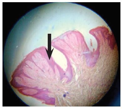
Figure 4: finger like projections
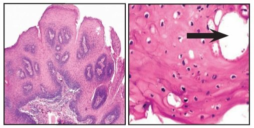
Figure 5: connective tissue stromafigure 6:koilocytes
DIAGNOSIS
On the basis of above clincal evaluation and thehistopathological examination,which reveals finger like projections, presence of Koilocytes and inflammatory cells etc.the final diagnosis was established as an Oral Squamous Papilloma.
ROLE OF HPV
Human Papilloma virus (HPV) is a member of “Papovavirus” group. It is a DNA virus containing a single molecule of double stranded DNA. The viruses are nonenveloped ranging from 45nm to 55nm.The class of HPVs is very large with more than 100 subtypes and these viruses are associated with many conditions of squamous epithelium.
LESIONS CAUSED BY HUMAN PAPILLOMA VIRUS

Figure 5: connective tissue stromafigure 6:koilocytes
DIFFERENTIAL DIAGNOSIS
Verruciform xanthoma has predilection for gingiva and alveolar ridge, while condyloma are larger than papilloma with broader base and appear pink to red.
TREATMENT
Surgical removal of the lesion including the base of mucosa into which the pedicle or stalk is inserted, is the treatment of choice for oral squamous papilloma’s either by routine excision or laser ablation. Other treatment modalities includes-
- Electro cautery
- Cryosurgery
- Intralesional injections of Interferon
- Laser ablation
- Topical podophyllin
- Imiquimod
PROGNOSIS
If the excision is done properly then recurrence is rare. As in our case,one year follow- up was performed, and there was no evidence of recurrence of the lesion.Recurrence is uncommon, except for lesions in patients with HIV. The recurrence rate is very low for the solitary type compared with multiple lesions.
REFERENCES
- Cawson’s Essentials of Oral Pathology and Oral medicine,7th edition by R.A Cawson and E.W Odell.
- Oral and maxillofacial pathology,3nd edition by Neville BW, Damm DD, Allen CM, Bouquot JE.
- Oral pathology, clinical pathologic correlations,4th edition by J.A Regezi, J.J Sciubba, Richard C.K Jordan.
- Contemporary Oral and maxillofacial pathology, 2nd edition by J. Phillip Sapp, Lewis R. Eversole, George P. Wysocki.
Disclaimer from Guident
The views and opinions expressed in articles published in Guident are solely those of the authors and do not necessarily reflect the views of the editors, the editorial board or the publisher, Ivory India. Guident does not endorse any product, technique or treatment described. Clinical content is intended for qualified dental and medical professionals and is not a substitute for individual clinical judgement. Guident, its editors and the publisher accept no liability for any consequence arising from the use of information published in the journal.
How to cite this article
Dr. Vikram Ahuja, Dr. Abhishek Kr. Pandey. Squamous Cell Papilloma. Guident. 2019 Sep;12(10). Available from: https://guident.in/articles/oral-surgery/squamous-cell-papilloma.html