3D Diagnosis and Endodontic Management of Mandibular Molar with Radix Entomolaris
Department Of Conservative Dentistry & Endodontics, K. D. Dental College & Hospital, Mathura
ABSTRACT
Thorough understanding of anatomical variant of permanent mandibular molars along with canal morphology leads towards successful outcome of root canal treatment. The aim of this paper is to review the prevalence of extra root and present cases of permanent mandibular first molars with an additional third root (radix entomolaris).
Key words: Mandibular Molar, Radix Entomolaris, Prevalence
INTRODUCTION
Success of endodontic therapy depends upon in-depth knowledge of morphology of root canal system and its variations. It is reported that many treatment failures in the mandibular molar were related to the inability to locate and clean the extra canal.1
Radix entomolaris (RE), first described by Carabelli, is an anatomical variant found in the permanent mandibular first molar. It refers to mandibular molars having an additional root located lingually.2
Incidence ranges from 0% - 43.7% in mandibular first molar. Highest prevalence is seen among the Mongolian and Eskimo traits. The prevalence of RE is also found to be high among Taiwanese (Chinese) population in a range from 21.1% to 33.33%, with a bilateral incidence ranging from 53.65% to 68.57%.3 Few studies reporting it to be more on left side while others on right side.4 Although, incidence of RE among the Indian population is found to be very low and only 0.2%.3 Tratman et al revealed 0.2 % of RE prevalence in Indians.5
In 1990, Carlsen & Alexandersen classified RE into four different types based on the location of its cervical part:6
- Type A: the RE is located lingually to the distal root complex which has two cone-shaped macrostructures.
- Type B: the RE is located lingually to the distal root complex which has one cone-shaped macrostructures.
- Type C: the RE is located lingually to the mesial root complex.
- Type AC: the RE is located lingually between the mesial and distal root complexes.
In 2004, De Moor et al. classified radix entomolaris on the basis of curvature of the root or root canal:7
- Type 1: a straight root or root canal.
- Type 2: a curved coronal third which becomes straighter in the middle and apical third.
- Type 3: an initial curve in the coronal third with a second buccally oriented curve which begins in the middle or apical third.
Song JS et al. (2010) further added two more newly defined variants of RE:8
- Small type: length shorter than half of the length of the distobuccal root.
- Conical type: smaller than the small type and having no root canal within it.
CASE SERIES
CASE 1
24 years old male patient reported to the department with the chief complaint of pain in lower right back tooth region since few days. History of present illness revealed the pain was spontaneous, moderate in intensity and throbbing in nature which was aggravated by hot drinks and sweets. On inspection 46 was grossly carious and tender on vertical Percussion. No abnormality was detected on palpation and periodontal probing was within the physiological limits. A heat test resulted in severe lingering pain. Periapical radiograph revealed radiolucency involving the distal pulp horn with widening of the periodontal ligament space (figure 1a). A faint outline of an additional root overlapping the mesial and distal roots was suggesting an extra root. CBCT was advised with consent.
Axial, coronal, and sagittal CBCT images (Carestream Health Care Inc., Internal version 3.5.15.0 at 5x5, 85KV, 8.00mA, 20.00s, 985 mGy.cm2) examined from the pulp chamber to the apex to find out the associated variations related to number of roots, the number of root canals, and the canal configuration. Extra root lingually in between mesial and distal root was observed as type A (Carlsen & Alexandersen 1990) and type 3 (De Moore et al 2004) (figure 1b and c). From the clinical and radiographic findings, a diagnosis of symptomatic irreversible pulpitis with apical periodontitis was made and nonsurgical endodontic treatment was advised.
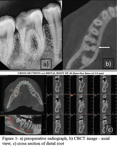
Fig 1: Pre- operative photograph
On the first visit, tooth was anesthetized with local anesthesia 2% lignocaine with 1:200,000 adrenaline (xylocaine; AstraZeneca Pharma Ind Ltd, Banglore, India), followed by rubber dam (dental dam kit, coltene/Whaldent) isolation. An endodontic access cavity had prepared with Endo access bur no. ESE014 (SS White, USA). It had modified from conventional triangular to trapezoidal in order to locate and access the orifice of additional roots. Visual inspection of dentinal map and exploration (DG-16 explorer - EXDF 166, Hu-Friedy) of pulp chamber floor suggested two mesial and two distal canal orifices. Root canals had negotiated with 10 sizes K file (Dentsply Maillefer, NiTiFlex, Ballaigues) and working length was determined by using electronic apex locator (Root ZX mini, J Morita company, Tokyo, Japan) and confirmed with digital radiograph (figure 2a). Cleaning and Shaping had done with crown-down technique with Hyflex CM file system (Coltene-Whaledent, Altstatten, Switzerland). Sequentially irrigation had performed using 5.25% Sodium hypochlorite, 17% EDTA (Dentsply Maillefer, Ballaigues, Switzerland) and normal saline. Corresponding gutta percha mastercone 25/4 (Dentsply Maillefer) had selected and radiograph was taken (figure 2b). Canals obturated with AH Plus sealer (Dentsply, Maillefer, and North America). Access cavity was restored with composite restoration (Tetric N Ceram, ivoclar vivadent) and all ceramic prosthesis was given (figure 2c). Follow up result was satisfactory (figure 2d).

Fig 1: Pre- operative photograph
CASE 2
A24 years-old male patient reported with a chief complaint of continuous pain and difficulty in chewing in lower- left posterior tooth region of jaw since few months. Pain was aggravated by hot, cold, sweets, and relieving only on taking medicines. Clinically, the lower left first molar was intact with deep pockets. Tooth was tender on vertical as well as horizontal percussion. Radiographically, vertical bone loss was seen along with extra root. CBCT was advised with consent. The presence of third additional root was also revealed on the distal side of molar as type B (Carlsen 1990) and type 2 (De Moor et al) via CBCT. The extra root was relatively straight and originated from distolingual aspect of the tooth. The tooth was unresponsive on cold and electric pulp testing. A diagnosis of pulp necrosis with symptomatic apical periodontitis in relation to lower left first molar was made. The tooth was anesthetized. Access opening was made, and two mesial canal and one middle mesial canal orifices, one distal canal orifice were initially located. Another orifice was located on distolingual part of the pulpal floor near lingual wall. The root canals orifices were enlarged using orifice opener for a straight line access and the shape of access cavity was modified from a triangular form to a more trapezoidal form. The root canals were explored with a K-file ISO number 15. Middle mesial canal was merging in middle third of ML canal. After working length determination biomechanical preparation was carried out using the Hyflex CM rotary files in all the canals with intermittent irrigation using 17% EDTA, 5.25% sodium hypochlorite, CHX and normal saline. Obturation of the root canals was performed with gutta percha points and AH plus sealer. The access cavity was then sealed with composite restoration.

Fig 1: Pre- operative photograph
CASE 3
24 years old female patient reported to the department with the chief complaint of pain in the lower left back tooth region since few days. History of present illness revealed the pain was continuous, moderate in intensity and aggravated by hot. On inspection 36 was grossly carious and tender on percussion. Cold test was negative. Periapical radiograph revealed proximal radiolucency involving the pulp with widening of the periodontal ligament space. A radiolucent outline was overlapping mesial and distal root. Type A RE (Carlsen & Alexandersen 1990) and type 2 (De Moore et al 2004) (figure-4a) root morphology was seen. From the clinical and radiographic findings, a diagnosis of pulp necrosis with symptomatic apical periodontitis was made and nonsurgical endodontic treatment was advised.
On the first visit, tooth was anesthetized under rubber dam isolation. An endodontic access cavity was prepared and modified to locate the orifice of additional roots. Visual inspection of dentinal map and exploration of pulp chamber floor suggested two mesial and two distal canal orifices (figure-4b). Root canals had negotiated with 10 sizes K file and working length was determined by using electronic apex locator and confirmed with digital radiograph (figure-4c). Cleaning and Shaping had done with crown-down technique. Sequentially irrigation had performed. Mater cone radiograph was taken (figure-4d). Obturation was done with Gutta percha and AH Plus sealer. Post obturation composite restoration was done (figure-4e).

Fig 1: Pre- operative photograph
CASE 4
32 years old female patient reported to the department with the chief complaint of difficulty in chewing and pain in lower right back tooth region since few days. Pain was continuous, moderate in intensity and aggravated by chewing food and taking hot drinks. On inspection, 46 was grossly carious and tender on percussion. Periapical radiograph revealed pulpal involvement along with widening of the PDL space. A radiolucent outline was overlapping mesial and distal root. From the CBCT and radiographic findings, Type AC RE (Carlsen & Alexandersen) and type 3 (De Moore et al) (figure-4a, b & c) root morphology was seen and a diagnosis of pulp necrosis with symptomatic apical periodontitis was made and nonsurgical endodontic treatment was advised.
On the first visit, tooth was anesthetized under rubber dam isolation. An endodontic access cavity had prepared and modified. Visual inspection of dentinal map and exploration of pulp chamber floor suggested two mesial and two distal canal orifices. Root canals had negotiated with 10 sizes K file and working length was determined by using electronic apex locator and confirmed with digital radiograph (figure-4d). Cleaning and Shaping had done with crown-down technique with Hyflex CM file system. Sequentially irrigation had performed. Mater cone radiograph was taken (figure-4e). AH Plus sealer was used for obturation and post obturation all ceramic crown prosthesis was given (figure-4 f, g & h).
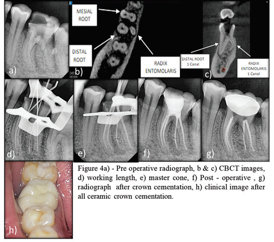
Fig 1: Pre- operative photograph
DISCUSSION
The aetiology for radix entomolaris is still unknown; it can be because of external factors during tooth formation or can be attributed to atavic gene or polygenic system.9 The radix entomolaris is located distolingually ranging from short, conical extension to normal mature root length with its coronal third partially or completely fixed to distal root. Generally, the radix entomolaris is smaller than mesio- and distobuccal roots and may contain pulpal tissue. Externally, the distal furcation is slightly lower (1 mm.) than the furcation between mesial and distal roots.10, 11
Clinically, tooth with additional distolingual root may present a more bulbous crown outline, an additional cusp, a prominent distolingual lobe or cervical prominence. Because the (separate) RE is mostly situated in the same buccolingual plane at the distobuccal root, a superimposition of both roots can appear on the preoperative radiograph and resulting in an inaccurate diagnosis. Additional radiographs taken from different horizontal projections, 20 degree from mesial and 30 degree from distal reveals the basic information about the anatomy of additional third root.12
Occasionally it may be missed because of its slender dimension or overlapping with distal root. Recently, cone-beam computed tomography (CBCT) has emerged as a useful tool to aid in the diagnosis of teeth with complex root anatomies. An accurate diagnosis of these can avoid complications or a ‘missed canal’ during root canal treatment.
Calcification, above the orifice of the RE and MM canal (as in case report 2), has to be removed for a better view and straight line access. However, to avoid perforation or stripping in the coronal third of a severe curved root, care should be taken not to remove an excessive amount of dentin on the lingual side of the cavity and orifice of the RE and MM canal. A straight canal initially in coronal third excessive curvature in apical part of root canal as in type 3 RE (case report 1) possibly may lead to ledge formation or transportation or other procedural errors. To avoid such errors glide path should be prepared and excessive use of flexible nickel titanium instruments and widening of coronal third with more centered preparation.
Access cavity modification should be done following the dentinal map. Various laws of access preparation should be considered. For that reason access cavity should be properly modified to trapezoidal or oval form rather than the conventional triangular form for ease of approach of the extra root during biomechanical preparation. Use of magnification devices (operative microscope, magnification loop) are also advocated during treatment.
CONCLUSION
Success of endodontic treatment begins with proper clinical and radiographic examinations. This paper may intensify the complexity of mandibular molar variation and intend to reinforce clinician’s awareness of the variable morphology of root canals. Diagnosis and endodontic management of mandibular molar with extra roots was made on thorough clinical and radiographic interpretation. CBCT is an essential tool for evaluation of anatomical variation. At the present time, CBCT is a modality for specific applications rather than a replacement for 2-D imaging modalities.
REFERENCES
- Kamble AP, Pawar RR, Mattigatti S, Mangala T M, Makandar S. Cone beam computed tomography as advanced diagnostic aid in endodontic treatment of molars wit multiple canals: Two case reports. J Conserv Dent 2017;20:273-7.
- Carabelli G. Systematisches Handbuch der Zahnheilkunde, 2nd ed. Vienna: Braumuller und Seidel, 1844:114.
- Pai V, Singh V, Vaitheeswaran MB, Thapa A, Kundabala M. Radix entomolaris: A case report. J Nepal Dent Assoc. 2010;11(2):162- 165.
- Abella F, Patel S, Durán-Sindreu F, Mercadé M, Roig M. Mandibular first molars with disto-lingual roots: review and clinical management. IntEndod J 2012;45:963-78.
- Tratman EK. Three-rooted lower molars in man and their racial distribution. Br Dent J 1938;64:264-74.
- Carlsen O, Alexandersen V. Radix entomolaris: identification and morphology. Scand J Dent Res 1990;98:363-73.
- De Moor RJ, Deroose CA, Calberson FL. The radix entomolaris in mandibular first molars: an endodontic challenge. IntEndod J 2004;37:789-99.
- Song JS, Choi HJ, Jung IY, Jung HS, Kim SO. The prevalence and morphologic classification of distolingual roots in the mandibular molars in a Korean population. J Endod 2010;36:653-57.
- Curzon ME, Curzon JA. Three-rooted mandibular molars in the Keewatin Eskimo. J Can Dent Assoc (Tor) 1971;37: 71-72.
- Reichart PA, Metah D. Three-rooted permanent mandibular first molars in the Thai. Community Dent Oral Epidemiol 1981;9: 191-192.
- Gu Y1, Zhou P, Ding Y, Wang P, Ni L. Root canal morphology of permanent three-rooted mandibular first molars: Part III - An odontometric analysis. J Endod 2011;37:485-90.
- Filip L. Calberson, Roeland J. De Moor, Christophe A. Deroose, The Radix Entomolaris and Paramolaris: Clinical Approach in Endodontics. J Endod 2007;33:58-63.
Disclaimer from Guident
The views and opinions expressed in articles published in Guident are solely those of the authors and do not necessarily reflect the views of the editors, the editorial board or the publisher, Ivory India. Guident does not endorse any product, technique or treatment described. Clinical content is intended for qualified dental and medical professionals and is not a substitute for individual clinical judgement. Guident, its editors and the publisher accept no liability for any consequence arising from the use of information published in the journal.
How to cite this article
Dr. Tanvi Paliwal, Dr. Rohit Paul, Dr. Ajay Nagpal. 3D Diagnosis and Endodontic Management of Mandibular Molar with Radix Entomolaris. Guident. 2019 Sep;12(10). Available from: https://guident.in/articles/endodontics/3d-diagnosis-and-endodontic-management-of-mandibular-molar-with-radix-entomolaris.html