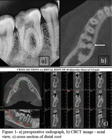
Radix Entomolaris
Department of Conservative Dentistry and Endodontics, Seema Dental College And Hospital, Rishikesh
INTRODUCTION
As stated by Barrett, “of all the phases of anatomic study in the human system, one of the most complex is that of pulp cavity morphology.1
A complete knowledge and potential variations in the dental anatomy from normal are essential to gain success in endodontic therapy. An awareness and understanding of the presence of unusual root canal morphology can thus contribute to the successful outcome of root canal treatment.2 Hence, successful endodontic treatment depends on the location of the canals; thorough chemomechanical debridement followed by three-dimensional (3D) obturation with a perfect fluid tight seal.
Carabelli in 1844 first mentioned a major anatomical variant of the two rooted mandibular first molar; a tooth with a third root that is a distolingual root named as the Radix Entomolaris (RE). If this root is placed buccally then it is called Radix Paramolaris (RP) which is even a rarer entity described by Bolk.3
Anatomical studies have reported an association between the presence of an RE in the first mandibular molar and certain ethnic groups. In the Eurasian and Indian populations, the frequency is <5%. The RE can be found on the first, second, and third mandibular molar, but least frequently on the second molar. The bilateral occurrence of the RE is reported around 50–67%.1
When found, complete diagnosis and treatment are necessary for Radix Entomolaris just like other canals. This article describes the diagnosis and management of a case with a Radix Entomolaris.
CASE REPORTS
CASE 1
21-year-old female patient reported with the chief complaint of pain in lower left back region since two weeks. The pain was aggravated on mastication.
The clinical examination revealed distoproximal caries w.r.t 36. Intraoral periapical radiograph of 36 revealed deep carious lesion involving the pulp with periapical radiolucency at the root apices and presence of an extra root in 36. Based on clinical and radiographic examination, diagnosis of symptomatic irreversible pulpitis with apical periodontitis was established.
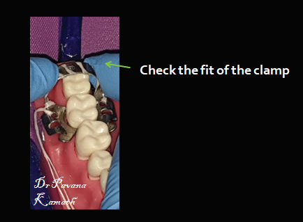
After adequate anesthesia and isolation with rubber dam, access cavity was prepared. Working length was determined using #15 k files followed by shaping and cleaning till # 25/0.6 in all canals. All the canals were irrigated with 2.5% sodium hypochlorite and 17% EDTA after use of each file. In the subsequent appointment canals were dried using paper points and obturation was done using guttapercha and MTA Fillapex sealer. Later, the access cavity was sealed with composite.
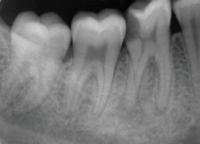
Pre-operative radiograph
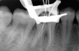
Working length radiograph
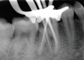
Master cone selection radiogrpaph
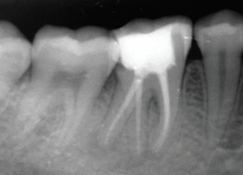
Radiograph showing obturation with permanent coronal seal
CASE 2
18-year-old male patient reported to the dental office with the chief complaint Pain in lower right back region of the mouth since 2-3 days. Clinical examination revealed unusual crown anatomy with and deep mesio occlusal caries with 46. Intraoral periapical radiograph of 46 revealed deep carious lesion approaching the pulp with thickened periodontal ligament space and presence of an extra root in 46. Based on clinical and radiographic examination, diagnosis of symptomatic irreversible pulpitis with apical periodontitis was established. After adequate anesthesia and isolation with rubber dam, access cavity was prepared. Working length was determined using #15 k files followed by shaping and cleaning till # 25/0.4 in all the canals. All the canals were irrigated with 2.5% sodium hypochlorite and 17% EDTA after use of each file. In the subsequent appointment canals were dried using paper points and obturation was done using guttapercha and MTA Fillapex sealer. Later, the access cavity was sealed with composite.

Pre-operative radiograph
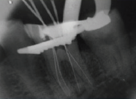
Working length radiograph
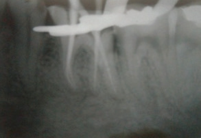
Master cone selection radiogrpaph
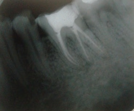
Radiograph showing obturation with permanent coronal seal
DISCUSSION
The presence of an RE or an RP is of paramount importance in endodontics. A proper diagnosis of these supernumerary roots can prevent complications or missing a canal during the root canal treatment. An inaccurate diagnosis may occur on the radiograph due to superimposition of the distobuccal root over the radix as they are mostly located in the same plane. To see the RE on the intraoral periapical radiograph, a second radiograph should be taken with a different angle.3
Thorough clinical examination of the tooth crown and inspection of the cervical morphology of the roots can give us a hint of an additional root. It can be identified by periodontal probing, and use of instruments like an endodontic explorer, JW-17 explorer, pathfinder and micro-opener. A technique can be used like, using sodium hypochlorite in the pulp chamber to see if bubbles are produced by remaining pulp tissue in the canal called as the “champagne effect”. Sometimes an additional cusp (tuberculin paramolaris) may hint an extra root. Extending the triangular access cavity to a rectangular or trapezoidal form along with the complete removal of roof of pulp chamber may help in finding the distolingual orifice. Visual aids such as a loupe, intraoral camera or dental microscope can also be useful.[4] Recently, conebeam computed tomography (CBCT) has emerged as a useful tool to aid in the diagnosis of teeth with complex root anatomies.5
A severe root inclination or canal curvature, particularly in the apical third of the root, can cause shaping aberrations such as straightening of the root canal or a ledge, with root canal transportation and loss of working length resulting. The use of flexible nickel-titanium rotary files allows a more centered preparation shape with restricted enlargement of the coronal canal third and orifice relocation. Nevertheless, unexpected complications such as instrument separation do occur, and are more likely to happen in an RE with severe curvature or narrow root canals. Therefore, after relocation and enlargement of the orifice of the RE, initial root canal exploration with small files (size 10 or less) together with radiographical root canal length and curvature determination, and the creation of a glide path before preparation, are step-by-step actions that should be taken to avoid procedural errors.2
CONCLUSION
Teeth are never alike. A number of variations occur which pose a challenge to a clinician. This particular variation Radix Entomolaris may be a challenge who do not have proper diagnostic aids and lack in proper knowledge of root canal anatomy. So thorough knowledge of root canal anatomy and awareness of the variations make the treatment more successful and if one exhibits proper skill, these cases can be done with ease.
REFERENCES
- Tu MG, Huang HL, Hsue SS, Hsu JT, Chen SY, Jou MJ et al. Detection of permanent three-rooted mandibular first molars by cone-beam computed tomography imaging in Taiwanese individuals. J Endod 2009;35:503-7.
- Calberson FL, Roeland J, Moor D, Deroose CA. The Radix Entomolaris and Paramolaris: Clinical Approach in Endodontics. J Endod 2007;33:58–63.
- Sarangi P, Uppin VM. Mandibular First Molar with a Radix Entomolaris: An Endodontic Dilemma. Journal of Dentistry 2014; Vol. 11, No. 1.
- Arun J, Mayuri BS, Nandini TN, Mallikarjun Goud K. Radix entomolaris and paramolaris: A case series. Int J Oral Health Sci 2015; 5:57-62.
- Parashar A, Gupta S, Zingade A and Parashar S. The Radix Entomolaris and Paramolaris: A Review and Case Reports with Clinical Implications. J Interdiscipl Med Dent Sci 2015; 3: 161
Disclaimer from Guident
The views and opinions expressed in articles published in Guident are solely those of the authors and do not necessarily reflect the views of the editors, the editorial board or the publisher, Ivory India. Guident does not endorse any product, technique or treatment described. Clinical content is intended for qualified dental and medical professionals and is not a substitute for individual clinical judgement. Guident, its editors and the publisher accept no liability for any consequence arising from the use of information published in the journal.
How to cite this article
Dr. Annil Dhingra, Dr. Ishita Agarwal. Radix Entomolaris. Guident. 2019 Sep;12(10). Available from: https://guident.in/articles/endodontics/radix-entomolaris.html