
Lingual Schwannoma a Rare Tumor in a Common Location
Department of Oral and Maxillofacial Surgery, Nair Hospital Dental College, Opposite Maratha Mandir Theatre, Mumbai
ABSTRACT
Background:Neurilemmoma’, also termed as Schwannoma is a benign, solitary, slow growing, well encapsulated tumor arising from the Schwann cells of the neural sheath of the peripheral nerves with uncertain etiology. Occurrence of these lesions is somewhat common in the head and neck region (25-48%), while it is rarely seen in the oral cavity (1%). It commonly affects individuals in the third to sixth decades of life.
Case Report: We present a case of a female in the 4th decade of her life who presented with a painless, rubbery nodule in the anterior two thirds of the right side of the tongue of 4 months duration. USG examination was confirmatory of a solitary nodule in the substance of the tongue. Excisional biopsy of the lesion was performed leaving the overlying mucosa intact
Conclusion: We report a case of lingual schwannoma of the tongue which was excised in toto and confirmed histologically as a Neurilemmoma of the tongue. The patient remained recurrence free in the subsequent annual follow up period.
Keywords: Tongue, Neurilemmoma, Schwannoma
INTRODUCTION
Peripheral nerve sheath tumors are of two types- ‘Neurofibroma’ and ‘Neurilemmoma’. Neurofibroma comprises of fibrobasts, neurites and Schwann cells in a collagenous matrix.
‘Neurilemmoma’, also termed as ‘Schwannoma’ is a benign, solitary, slow growing, painless (69%), well encapsulated tumor arising from the Schwann cells of the neural sheath of not only the peripheral nerves, but also of the cranial (except olfactory and optic), spinal and autonomic nerves.1
The Schwann cells form the myelin sheath which wraps around the extracranial nerves and large nerve fibres and helps in faster impulse conduction. Schwannomas are formed when proliferating Schwann cells form a tumorous mass involving the peripheral sensory and motor nerves. The source of myelin sheath of the nerves changes from oligodendrocytes to Schwann cells upon exit of the nerves through the brain and spinal cord.1
The incidence of extracranial schwannomas seen in the head and neck region accounts to around 25 to 48%, however the incidence in oral cavity falls to merely 1% of all the benign oral cavity tumors. The peripheral variety is commonly seen in the tongue, followed by the buccal mucosa, floor of the mouth, palate and mandible.2 By virtue of its occurrence, it frequently occurs in adults in their 3rd to 6th decades of life, which is in contrast to the neurofibromas and does not display any gender or racial predilection.1,2
Upto 50% of these tumours are in relation to a nerve; however, in the tongue it is sometimes difficult to distinguish whether it arises from the hypoglossal, glossopharyngeal or the lingual nerve, considering their close proximity.2
The presence and severity of symptoms are directly associated to the size and location of these lesions. Usually these lesions are considered solitary, slow-growing and painless ones, which seldom cause ulceration; albeit occasional pain and discomfort are reported.3
A low recurrence rate has been observed after excision of the lesion and rate of malignant transformation reported is around 8% to 13.9%.2,3
We report such a rare case of lingual schwannoma in a woman in her 4th decade of life and discuss in brief the differential diagnosis and review of literature.
CASE REPORT
A 47 year old lady reported to the Dept. of Oral and Maxillofacial Surgery for evaluation of a growth on the right side of the tongue. The history reported by the patient revealed that the lesion was present in the same location from the beginning and had shown no sudden increase in size and shape. The lesion had neither caused difficulty in chewing, swallowing, impairment of taste perception nor interference with phonation or tongue movements.
On extraoral examination, the patient did not have any associated cervical lymphadenopathy (fig 1). Intraoral examination revealed a solitary sub-mucosal nodule of around 1.5 x 2 cms in dimensions in the right anterior two thirds of the dorsum of the tongue with no changes in the overlying mucosa (fig 2). The mass was firm/rubbery, non-tender and did not show any pulsations/bruit on palpation.
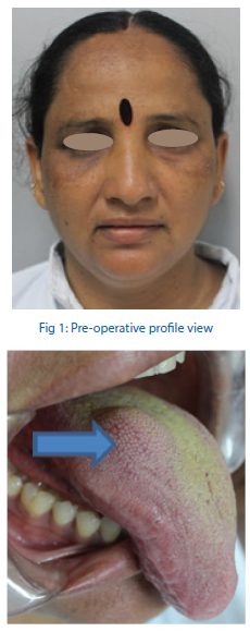
Fig 2: Pre-operative intraoral view of the mass over the dorsum of the right side of the tongue
USG was suggestive of a well-defined, hypoechoic lesion in the dorsum of the anterior two-thirds of the right side of the tongue with no internal vascularity and with dimensions of 15x14x13mm (fig 3).
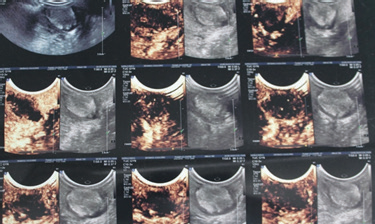
Fig 3: USG showing a well-defined hypoechoic lesion on the right anterior half of the dorsum of the tongue
On the basis of clinical and USG findings, a provisional diagnosis of Neurilemmoma was made. The differential diagnosis included traumatic fibroma, neurofibroma, benign tumor of salivary origin, muscle origin (leiomyoma), rhabdomyoma.
Taking into consideration the size of the lesion, an excisional biopsy was planned. The patient was taken up for the excisional procedure under GA. Tongue traction suture was placed to facilitate retraction of the tongue anteriorly and easy accessibility for the procedure. The lesion was outlined with methylene blue marking ink (fig 4).
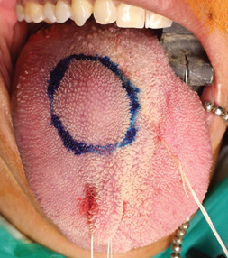
Fig 4: Intraoperative view of the marking of the lesion
A mucosal incision was made as shown in Fig 5. Submucosal blunt dissection was carried out to expose the lesion. It was readily shelled out in toto without rupturing the mass or the overlying mucosa (Fig 6). Hemostasis was achieved (fig 7). The surgical site was closed layerwise using horizontal mattress suturing technique with 3-0 VICRYL (fig 8).
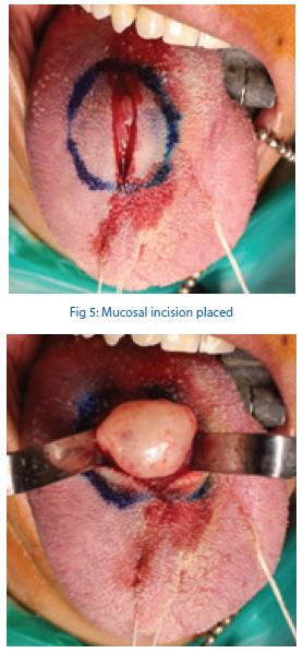
Fig 6: Shelling out of the encapsulated mass in toto without rupturing it

Fig 7: The soft tissue bed after excising the tumour

Fig 9: The excised firm and rubbery mass measuring around 2cms

Fig 10: Histopathology report suggestive of Neurilemmoma
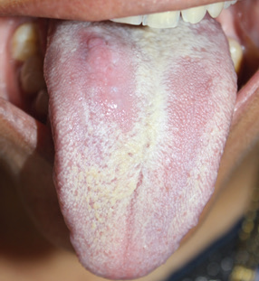
Fig 11: Uneventful healing of the surgical site
DISCUSSION
Schwannoma’ is a benign, solitary, slow growing, painless (69%), well encapsulated tumor arising from the Schwann cells of the neural sheath of not only the peripheral nerves, but also of the cranial (except olfactory and optic), spinal and autonomic nerves.1 Given its origin from the nerve tissue, lingual schwannomas are still painless lesions3 They are frequently noticed because of their location and related consequences of dysphagia, dysphonia.4
The exact etiology of schwannoma still remains ambiguous.2, 4 The incidence of extracranial schwannomas seen in the head and neck region accounts to around 25 to 48% (where it is commonly seen in relation to vestibulocochlear nerve), however the incidence in oral cavity falls to merely 1% of all the benign oral cavity tumors.4 The peripheral variety is most commonly seen in the tongue, followed by the buccal mucosa, floor of the mouth, palate, gingiva and vestibule.2
By virtue of its occurrence, it frequently occurs in adults in their 3rd to 6th decades of life, which is in contrast to the neurofibromas.1,2
The recent review of the literature4 reported no predilection for gender, and a significantly higher incidence of occurrence in the second and fourth decades. The average size reported was 2.4 cm with a typical location in the anterior third of the tongue (66% vs 34% in the posterior two thirds).
Patients with small tumours (less than 2 cm) in the anterior tongue were usually asymptomatic, whereas those with larger tumours (more than 3 cm) or tumours located in the posterior thirds of the tongue were more likely to experience symptoms like snoring and dysphagia.4,5
Less than 50 cases of lingual schwannomas have so far been reported in the English literature.4, 5,6
Wright and Jackson7 have reported 146 cases of schwannoma of the oral cavity soft tissue. They concluded that schwannomas rarely occured in the oral cavity and produced symptoms varying from hypoesthesia to vague paresthesia. The final diagnosis was always made after a definitive histological examination.
Imaging options for this lesion include USG and MRI.8,9 USG is a reliable, non-invasive and safe option for delineating the extent of the lesion with spot on accuracy. Some case reports8 also considered use of CT scan, but they concluded that it showed many artefacts and hence led to an inaccurate final picture.
MRI however, remains the gold standard investigation for such lesions. It has a better contrast compared to a CT, allows for a better delineation and appreciation of the extent, the dimensions and location of the lesion. It does carry the risk of more radiation exposure and cost as compared to USG.9
We, however, resorted to the non-invasive, less expensive and least risk modality of USG and we found it to be equally reliable for knowing the size, location and extent and relation to the surrounding tissues.
Histologically these lesions are characterised by a thin, fibrous capsule and tumor like proliferation formed by 2 types of tissue arrangements- Antoni Type A and Type B. Antoni type A tissues are seen to be closely packed forming bundles with elongated palisaded nuclei, with presence of amorphous Verocay bodies; whereas, Antoni type B tissues have less number of cells and poor organization.10
Immunohistochemistry studies have shown strong reactivity with S-100 protein which provides a clue for diagnosis.11 However, it is not always necessary as H&E stained sections are able to conclusively confirm the diagnosis.
Lipomas, hemangiomas, lymphangiomas, traumatic fibroma, dermoid and epidermoid cysts, leiomyoma, eosinophilic granuloma, benign salivary gland tumour, rhabdomyoma and granular cell tumour are included in the differential diagnosis list.7 Clinically, the lesions may be indistinguishable from the other encapsulated benign tumors, necessitating biopsy and histological examination to formulate a correct diagnosis.10,11
Standard treatment reported in literature for schwannoma includes transoral surgical excision in 93.18% cases which is usually uncomplicated.3 Lesions in the antero-lateral part(s) of the tongue are treated transorally, while lesion in the posterolateral border/ large lesions (>4cms) which are inaccessible through intraoral route are treated through the transcervical route using submandibular or lip split approach described in literature.5 Recently, CO2 laser excision has been used in excising schwannomas of the posterolateral tongue base.5
Schwannomas being encapsulated can be excised in toto without leaving any remnants behind. The recurrence rate of these lesions is very rare and malignant transformation is around 8% to 13.9%.2,3,7
Less than 15 paediatric cases of lingual schwannomas are reported in literature; the youngest of them being a 7 year old male child.12
CONCLUSION
Lingual schwannoma is a benign,solitary, slow growing, painless lesion. It is a relatively rare tumour of the head and neck region but seen to occur in a not so common location like the tongue. Considering the rarity of this lesion, a careful consideration is thereby warranted for clinically distinguishing the lingual swellings. MRI remains the gold standard investigation of choice for delineating the size, extent and the relation of the tumour to the surrounding tissues. The definitive diagnosis requires a histopathological evaluation. Immunohistochemical marker like S-100 provides valuable clue in the correct diagnosis of this lesion. Recurrence after complete excision is reportedly rare with a small range of malignant transformation.
REFERENCES
- N. Zachariades. “Schwannoma of the oral cavity: review of the literature and report of a case,” Journal of Oral Medicine, vol. 39, no. 1, pp. 41–43, 1984.
- K. Hwang, S. G. Kim, S. I. Ahn, and S. I. Lee. “Neurilemmoma of the tongue,” Journal of Craniofacial Surgery, vol. 16, no. 5, pp. 859–861, 2005.
- H. K. Williams, H. Cannell, K. Silvester, and D. M.Williams.“Neurilemmoma of the head and neck,” British Journal of Oral and Maxillofacial Surgery, vol. 31, no. 1, pp. 32–35, 1993.
- M. Cohen and M. B. Wang. “Schwannoma of the tongue: two case reports and review of the literature,” European Archives of Oto-Rhino-Laryngology, vol. 266, no. 11, pp. 1823–1829, 2009.
- Lee et al. Schwannoma of the tongue: a case report with review of literature. Maxillofacial Plastic and Reconstructive Surgery (2017) 39:17 DOI 10.1186/s40902-017-0116-2.
- R. B. Lira, J. G. Filho, G. B. Carvalho, C. A. Pinto, and L. P. Kowalski, “Lingual schwannoma: case report and review of the literature,” Acta Otorhinolaryngologica Italica, vol. 33, no. 2, pp. 137–140, 2013.
- Wright BA, Jackson D. Neural tumors of the oral cavity. A review of the spectrum of benign and malignant oral tumors of the cavity and jaws. Oral Surg Oral Med Oral Pathol 1980;/49:/509 22.
- Cohen L, Schwartz A, Rockoff S. Benign schwannomas: pathologic basis for CT inhomogeneities. AJR 147:141–143.
- La'porte S, Juttla J, Lingam R. Imaging the floor of the mouth and the sublingual space. Radiographics 31:1215–1230.
- C. Fisher, Diagnostic Pathology: Soft Tissue Tumors, 2nd edition, Amirsys, 2011.
- M.R. Lindberg, Diagnostic Pathology: Soft Tissue Tumors, 2nd edition, Amirsys, 2016.
- Bhola N, Jadhav A, Borle R, Khemka G, Bhutekar U, Kumar S. Schwannoma of the tongue in a paediatric patient: a case report and 20-year review. Case Rep Dent. 2014;2014:780762. doi:10.1155/2014/780762.
Disclaimer from Guident
The views and opinions expressed in articles published in Guident are solely those of the authors and do not necessarily reflect the views of the editors, the editorial board or the publisher, Ivory India. Guident does not endorse any product, technique or treatment described. Clinical content is intended for qualified dental and medical professionals and is not a substitute for individual clinical judgement. Guident, its editors and the publisher accept no liability for any consequence arising from the use of information published in the journal.
How to cite this article
Dr. Neelam N. Andrade, Dr. Harsh K. Desai, Dr. Yashpal Choudhary P.G. Student, Dr. A. L. Nair Road. Lingual Schwannoma a Rare Tumor in a Common Location. Guident. 2019 Dec;13(1). Available from: https://guident.in/articles/oral-surgery/lingual-schwannoma-a-rare-tumor-in-a-common-location.html


