Management of Bisphosphonate Related Osteonecrosis of the Jaw an Insight!
Department of Prosthodontics, K. D. Dental College & Hospital, Mathura
ABSTRACT
Bisphosphonates have been associated with a serious adverse reaction known as bisphosphonate-related osteonecrosis of the jaws (BRONJ). The purpose of this review was to assess the outcomes and evaluate the impact of bisphosphonates on dental implants and the risk of developing BRONJ after dental implant surgery followed by its management.
BRONJ manifests as exposed, nonvital bone involving the maxillofacial structures. Bisphosphonate-related osteonecrosis of the jaw (BRONJ) is thought to be caused by trauma to dentoalveolar structures that have a limited capacity for bone healing due to the effects of bisphosphonate therapy.
INTRODUCTION
Since the advent of bisphosphonates (BPNs) to medical practice they have been increasingly used for an array of skeletal disorders. The BPNs inhibit bone resorption and have recently attracted considerable attention because of their ability to prevent osteoporosis in addition to their usefulness in metabolic bone diseases and hypercalcaemia. they are most effective antiresorptive drugs. Chronologically and according to potency, the BPNs can be grouped in 3 generations
First generation bpn
1. Etidronate
Secound generation bpn
1. Pamidronate
2. Alendronate
Third generation bpn
1. Risedronate
2. Zoledronate
The BPNs are useful in conditions characterized by enhanced bone turnover.
- Osteoporosis: the second and third generation BPNs are effective in preventing and treating postmenopausal osteoporosis as well as idiopathic and steroid induced osteoporosis in both men and women
- Paget’s disease: the disease due to abnormal osteoclast function producing disordered bone architecture is benefited by BPNs. They may also have adjuvant value in the treatment of attrition or tooth decay due to osteoporosis particularly in postmenopausal women.
- Osteolytic bone metastasis: parenteral pamidronate/zoledronate arrest osteolytic lesions and reduce bone pain (most commonly secondary to prostate cancer)
- Hypercalcaemia of malignancy: pamidronate and zoledronate injected iv normalize plasma calcium level in medical emergency.
CONCERNS ASSOCIATED WITH BISPHOSPHONATE THERAPY
Among all potential adverse effects associated with the use of bisphosphonates, osteonecrosis of jaws (ONJ) has received the greatest attention. As reviewed by Woo et al,111 nearly all ONJ cases (94%) have been described in patients receiving high doses of IV bisphosphonates (primarily zoledronic acid and pamidronate) for oncological conditions.
Since a 2003 report by Marx, in which the author described a case of avascular necrosis of jaw in a patient treated with pamidronate (Aredia) and zoledronate (Zometa), numerous cases of BRONJ have been reported worldwide, including in India.
PATHOPHYSIOLOGY OF BISPHOSPHONATES
The BPNs have strong affinity for calcium phosphate; therefore exert selective action in calcified tissue. They have also been shown to affect isoprenoid lipid synthesis. The net result is:
- Accelerated apoptosis of osteoclasts reducing their number
- Disruption of cytoskeleton and ruffled border of osteoclasts.
In addition, BPNs appear to affect osteoclast precursors and inhibit their differentiation by suppressing IL-6. Interface with mevalonate pathway may also impart anti-tumor action on bony metastasis.
BISPHOSPHONATE – RELATED OSTEONECROSIS OF THE JAW (BRONJ)
The Advisory Task Forces of the American Association of Oral and Maxillofacial Surgeons (AAOMS) and the American Society for Bone and Mineral Research (ASBMR), have defined BRONJ as:
“Exposed, necrotic bone in the maxilla or mandible that has persisted for more than eight weeks, despite adequate treatment, in a patient with current or previous history of bisphosphonate use, without local evidence of malignancy and no prior radiotherapy to the affected region.”
As illustrated in the 2017 position paper on antiresorptive agent-related osteonecrosis of the jaw (ARONJ) (PP 2017) -compiled by the Japanese Allied Committee on Osteonecrosis of the Jaw , BRONJ is definitively diagnosed when the following criteria is met
- Former use of Bisphosphonates should be present
- Any history of radiation therapy to the jaw must be excluded. Bone lesions associated with BRONJ must be differentiated from cancer metastasis through a histological examination.
- An exposure of alveolar bone in the oral cavity, jaw and/or face is continuously observed for longer than 8 weeks after first detection by a medical or dental expert, or the bone is palpable for longer than 8 weeks.
These criteria do not apply for stage 0 BRONJ
The AAOMS 2014 proposed that patients presenting ONJ-like clinical manifestations but without alveolar bone exposure must be diagnosed as a stage 0 ONJ, and that these cases must be treated as pre-ONJ.
DIAGNOSIS
Mainly the diagnosis is based on the clinical and the radiographic criteria.
The following procedures have been valuable in the diagnosis of bisphosphonate-related osteonecrosis of the jaw (BRONJ):
Imaging studies
Panoramic radiographs (OPG) of the mandible of patients suffering from BRONJ give away areas of sequestration, pathological fractures, destruction or sclerosis. Delayed or persistent tooth sockets post extraction are also likely to be revealed in such patients. Bone scan with Tc-99 m methylene diphosphonate frequently discloses intense uptake in and/or around the affected area, indicating active bone turnover.
A recent studies evaluating the features of bisphosphonate –related osteonecrosis of the jaw (BRONJ) using magnetic resonance imaging (MRI) and computed tomography(CT) demonstrated characteristic findings.
CT scans revealed increased medullary density, periosteal reaction and bone sequestration. MRI revealed a low signal in T1 and T2 images with exposed bone. Reason behind which is likely due to decrease in water content. Unexposed, diseased bone showed hypointensity in T1 images and hyperintensity in T2 images. These ï¬ndings suggest an increase in water content.
Tissue biopsy is not always necessary and should be performed only to rule out a primary malignancy, benign bone lesion, osteomyelitis or metastatic lesion if suspected.
At microscopic examination, An inflammation of the mucosa with massive presence of plasma cells and granulocytes in the connective tissue was found in the samples taken from above the necrotic bone. The histo-architecture of the connective tissue and of the mucosa appears modified: stratification is visible only in some areas and the papillomatosis is clearly noticeable on the slide. The most superficial layers are made up of few mature cells and are characterized by phenomena of ulceration and fray. Also, a phenomenon of interstitial edema is evident.
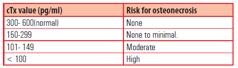
A blood test, the serum C terminal telopeptide test (CTX) is conducted on an empty stomach, in order to evaluate the risk of osteonecrosis in patients who have been administered bisphosphonates for longer than 3 years.
MANAGEMENT
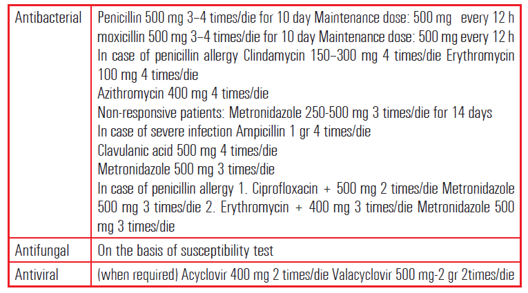
1. NON SURGICAL MANAGEMENT: Antimicrobial rinses ∙ Systemic antibiotics (Table 1) ∙ Systemic or topical antifungals ∙ Discontinuation of bisphosphonate therapy38 ∙ No dental therapy or minimally invasive dental therapy (root canal therapy instead of extractions)Systemic Antibiotics for management of BRONJ
2. SURGICAL MANAGEMENT: surgical treatment range from conservative including:debridement, bone curettage, and sequestrectomy and radical methods that include: marginal or segmental resection
The surgical resection of necrotic jawbone has traditionally been considered palliative rather than curative, as it has been offered primarily to patients with advanced disease who have not responded to nonsurgical treatment
3. ADJUNCTIVE THERAPY: A regimen of HBO treatment for 2 months as per the Robert Marx protocol. This protocol uses HBO and aggressive surgery in a progressively staged manner to treat osteoradionecrosis.
STAGING AND TREATMENT OF BRONJ
Current American Association of Oral and Maxillofacial Surgeons Guidelines on bisphosphonate-related osteonecrosis of the jaw Staging and Treatment (Ruggiero et al, 2009)
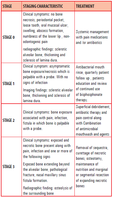
Initially, BRONJ was difficult to cure, and more conservative non-surgical treatment options for e.g. local irrigation were often carried out. various Surgical procedures range from conservative to radical, and studies suggests that surgical treatment is more effective than non-surgical treatment. There is now enough evidence to advocate that nonsurgical treatment has often been unsuccessful to provide positive outcomes in patients with advanced BRONJ whereas radical surgery seems to offer more predictable and curative results However, surgical treatment for early-stage BRONJ remains controversial.
Prevention of bronj
Management of oral hygiene, patient education, and providing proper dental treatment are all critical for preventing BRONJ.
Nonetheless , whether a preventative drug holiday is advantageous in the time of tooth extraction or dental implant therapy for patients receiving BPs remains controversial. The PP 2017 proposes that discontinuation of BPs treatment for approximately 2 months should be considered in patients at high risk for BRONJ who have received BPs for more than 4 years.
BISPHOSPHONATES AND DENTAL IMPLANTS
There have been several case reports that have evaluated implant failures in patients receiving intravenous bisphosphonate therapy contrasting earlier studies where an improvement in implant osseointegration using bisphosphonates was observed.There is mixed evidence regarding the association between oral bisphosphonate therapy and dental implant failure in the available literature related to this topic. Some studies found an association between the two, while other studies did not.
THE MARX PROTOCOL FOR DENTAL IMPLANT CANDIDATES ON BPNs
Patient is on BPNs for more than 3 years- Consult the physician to discontinue bisphosphonate 3 months before and three months after surgery
- CTx levels must be > 150pg/ml before planning implant surgery.
- Detailed informed consent.
- CTX levels must be > 150pg/ml.
- Proceed with surgery after informed consent for BRONJ.
- If CTx <150pg/ml- consult physician for a drug holiday and monitor every three months till CTx levels are >150pg/ml.
- If CTx <150pg/ml- physician approved drug holiday and monitor every three months till CTx levels are >150pg/ml.
- CTx levels must be > 150 pg/ml to proceed with informed consent for BRONJ.
CONCLUSION
BRONJ, which is regarded as a true side effect of BP treatment, is difficult to avoid using preventive dental measures alone. However,preventive dental measures are indispensable for reducing the risk of secondary infection and disease progression. In patients taking low doses of BPs, dental implant surgery is not contraindicated if there are no other risk factors, such as combined use of corticosteroids or concomitant diabetes. However, the risk of BRONJ due to peri-implantitis must be explained when obtaining patient consent.
REFERENCES
- J.L.López-Cedrún, J.F.Sanromán, A.GarcÃa, M.Peñarrocha, J.F.Feijoo, J.Limeres, P.Diz. Oral bisphosphonate-related osteonecrosis of the jaws in dental implant patients: a case series. British Journal of Oral and Maxillofacial Surgery Volume 51, Issue 8
- Mendes, V., dos Santos, G. O., Calasans-Maia, M. D., Granjeiro, J. M., & Moraschini, V. (2018). Impact of bisphosphonate therapy on dental implant outcomes: An overview of systematic review evidence. International Journal of Oral and Maxillofacial Surgery. doi:10.1016/j.ijom.2018.09.006
- Kishimoto, H., Noguchi, K., & Takaoka, K. (2019). Novel insight into the management of bisphosphonate-related osteonecrosis of the jaw (BRONJ). Japanese Dental Science Review, 55(1), 95–102. doi:10.1016/j.jdsr.2018.09.002
- Matthew T. Drake, MD, PhD, Bart L. Clarke, MD, and Sundeep Khosla, MD. Bisphosphonates: Mechanism of Action and Role in Clinical Practice. Mayo Clin Proc. 2008 Sep;83
- Saldanha, S., Shenoy, V. K., Eachampati, P., & Uppal, N. (2012). Dental implications of bisphophonate-related osteonecrosis. Gerodontology, 29(3), 177–187. doi:10.1111/j.1741-2358.2012.00622.x
- Sara Di Lorenzo, Alberto Trapassi, Bartolo Corradino, and Adriana Cordova. Histology of the Oral Mucosa in Patients With BRONJ at III Stage: A Microscopic Study Proves the Unsuitability of Local Mucosal Flaps
- Essentials of pharmacology for dentistry ; KD Tripathi
Disclaimer from Guident
The views and opinions expressed in articles published in Guident are solely those of the authors and do not necessarily reflect the views of the editors, the editorial board or the publisher, Ivory India. Guident does not endorse any product, technique or treatment described. Clinical content is intended for qualified dental and medical professionals and is not a substitute for individual clinical judgement. Guident, its editors and the publisher accept no liability for any consequence arising from the use of information published in the journal.
How to cite this article
Dr. Gitika Singh, Dr. Manesh Lahori, Dr. Manisha Sharma. Management of Bisphosphonate Related Osteonecrosis of the Jaw an Insight!. Guident. 2019 Dec;13(1). Available from: https://guident.in/articles/general/management-of-bisphosphonate-related-osteonecrosis-of-the-jaw-an-insight.html