Comparative Evaluation of Microleakage at the Implant Abutment Interface Using Prefabricated and Customized Abutments
Department of Prosthodontics School of Dentistry, D.Y. Patil University Nerul Navi Mumbai
ABSTRACT
Prevention of microleakage at the implant abutment connection is a major challenge for the construction of modern two- stage implant systems. Implant manufacturers aim to reduce the leakage by increasing the stability of the implant abutment connection. This study was done to evaluate and compare the microleakage through the implant abutment interface using prefabricated and customized abutments. Materials and Method: The implants to be studied were divided into 2 groups of 10 each. They are placed in a Staphylococcus rich media and the microleakage at the interface is evaluated using serial dilution and the colonies are confirmed using grams staining and coagulase test. Results: The results showed a significant amount of microleakage in Group A and Group B when compared to the control group. But no statistically significant differences were seen when comparing the microleakage between Group A and Group B.
Keywords:Implant- Abutment, Prefabricated, Customized, microleakage.
INTRODUCTION
The need for additional retention or the desire to eliminate removable dentures, are common indications for a more permanent solution, such as dental implants.
Loading of the implant abutments may affect their adaptation and in consequence result in microscopic gaps that causes leakage of microorganisms and acts as a bacterial reservoir that in turn causes bone resorption at the crestal areas.1 Several in vitro studies have shown bidirectional fluid and bacterial leakage into and out of implant abutment assemblies.2
Thus, this study was done to evaluate and compare the microleakage through the implant abutment interface using prefabricated and customized abutments.
MATERIALS AND METHOD
The study was divided into the following steps:
A. Preparation of the samples
B. Preparation of the culture test specimen
C. Evaluation and verification of microleakage
A. PREPARATION OF THE SAMPLES: (Fig 1-3)
The specimens to be studied are divided according to their abutment type. Each group consists of 10 samples.
GROUP A: 10 dummy implant abutment assemblies with prefabricated abutments. (Fig 1)
The prefabricated abutments were milled in an industrial mill in the centralized milling centre by the manufacturers of the implants.
GROUP B: 10 dummy implant abutment assemblies with customized abutments. (Fig 2)

The customized abutments were made using pre- milled (ready-made) internal fixtures (hex) with a metal block on top. This metal block was then milled in the laboratory using the laboratory milling machine (Yenadent).
All the components, namely the implants, abutments, Internal hex driver for the implant, Torque Ratchet, Pliers, Tweezers and Artery forceps were packed into autoclavable pouches, sealed and autoclaved.
The implants were retrieved from the sterile pouch with the help of sterile pliers. The prefabricated and customized abutments were carefully connected to the implants and tightened using a torque ratchet in a Biosafety Level 2 cabinet.
The prefabricated abutments were tightened onto their respective 10 implants with a torque of 15Ncm. (According to manufacturer’s instructions). The customized abutments were tightened onto their respective 10 implants with a torque of with 35Ncm. (According to manufacturer’s instructions) (Fig 3)

Fig. 3: Tightening of the customized abutment on the implant
These assemblies were used to test the microleakage through the implant abutment interface. The prepared samples were then sealed into the autoclavable pouches again and autoclaved.
B. PREPARATION OF THE CULTURE TEST SPECIMEN: (Fig 4-6)
To prepare the test specimen, a pus culture in nutrient agar plate was incubated overnight at 37°C (Fig 4).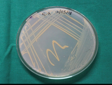
Fig. 4: The test specimen of Staphylococcus aureus
The culture was confirmed by Gram staining, catalase and coagulase test. An inoculating loop was heated red hot on a Bunsen burner, allowed to air cool and then used to pick up bacterial colonies from the agar plate.
This inoculating loop with bacterial colonies was inserted in sterile test tubes filled with 4ml of Sterile Luria broth at room temperature and compared to a 0.5 McFarland Standard to adjust the turbidity of bacterial suspension. (Fig 5)
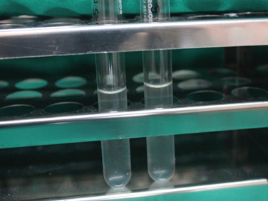
Fig. 5: McFarland Standard test
21 such test tubes filled with Sterile Luria broth were prepared to test the samples. Of these, 1 test tube was kept aside as a Positive control group in which no implant abutment assembly was placed. 10 samples each of Group A and Group B were placed in the remaining test tubes.
The Group A, Group B, Positive control group and a nutrient agar plate streaked with Staphylococcus aureus culture were placed in incubator at 37°C for 15 days and checked intermittently. (Fig 6)
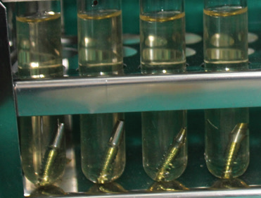
Fig. 6: Samples before incubation
C. EVALUATION OF MICROLEAKAGE: (Fig 7- 19)
After 15 days of incubation, (Fig 7) the samples of Group A and Group B were removed from the test tube using sterile artery forceps (Fig 8, Fig 9) and immersed in 70% alcohol for 3 minutes to prevent external contamination and dried with sterile gauze. This procedure ensures the sterility of the outer surface of the samples without affecting bacterial viability on the inside.
(Fig 10, Fig 11)
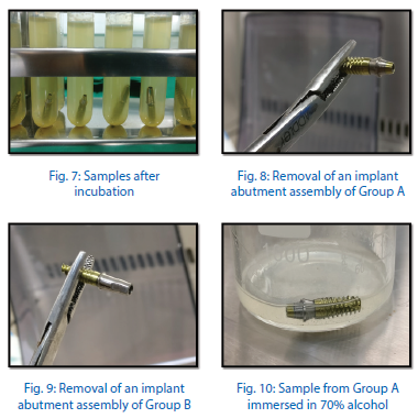
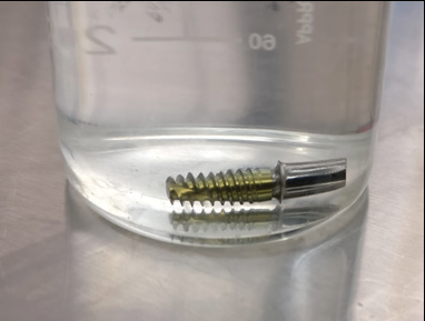
Fig. 11: Sample from Group B immersed in 70% alcohol
In a Biosafety Level 2 cabinet, the abutments were carefully disassembled with a sterile hex driver (Fig 12, Fig 13) and the inner surface of the implants were sampled for microbial contamination using sterile paper points (Fig 14) which were then dipped in saline to prepare the dilutions for the enumeration.
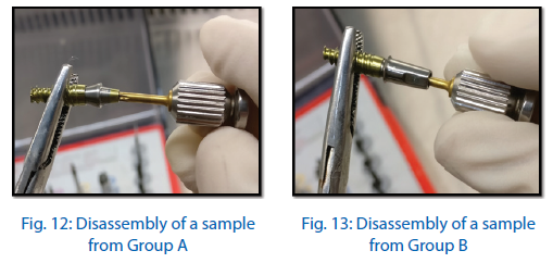
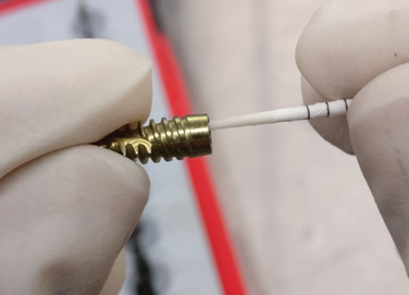
Fig. 14: Swab being taken from the inner surface of the implant
Samples from each specimen had 3 serial dilutions carried out. (Fig 15) Serially diluted test samples were then subjected to enumeration surface spreading technique, on the surface of three sterile Nutrient Agar plates.
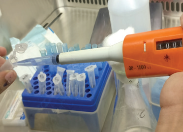
Fig. 15: Serial dilution
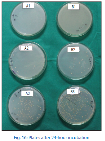
After incubation of the nutrient agar plates at 37°C for 24 hours, the growth of the colonies in the culture medium were evaluated. (Fig 16)
Colonies of bacteria from the nutrient agar plates were picked up using the inoculating loops (heated red hot on a Bunsen burner and air cooled) and placed on the sterile glass slides for Gram staining. (Fig 17)
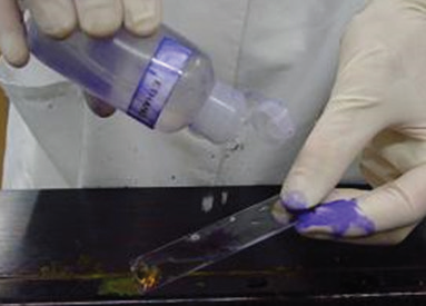
Fig. 17: Gram staining
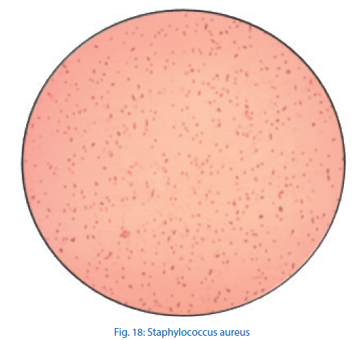
The slides were then observed under a compound microscope. The microscopic appearance of Staphylococcus species were cocci in grape like clusters. (Fig 18) The number of colonies in each serial dilution for each specimen was calculated and tabulated for further statistical analysis.
After microscopic examination, a coagulase test was carried out to verify and confirm the presence of Staphylococcus aureus in the inner part of the assembly.
In the slide coagulase test, a drop of saline is taken on a clean slide. A colony of microorganisms is picked on an inoculation tube and got in contact with the saline. A reaction to that with clumping shows a positive coagulase test. (Fig 19)
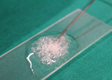
Fig. 19: Coagulase test
RESULT
Table 1. Shows the 3 serial dilutions carried out for the samples. This was done to ensure a more even and equally distributed sample and minimize error.
SERIAL DILUTION
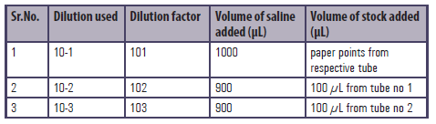
Table 1
Table 2 shows the positive control group, that is the total number of micro- organisms present in the staphylococcus culture. This too was serial diluted and the CFU/ml was evaluated. Colony forming Units (CFU) is a unit used to estimate the number of viable bacteria cells in a sample. Viable is defined as the ability to multiply via binary fission under the controlled conditions.

Table 2
Table 3 and 4 show the microleakage in Group A and Group B respectively. Table 5 shows the Mann Whitney U test for test of significance.
MICROLEAKAGE IN GROUP A
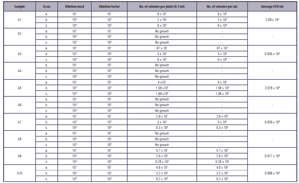
Table 3
MICROLEAKAGE IN GROUP B
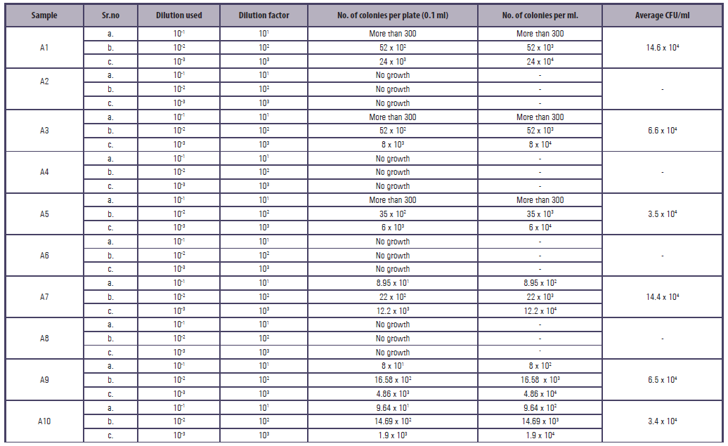
Table 4
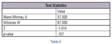
Table 4
Graph 1 shows the comparative value of microleakage between Group A and Group B where as Graph 2 shows the value of microleakage of Group A and Group B when compared with the Control Group.
Mean Comparative Value Graph Comparing Microleakage Between Group A And Group B

Table 4
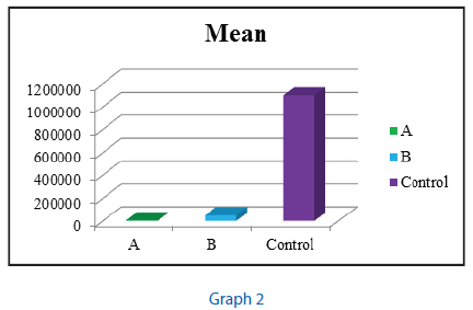
Table 4
The results showed a significant amount of microleakage in Group A and Group B when compared to the control group.
But no statistically significant differences were seen when comparing the microleakage between Group A and Group B.
DISCUSSION
Over the years, two-piece implants have proven their versatility in resolving aesthetic and functional problems. Recent advances in implant-supported prosthodontic restorations have dominated the interest of most prosthodontists. This interest has led to a dramatic increase in the number of implant systems available to the dentist. To make the critical selection of an implant a set of criteria for success based on scientific investigations is essential. One of the important criteria for success of implants is prevention of bone loss and maintenance of peri-implant health.3
In a study by Ohara Nemoto Y,4 the results showed that Staphylococcus aureus was the most frequent species found in the oral cavity and is a significant cause of chronic biofilm infections around medical implants in healthy adults.
To prevent cross contamination the study was conducted in a Biosafety Level 2 cabinet. Several different types of Biosafety Levels exist, differentiated by the degree of biocontainment required.
Biosafety Level 2 Practices, safety equipment and facilities appropriate for work done with a broad spectrum of indigenous moderate-risk agents present in the community and associated with human disease in varying severity. Its features are that:
a. the presence of a laboratory personnel that has specific training in handling pathogenic agents and directed by competent scientists
b. access to the laboratory is limited when work is being conducted
c. extreme precautions are taken with contaminated sharp items
d. certain procedures in which infectious aerosols or splashes may be created are conducted in biological safety cabinets or other physical containment equipment. A Level 1 or Level 2 biological safety cabinet is highly recommended for work involving these agents.
Although the gingiva at teeth and the peri-implant mucosa at titanium implants have many common features, but there are however, also important differences between the two tissues. From the cementum, collagen fibers project laterally perpendicular to the root. Implants lack cementum and the orientation of the attachment fibers in the supracrestal soft tissue compartment is parallel to the implant surface. In the natural dentition, the junctional epithelium provides a seal at the base of the sulcus against bacterial penetration.5,6 The other line of defense present in the natural dentition and absent in implants is the periodontal ligament. Since no cementum or fibers are present on the surface of an implant, infection has the potential to spread directly into the osseous structures resulting in bone loss and ultimately implant failure. Thus, for the maintenance of osseointegration and long-term success of implants, the presence of a leak proof peri-implant soft tissue cuff is desired. This requires the formation of a biological seal dependent on the tight contact between the epithelium and adjacent connective tissue with the implant surface.7
The soft tissue attachment around dental implants or abutments consists of an epithelial zone and a connective tissue zone, represented by the JE and the gingival connective tissue, respectively. The height of the suprabony connective tissue can vary between 1.3 and 1.8 mm.6
Schierano et al41 studied collagen fiber orientation around the implant stem as it exited the bone; collagen bundles were abundant all around the implant, with a maximum density between 200 and 800 μm from the abutment surface.
According to Quirynen M, De Soete M and Van Steenberghe D[8], gaps in the implant abutment interface act as a trap for bacteria, favoring the development of biofilm with varying composition and have an impact on periodontal tissues. Microbial biofilm is infectious in nature and can results in nosocomial infections. Periodontal disease affects tissues that support teeth and is characterized by loss of periodontal ligament insertion, and resorption of adjacent alveolar bone. In implants, as the microbial colonization in the gap increases, it causes an initiation of the inflammatory process which may result in bone resorption.9
Studies by Lindhe J10, Persson LG11, and Cosyn J et al5, suggested that periodontal pathogens and/or their metabolic products might be involved in peri-implant bone losses and similarities have been found in the microbiota of samples collected from the internal parts of implants and from their related peri-implant sulci.
In a study conducted by Harder et al,12 the tightness of the internal conical connections of two implant systems was evaluated and it was concluded that the connections appeared to be unable to prevent endotoxin leakage proving that microscopically, gaps are generally always present.
The data was collected and tabulated for the 10 samples and their respective serial dilutions and averages were calculated.
Prefabricated abutments showed only 0.662 X 10⁴CFU/ml of micro-organisms since the prefabricated abutments are precision milled abutments that have predefined and reproducible contact zones that produce a stable connection between the abutment seat and the implant.
However, Customized abutments showed 4.9 X 10⁴CFU/ml of micro-organisms possibly due to the increased micro gap due to the milling procedure.
Samples from Group A, i.e. prefabricated abutments show lower microleakage when compared to Group B but it was not statistically significant.
Though customized abutments have their advantages over prefabricated abutments in terms of aesthetics, margin placement and gingival contour, there seems to be no statistically significant difference in the amount of microleakage between the two.
The prefabricated abutments are precision milled abutments that have predefined and reproducible contact zones that produce a stable connection between the abutment seat and the implant. The abutment interface is designed to create a barrier for preventing bacterial entry. These abutments are made with advanced production equipment and milling strategies that allow for consistent fit and outstanding surface finish. The design and optimized industrial milling process minimises the rotational tolerance.
Industrial milling machines are bigger and heavier and hence their spindle is more stable when compared to the smaller, lighter laboratory milling machines. This could be the reason why the microleakage in the prefabricated abutments was lesser than that of the customized abutments.
In a similar study carried out by Declan et al,13 the adaptation of abutments to implants was closer and the amounts of contacts were larger for assemblies with premachined and laboratory modified premachined abutments than those with cast abutments hence the microbial leakage maybe less in assemblies with premachined and laboratory modified abutments than those with castable abutments.
REFERENCES
- Aloise JP, Curcio R, Laporta MZ, Rossi L, Madeira A, Da Silva I, et al. Microbial leakage through the implant–abutment interface of morse taper implants in vitro. Clin Oral Implants Res. 2010;21(3):328-35. https://doi.org/10.1111/j.1600-0501.2009.01837.x
- Faria R, May LG, De Vasconcellos DK, Volpato C, Bottino MA. Evaluation of the bacterial leakage along the implant abutment interface. J Dent Implant. 2011;1(2):51-7.
- Ericsson I, Persson LG, Berglundh T, Marinello CP, Lindhe J, Klinge B. Different types of inflammatory reactions in peri – implant soft tissues. J Clin Periodontol 1995;22:255-61.
- Ohara-Nemoto, Y., Haraga, H., Kimura, S., & Nemoto, T. K. (2008). Occurrence of staphylococci in the oral cavities of healthy adults and nasal oral trafficking of the bacteria. Journal of Medical Microbiology, 57(1), 95–99. https://10.1099/jmm.0.47561-0
- Cosyn J, Van Aelst L, Collaert B, Persson G.R. and De Bruyn H. The Peri-Implant Sulcus Compared with Internal Implant and Suprastructure Components: A Microbiological Analysis. Clin Implant Dent Relat Res 2011;13(4):286-95. https:// 10.1111/j.1708-8208.2009.00220.x.
- Berglundh T, Lindhe J. Dimension of the periimplant mucosa. Biological width revisited. J Clin Periodontol 1996;23(10):971-3.
- Gargiulo AW, Wentz FM, Orban B. Dimensions and relations of the dentogingival junction in humans. J Periodontol 1961;32:261-7.
- Quirynen M, De Soete M. and Van Steenberghe D. Infectious risks for oral implants: a review of the literature. Clin Oral Implants Res 2002;13(1):1-19.
- Jansen VK, Conrads G, Richter EJ. Microbial leakage and marginal fit of the implant-abutment interface. Int J Oral Maxillofac Implants 1997; 12:527-40.
- Lindhe J, Berglundh T, Ericsson I, Liljenberg B and Marinello C. Experimental breakdown of peri-implant and periodontal tissues. A study in the beagle dog. Clin Oral Impl Res 1992;3(1):9-16.
- Persson LG, Lekholm U, Leonhardt A, Dahlen G and Lindhe J. Bacterial colonization on internal surfaces of Branemark system implant components. Clin Oral Impl Res 1996;7(2):90-5.
- Harder S, Dimaczek B, Acil Y, Terheyden H, Freitag-Wolf S and Kern M. Molecular leakage at implant abutment connection - in vitro investigation of tightness of internal conical implant-abutment connections against endotoxin penetration. Clin Oral Investig 2010;14(4):427-32. http://dx.doi.org/10.1007/s00784-009-0317-x.
- Byrne D, Houston F, Cleary R, Claffey N. The fit of cast and premachined implant abutments. J Prosthet Dent. 1998;80(2):184-92. https://doi.org/10.1016/S0022-3913(98)70108-8
Disclaimer from Guident
The views and opinions expressed in articles published in Guident are solely those of the authors and do not necessarily reflect the views of the editors, the editorial board or the publisher, Ivory India. Guident does not endorse any product, technique or treatment described. Clinical content is intended for qualified dental and medical professionals and is not a substitute for individual clinical judgement. Guident, its editors and the publisher accept no liability for any consequence arising from the use of information published in the journal.
How to cite this article
Dr. Naina Swarup, Dr. Gaurang Mistry, Dr. Omkar Shetty. Comparative Evaluation of Microleakage at the Implant Abutment Interface Using Prefabricated and Customized Abutments. Guident. 2019 Dec;13(1). Available from: https://guident.in/articles/research/comparative-evaluation-of-microleakage-at-the-implant-abutment-interface-using-prefabricated-and-customized.html