Management of Pan-Facial Trauma and Orbital Floor Reconstruction with Iliac Crest Bone Graft
Department of Dentistry, SIPS Superspeciality Hospital & Trauma Centre, Lucknow (U.P) INDIA
ABSTRACT
Pan facial fracture being the most complicated, as it involves multiple fracture of lower third, middle third and upper third of face and so the management of such fracture is extremely complicated. The pattern of repairing the pan facial fracture is most challenging and requires great experience and knowledge. All the vertical and horizontal pillars have to be made to restore and rehabilitate the facial anatomy, aesthetics and occlusion.9 The diagnosis of these fractures should be established with thorough clinical examination and careful radiological investigations so that a 3-dimensional view of the fractured bones can be obtained. This is important in order to plan a proper surgical treatment to reconstruct the
face in terms of function and aesthetics.4
This article presenting a case of compound and comminuted orofacial trauma associated with soft tissue lacerations. This is an unusual case of unilateral (Left side) fracture of Maxilla, Zygomatic and Orbital floor, in which a complete block of these bones are separated, which is extremely rare. As it was basically an occlusion guided surgery, the fracture is well managed, and stabilized and orbital floor is reconstructed using iliac crest graft by a combined team of Plastic surgeon and Dental surgeon.
Keywords
Panfacial, trauma, fracture, reconstruction, maxilla, orbital, zygomatic, occlusion.
INTRODUCTION
Pan facial fractures are common facial fractures that can result in severe esthetics and functional sequale. Surgical correction of these fractures is a delicate approach and challenging for the surgeon, because of lack of an occlusal guide and involvement of various structures of the face. The approach of such complex injuries must be individualized and basic principles of diagnosis, preoperative planning, wound care and post operative management must be applied.2 Although there is no clear definition & classification of Panfacial fractures in the literature but the term usually refers to simultaneous fracture of several bones of the face with severe degree of fragmentation which makes it difficult to restore the facial architecture. Panfacial fractures involve fractures of mandible, maxilla, zygomatic complex, most often nasoorbitoethmoid (NOE) & frontal bone. They are associated with malocclusion, dish face deformity; enopthalmos, diplopia, CSF leak & soft tissue injuries. Markowitz & Manson describe frontal and palatoalveolar fractures as extended injuries of Panfacial fractures. The surgeon’s task is not only to ensure a complete anatomical esthetics and functional repair of the face but restoring to its original 3- dimensional shape.6-7
The zygomatic bone or zygoma is a strong buttress of the lateral portion of the middle third of the facial skeleton lying between the zygomatic processes to the frontal bone and the maxilla. Due to its prominent position, it is frequently fractured, alone or along with other bones of the midface. Direct blows usually first strike on the most prominent part which is the malar eminence. This causes disruption at the relatively weaker part which is the zygomatic arch, the frontal process, and the zygomaticomaxillary suture. A number of classifications for zygomatic fractures have been developed based on the anatomy and displacement of the fracture. The most widely used classification of zygomatic fracture is those proposed by Dingman and Rowe and Killey’s.4 Studies on the incidence of zygoma fractures have shown that road traffic accidents and assaults are the commonest causes of zygomatic complex fractures. Tetrapod fractures (43.7%) were the more frequently.
reported compared with isolated zygomatic arch fractures (34.53%). Tetrapod is term used to describe zygomaticomaxillary complex fracture. Tetrapod means ‘‘four processes’’.95 Maxillary fracture is defined as the separation of parts or the entire tooth-bearing part of the maxilla from the residual midface or the neurocranium.4
Anatomically, the anteromedial 3rd of the floor of orbit is formed by orbital surface of maxilla, where as the postero-medial part is formed by palatine bone and lateral 3rd is formed by zygoma. The medial wall of the orbit is formed anterior to posterior, by frontal process of the maxilla behind its lacrimal crest, lacrimal bone, orbital plate of ethmoid and body of sphenoid. The ‘blow out’ fracture of orbit usually involves the medial wall and floor of the orbit being the thinnest. Absolute indications for reconstruction of orbital floor are diplopia with muscle entrapment, enopthalmos and large defect. Many materials have been described for the reconstruction of orbital floor defects with varying rate of success. They include autogenic, allogenic and alloplastic material.3 This article reports management of a case of orbital floor fracture using autogenous iliac crest bone graft to reconstruct the defect.
The diagnosis of fracture of zygoma and maxilla can usually be made with thorough clinical examination and adequate radiological evaluation. Plain radiograph commonly used in midfacial fracture is occipito-mental or Water’s view which can clearly demonstrate the bone discontinuity in the zygomaticomaxillary buttress and the inferior orbital rim. The submentovertex view more clearly detects fractures of the zygomatic arch. CT scan and its three-dimensional applications are indicated for visualization of the orbit if the orbital portion of the zygomatic fracture is suspected. Once the diagnosis is established a surgical treatment plan can be made. The current principle of treatment of maxillofacial fracture is open reduction and rigid fixation and in a complex fracture of the zygoma and the midface a certain surgical plan should be followed in order to have good result.4
EPIDEMIOLOGY OF PANFACIAL FRACTURE
The more the velocity of trauma more the destruction and fragmentation of bone occurs, as the facial framework involve multiple bones. Therefore high energy injuries are the commonest cause of pan facial injury (road traffic accident, gunshot injuries). The incidence for such injury is 4-19% for all facial fractures.CASE REPORT
A 23 years old male patient, a motorcycle accident victim, was brought by an ambulance to the emergency department of SIPS Superspeciality Hospital & Trauma Centre with open wound & bleeding in left side in infraorbital, cheek and zygomatic region and soft tissue lacerations in left side of the face. Airway was clear and all the vital signs were normal. Patient was stabilized and thoroughly examined to rule out any multisystem injury. Lip lacerations were sutured using 3-0 vicryl under LA (1:80000).
On clinical examination there was crushed lacerations with loss of skin and soft tissue with ragged margins and comminuted and compound fracture of maxilla, zygomatic and orbital floor. On intraoral examination, there was deranged occlusion with floating maxilla from canine to molar region in left side. The patient was submitted to face high resolution CT scan with 3-D reconstruction for better characterization of visible fractures and exclusion of associated lesions. Clinical and radiographic analysis revealed fracture involving maxilla, zygomatic and orbital floor of left side of face and a complete block of these bones was separated.
RADIOGRAPHIC EVALUATION
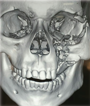
Fig. 1
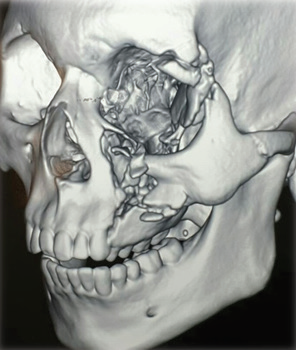
Fig. 2

Fig. 3
CT scan with 3-D reconstruction
Patient relatives were informed about need for surgical intervention and informed written consent was obtained. All the routine blood investigations were done which are required for surgery to be done under general anaesthesia. Reduction of fractures and internal fixation and reconstruction of orbital floor with the help of autogenous bone graft under general anesthesia was planned.Patient was taken to OT and nasal intubation was done for induction of GA. As, occlusion was main guide of surgery, first of all, upper and lower dental arch was immobilized with Ehrich arch bar and Maxillomandibular [MMF] was done to achieve a good occlusion.
Two Intrafragmentary screws of 6mm in length and 1.5mm thick were fixed. One screw is fixed between first and second molar region and second screw was fixed at the junction of frontonasal and maxillary process.

Intrafragmentary screw fixation
Axial view CT scan of skull reveled fracture of left orbital floor along with herniation of periorbital soft tissue into the antrum. As the complete orbital floor was damaged, there was no need to give any incision. The infraorbital nerve was identified and protected. The herniated periorbital content including inferior rectus muscle and fat was disengaged from the bony defect and is lifted up and keeping back in correct position. Autogenous anterior iliac crest bone graft was harvested from the inner table of right iliac crest with the help of osteotome. Some cortical portions of the bone was harvested and on the basis of measurement of defect done with the scale, the graft was shaped to match the contour and size of the defect of the orbital floor. Closure of donor site was performed in layers and the graft pieces was fixed with the help of sutures and a mesh form structure was given according to the curvature and anatomy to adapt in the proper framework on the orbital floor. A spatula was used to protect the globe and to restrict the herniation of the fat in the recipient site. The graft was placed over the defect and fixed and tied with the help of wire and spatula was removed. Symmetry of globe was checked after achieving the desired result and length and width of face is ensured from the contralateral side.
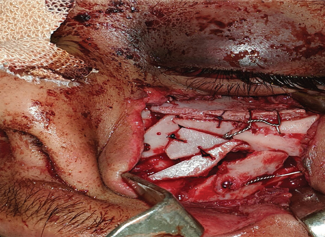
Fig. Iliac crest bone graft reshaped and fixed with the sutures and placed over the defect forming a mesh type structure and tied with the help of wire.
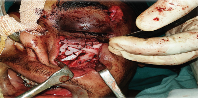
Orbital floor reconstruction
By the end of procedure, a satisfactory reduction of the bone fragment was achieved without its vascular compromise. The closure was done in layers with 3-0 vicryl. The patient recovered uneventfully after which the patient was shifted to post-operative ward. Post- operative medications were advised including antibiotics and analgesics. Oral hygiene was maintained using chlorhexidine irrigation. Extraoral sutures were removed after a week. Patient was advised to take soft diet for about one month. Post operative stability and functions were satisfactorily with an imperceptible scar. Ehrich arch bar was removed after 3 weeks of surgery. The post operative recovery was uneventful with good mouth opening and perfect occlusion with no cosmetic deficit. Post operative radiograph showed well established graft in place.
Patient was reviewed at regular intervals, initially once every week for 2 months, followed by every 2 weeks for 3 months and subsequently once in every month postoperatively. At 1 month postoperative review, the patient did not complain of any pain nor difficulty in eating and he was completely satisfied with his appearance.
POST-OP PROFILE

(A) Straight profile (B) Lateral profile

(C)
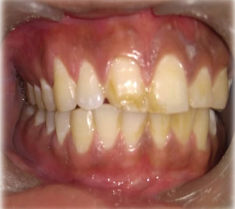
Right side
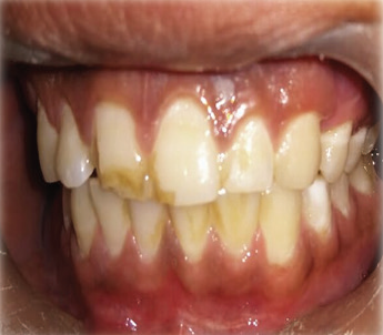
Left side
Post-op occlusion
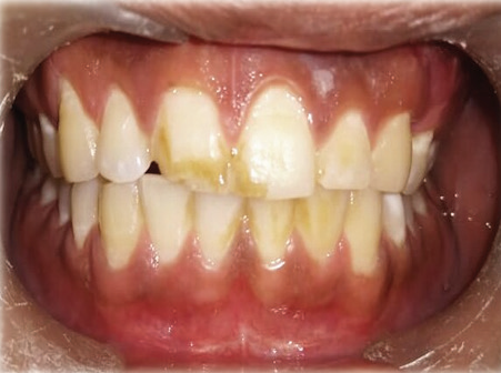
Complete Occlusion
POST-OP PNS VIEW
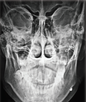
Fig. 1
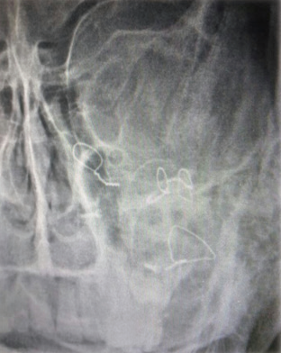
Fig. 2
POST-OP 3-D RECONSTRUCTION
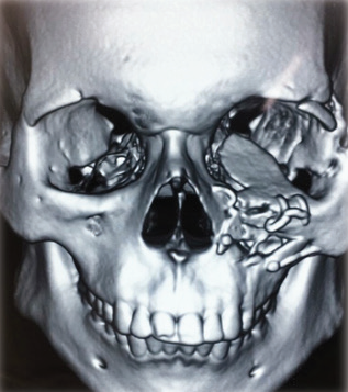
Fig. 1
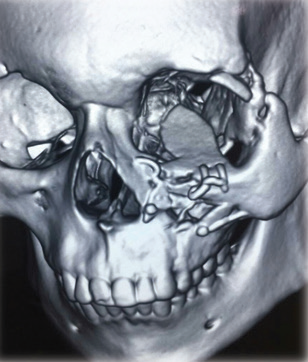
Fig. 2
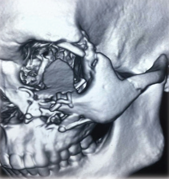
Fig. 3
DISCUSSION
Midface orofacial trauma may result in potentially disfiguring injuries with important functional impact, as it commonly affects dental occlusion. The initial approaches to complex facial trauma include the confirmation of a patent airway, hemodynamic stability and hemorrhage control, along with a carefully collected history and complete physical examination. Imaging studies play an important role in the characterization of facial traumatic injuries, particularly in the presence of facial bone fractures. High-resolution CT is often the most valuable exam to diagnose and characterize facial fractures in the emergency department, and may also prove valuable to diagnose associated soft tissue injuries.
Principles of treatment in this type of injuries include exploration, debridement of devitalized tissue, generous wound irrigation and reduction of fractures achieving dental and bone stabilization. Although the maxilla is commonly involved in facial trauma. The management of maxillary fractures requires achievement of dental stability and previous occlusion as well as restoration of height and projection of the midface.2
The management of Panfacial fracture is extremely complex. The goal of treatment, as with all facial fractures is to restore both the function and pre injury three dimensional facial contours. To achieve this goal, various management schemes have been proposed including ‘’bottom to top’’, ‘’top to bottom’’, ‘’inside-out’’, or ‘’outside-in’’.7 The horizontal and vertical buttresses make the framework of face and also help in transmission of mastication force to the base of skull. The facial buttresses absorb the forces and prevent its transmission to brain. The buttress of face are like pillars and hence need to be reduce and stabilize properly for complete rehabilitation of facial structure and profile. Proper alignment of facial skeleton provides functional and anatomic stability to middle third of face.
The current method of treatment in maxillofacial, including midface, fracture is open reduction and internal fixation with miniplates and screws which offers stable reduction of the fracture fragments. This would consequently allow for early mobilization of the jaw thus early and optimal recovery of function and esthetic.4
Patients with pan-facial trauma should be taken care according to Advanced Trauma Life Support (ATLS) guidelines as stated by Robert Marciani. After clinical examination, confirming the diagnosis with the help of imaging techniques followed by surgical intervention, we have done the same in this case report. To avoid the post operative deformity and undesirable aesthetics, early surgical intervention is must to perform. The patient was operated after 2 days of trauma, on complete clearance from medicine, neurosurgery and other specialized department.9
Occlusion was achieved by Maxillomandibular fixation also brings maxilla in its proper position. Interfragmentary screws were used for stabilization and fixation of zygoma and iliac crest bone graft is used for the reconstruction of orbital floor. As there were so many crushed fragments of bone, the fixation with miniplates was not possible that’s why it is planned to fix them with the help of wire.
The commonly used surgical approaches to reconstruct the floor of the orbit are transconjunctival, subciliary and infraorbital. The basic objective of reconstruction of orbital defect is to restore orbital volume, function and aesthetics. To reconstruct the defect of the orbital floor, various autogenous as well as allogenic and alloplastic material have been used with their merits and demerits. In the present case, autogenous iliac cortical bone graft was used to repair the defect as it maintains the volume better in comparison to the cancellous one irrespective of their origin and there is least chance of contamination of the graft. In the present case, the autogenous iliac crest cortical bone graft served the basic goal of reconstruction of the orbital defect satisfactorily.3 Autogenous bone grafts are the profound materials of choice to avoid the graft-host reaction but have a drawback of displacement problems, secondary field of surgery, donor site morbidity & unpredictable resorption of graft. Infection,extrusion,tissue reaction,residual diplopia,foreign body reaction is associated with allogenic & alloplastic implants. The rationale of using iliac crest bone graft is its relative resistance to infection, incorporation by the host in the new bone, Lack of host response against the graft & lack of concern for late extrusion. Although there are multiple sites of autogenous grafts, the anterior iliac crest bone remains the most common site& is a favorable reconstructive material as enough bone is always available & also bone can be harvested simultaneously with orbital exploration. It provides the gold standard framework for facial skeleton & orbital wall. Other autogenous grafts are calvarium,tibia,ulnar,mandibular symphysis,ribs,coronoid process & so on. Medpore is also used for the same. The desirable characteristic of a bone grafts is sufficient volume, minimal donor site morbidity,obtaining intramembranous bone with high cortical component, proximity to the residual site, ease of harvesting& achieving of reproducible a good results & minimal resorption rate.10
The midface fractures has tendency to quick and spontaneous healing especially in non-displaced fractures where the maxilla is immobile or only slightly mobile. However, it may be disadvantageous in such clinical situation as when the trauma team needs several days to stabilize the patient and the maxillofacial surgeon cannot intervene early enough, the fractures may have begun to consolidate in displaced positions. Fourteen days is the upper limit of primary facial reconstruction in midfacial fractures. It is suggested that reduction and fixation up to 3 weeks after trauma produces satisfactory results. After this time, bone healing and resorption begin to take place, and this period has been referred to as a gray time zone. This applies to the case presented here. As the bony fracture in the zygoma and the maxilla were still two days old then it is reasonable that bone consolidation was not found during the surgery and the displaced zygomatic complex could be reduced without much difficulty.4
CONCLUSION
Pan facial trauma can be complex and the management of such complex injuries often demands an individualized approach. Careful diagnosis using adequate imaging studies, adequate preoperative planning, and proper wound care and infection prevention are common principles important for the successful treatment of these patients. Diagnostic-therapeutic management inpatients following Panfacial trauma of the facial skeleton should include teamwork of specialists in the field of maxillofacial surgery, ophthalmology, neurosurgery, plastic surgery, dental surgery and anesthesiology. Depending on the patient’s general condition therapeutic management should be individual depending on the extent of injuries, fractures should be treated according to 4E rule (early, everything, entirely, equally).Thorough anatomical knowledge and expertise of the maxillofacial surgeon is must for managing a case of pan facial trauma using either of the approaches. To conclude; a minimally invasive approach should be used to treat the Panfacial fractures.Early surgical intervention to reduce and fix the fractures after stabilizing the trauma patient yields good postoperative results. The surgical approach to facial fracture management should focus on attaining proper occlusal, vertical and horizontal relationships of the facial frame along with restoration of orbital and oral cavities.
REFERENCES
- Amanda Lucio do O Silva, Isabella Jardelino Dias, Rafael Grotta Grempel, Hecio Henrique Araujo de Morais, Nadja Maria da Silva Oliveira Brito, Daliana Queiroga de Castro Gomes. Prototyping for the treatment of late zygomaticorbital fracture: A case report. International Journal of Surgery Case Reports. 38 (2017) 91–94.
- Ines Insua-Pereira, Pedro Ferreira, Joana Costa, Sergio Teixeira and Alvaro Silva (2018).Maxillary and Palatal Fracture With Near-Avulsion of Bone Fragment-Case Report. Crimson publishers Modern research in dentistry. 2 (5):1-3
- Lt Col TK Bandyopadhyay, Brig BL Sapru (Retd) VSM. Management of an Isolated Orbital Blow-out Fracture. MJAFI 2004; Vol 60, No. 4: 392-394.
- David B. Kamadjaja and Coen Pramono D. Management of zygomatic-maxillary fracture (The principles of diagnosis and surgical treatment with a case illustration).Dent. J. (Maj. Ked. Gigi), Vol. 41. No. 2 April-June 2008: 77-83.
- Manikandhan Ramanathan and Mathew Pynumootil Cherian. Isolated Bilateral Zygomatic Complex and Arch Fracture: A Rare Case Report. Craniomaxillofacial Trauma Reconstruction 2010; 3 (4):185–188.
- Usha Asnani, Smita Sonavane, Fawaz Baig, Srivalli Natrajan. Panfacial Trauma - A Case Report. International Journal of Dental Clinics 2010: 2 (2): 35-38 Vol. 2, Issue 2, April- June 2010.
- Mall BB, Kandel L, Mishra R, Kandel S. Panfacial trauma- A flexible surgical approach. Journal of universal college of Medical sciences (2014) Vol. 02 No. 03 Issue-7 (42-44).
- Aneta Neskoromna-Jedrzejczak, Katarzyna Bogusiak, Aleksander Przygonski, Dariusz Timler. Penetrating trauma to the facial skeleton by Pickaxe– Case report. POLSKIPRZEGLAD CHIRURGICZNY 2016, 88, 1, 48–53.
- Dr. Anirudha Singh, Dr. Vikas Kunwar Singh, Dr. Ruchika Tiwari, Dr. Gaurang Thanvi. Journal of Medical science and clinical research. Vol. 6, Issue 5, Page 691-695, March 2019.
- Dr. Rahul Tiwari, Dr. Philip Mathew, Dr. Bhaskar Roy, Dr. Yasmin Jose, Dr. Kritika Sherawat, Dr. Kaushal Charan Pahari, Dr. Heena Tiwari. IOSR Journal of Dental and Medical Sciences (IOSR-JDMS) e-ISSN: 2279-0853, p-ISSN: 2279- 0861.Volume 16, Issue 7 Ver. 6 (June. 2018), Page: 9-11.
Disclaimer from Guident
The views and opinions expressed in articles published in Guident are solely those of the authors and do not necessarily reflect the views of the editors, the editorial board or the publisher, Ivory India. Guident does not endorse any product, technique or treatment described. Clinical content is intended for qualified dental and medical professionals and is not a substitute for individual clinical judgement. Guident, its editors and the publisher accept no liability for any consequence arising from the use of information published in the journal.
How to cite this article
Dr. Sumit Malhotra, Dr. Vikram Ahuja, Dr. Abhishek Kr. Pandey. Management of Pan-Facial Trauma and Orbital Floor Reconstruction with Iliac Crest Bone Graft. Guident. 2019 Jul;12(8). Available from: https://guident.in/articles/oral-surgery/management-of-pan-facial-trauma-and-orbital-floor-reconstruction-with-iliac-crest-bone-graft.html