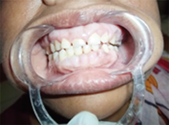
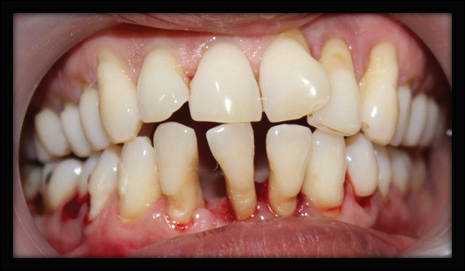
Vestibular Incision Subperiosteal Tunnel Access a Novel Technique for the Management of Multiple Recession Defects.
Department of Periodontology, Yenepoya Dental College, Mangalore, India
INTRODUCTION
In routine dental practice “gingival recession (GR)” defects are frequently encountered complain because of functional and esthetic concern of the patients. GR is defined as the “displacement of marginal tissue apical to the cementoenamel junction (CEJ)1 Recession defects can be isolated or multiple. First line of treatment includes removal of etiological factors followed by surgical correction. Several periodontal plastic surgical procedures have been documented for the treatment of Miller’s Class I and II recession defects and negotiate patient’s discomfort due to hypersensitivity, food lodgement, fear of tooth loss, etc.2The treatment of recession defects associated with multiple teeth poses greater challenge to clinician as avascular root surface area is more extensive. Also, thin biotype, decreased keratinized tissue width (KTW), root prominence and root proximity make the choice of surgical treatment difficult as compared to localized gingival recession type defects Different tunnel techniques that can maintain the better blood supply and maintain critical papillary integrity have been attempted for management of recession defects.3
Various tunnel techniques were described to maintain the recipient site esthetics and to prevent relapse of GR. These procedures, while maintaining the critical papillary integrity and avoiding vertical releasing incisions, allow management of multiple GR defects at the same time. The technique sensitivity with these tunnel techniques and increased trauma to the sulcular epithelium led to unfavorable healing outcomes, so a newer approach, known as the vestibular incision subperiosteal tunnel access (VISTA) technique, is used to avoid some of the potential complications occurring with other intrasulcular tunneling techniques.4
Various adjunctive agents, such as recombinant human growth factors and platelet-rich plasma, were used to accelerate healing and further enhance clinical outcomes. Platelet-rich fibrin (PRF), a second-generation platelet concentrate, is obtained from autologous blood with simplified processing without the need for biochemical blood handling. It has become a focus of studies because of its potential to accelerate healing. The processing of PRF involves the sequestration and concentration of platelets in autologous whole blood through centrifugation and natural polymerization. The strategy is to amplify and accelerate the effects of growth factors contained in the platelets.2
The purpose of this case report is to evaluate clinically, the efficacy of the novel and minimally invasive VISTA in combination with PRF in the treatment of multiple gingival recession defects.
CASE REPORT
A 35 year old female patient with a chief complaint of poor esthetics and hypersensitivity attributed to Gingival recession reported to the Department of Periodontology, Yenepoya Dental College Mangalore. On examination Miller’s Class I recession was noticed in relation to 11, 12. Recession depth was 2mm and width was 3mm in relation to 11.( Fig 1-3) In relation to 12 both recession depth and width was found to be 2mm. Thorough scaling and root planing was performed as a part of phase 1 therapy. Patient was recalled after 15 days for review. It was decided to subject the sites for root coverage procedure using combination of VISTA and PRF.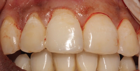
Fig 1: Pre- operative photograph

Fig 2: Pre operative Recession width-11
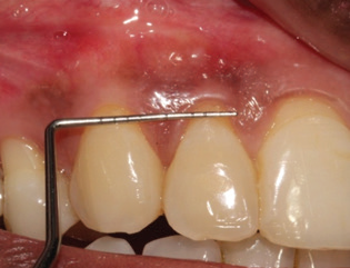
Fig 3: Pre operative Recession width-21
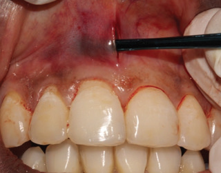
4: Subperiosteal tunnel incision
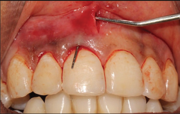
Fig 5: Subperiosteal pouch

Fig 6: PRF prepration
Under local anaesthesia, a vestibular access incision was given in midline of the maxillary labial frenum. The incision was continued through the perioosteum creating a subperiosteal tunnel. Through the vestibular access incision the periosteal elevator was introduced and inserted between the periosteum and bone to elevate the periosteum, creating the subperiosteal tunnel. The incision was extended one or two teeth beyond the teeth requiring root coverage and beyond mucogingival junction to mobilize gingival margin and allow coronal repositioning. Additionally, the subperiosteal tunnel was extended interproximally under each papilla as far as the embrasure space permits, without making any surface incisions through the papilla. ( Fig 4-5).
PRF Procurement
The preparation of PRF was performed as per the protocol developed by Choukroun et al.5 Intravenous blood (10mL; antecubital site) was centrifuged using a tabletop centrifuge at 3,000 rpm for 10 minutes. ( Fig 6) After centrifugation, the PRF clot was removed from the test tube using a sterile tweezer, then separated from the red blood cell base (leaving a small RBC layer) using a sterile scissor. The PRF membrane was prepared by placing the PRF clot into PRF-BOX (Fig 7).

Fig 7: PRF membrane
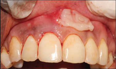
Fig 8: Placement of prf membrane
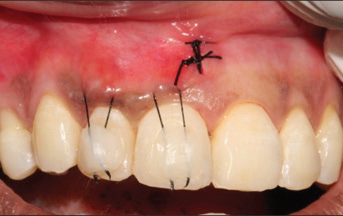
Fig 9: Coronally advanced suture placed
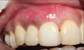
Fig 10: Post operative 10 days
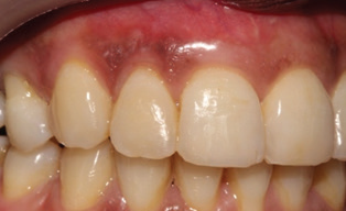
Fig 11: Post operative 3 months

Fig 12: Pre - operative

Fig 13: Post operative 6 months
A freshly prepared platelet rich fibrin, was trimmed according to the desired dimension of the recipient site and extended 3-4 mm beyond the area to be covered. (Fig 8)The membrane and mucogingival complex were then advanced coronally and stabilized in the new position with a coronally anchored suturing, by placing a horizontal mattress suture at approximately 2 to 3 mm apical to the gingival margin of each tooth (or within the band of keratinized gingiva). The suture was then tied to position the knot at the mid coronal point of the facial aspect of each tooth, which was secured with help of composite resin to prevent apical relapse of the gingival margin during initial stages of healing. (Fig 9) Patient was advised post operative instructions and strict oral hygiene maintenance. Patient was prescribed analgesics for 3 days. Suture removal was done after 10 days. (Fig 10). Patient was recalled for 3 and 6 month follow up (Fig 11,13).
DISCUSSION
Periodontal therapy has historically been directed primarily at the elimination of disease and maintenance of a functional healthy dentition and supporting tissues. However, more recently periodontal therapy, consistent with dental therapy in general, is increasingly directed at esthetic outcomes for patients, which extend beyond tooth replacement and tooth color to include the soft tissue component framing the dentition.6,7
There are numerous periodontal plastic surgical techniques undertaken for root coverage that restore the periodontal tissues to healthy, esthetic and functional state. It is very important to determine the appropriate technique depending upon the clinical situation to yield satisfactory, successful and predictable results in management of gingival recession.
Gingival recession is not always localized. Many times multiple teeth are involved. Esthetic appearance of anterior maxillary teeth while smiling is the main indication for root coverage.8
Treatment of gingival recessions has become an important therapeutic issue due to the increasing number of cosmetic requests from patients. Patient’s esthetic demands, due to exposure during smiling or function, of portions of the root surface are the main indication for root coverage procedures. Thus, complete root coverage up to the cementoenamel junction is the goal to be achieved when the patient complains about esthetic appearance of teeth. Furthermore, even if complete root coverage is surgically accomplished; the result may not be completely satisfactory in the case of excessive thickness of gingiva or poor blending of the area. This happens very frequently when free or connective tissue graft is harvested from the palate and utilized for root coverage.7
Although considered the current gold standard, the CTG presents a number of disadvantages, including the need for harvesting at a distant donor site, limited tissue availability, and increased potential for post harvesting morbidity. These disadvantages are even more problematic in patients with multiple contiguous gingival recession defects, since optimizing esthetic results in part depends on simultaneous treatment of all contiguous recessions.8
VISTA is modified CAF introduced by Zadah et al. in 2011 for coverage of Class I multiple maxillary recession defects9 One important feature that makes the VISTA technique stand out of the rest is the difference in the approach used in the formation of the tunnel flap. In the VISTA technique there is a wide dissection of the soft tissues in order to eliminate possible pull. This allows for a maximal move of the tissues over the cementoenamel junction (CEJ) while the stitching technique effectively prevents possible apical movement of the marginal gingiva during the initial healing period. The elimination of the pull on the gingival tissues is essential in achieving good final results when treating gingival recessions.10
In the present case report we have achieved 100% root coverage, and consisting results where observed till the end of 6 months similar results where observed in a report published by Zadeh et al9 using combination of VISTA technique with Bioguide collagen membrane.
Singh AK et al11 used platelet rich fibrin reinforced periosteal pedicle graft with vestibular incision subperiosteal tunnel access technique for the coverage of exposed root surface and attained 84.6% of the root coverage. Significant improvement during the early periodontal healing phase with 96% root coverage is seen with VISTA technique along with PRF in a recent 12 month study by Chatterjee A et al7 Ready SP et al3 used PRF with VISTA technique and obtained 100% root coverage in Millers class I recession. In a study by Monica GS et al 12, 98.7% root coverage was obtained after 4 months.
Grafting serves as a scaffold to support wound healing and provide better root coverage. Thus, this technique was further reinforced by PRF-membrane to increase and improve the width and thickness of keratinized gingiva for the treatment of Classes I and II recession defects.13 The usage of this autogenous biological material eliminates completely the danger of adverse reactions to foreign (non-autogenous) materials. Use of PRF in these case reports has several advantages over other membranes or grafts like they play multiple vital roles in early wound healing through angiogenesis, cementogenesis, osteoblast differentiation, mitosis, chemotaxis, development, and maturation of a normal vasculature, cost effective and eliminates any chances of immune reaction.10,12
CONCLUSION
Multiple contiguous gingival recession defects, however, pose significant functional and esthetic problems to large numbers of the population. VISTA is a novel approach for managing multiple gingival recession defects in the esthetic zone, Available literature suggests that results are promising when attempted in combination with PRF. However, further studies with large samplesize and longer duration are advised to determine the success and predictability of this technique.
REFERENCE
- American Academy of Periodontology. Glossary of Periodontal Terms. 3rd ed. Chicago: American Academy of Periodontology; 1992.
- (Root-coverage procedures for the treatment of localized recession-type defects: a Cochrane systematic review. Chambrone L, Sukekava F, Araújo MG, Pustiglioni FE, Chambrone LA, Lima LA J Periodontol. 2010 Apr; 81(4):452-78.)
- Reddy S et al. Vestibular incision subperiosteal tunnel access (VISTA) with platelet rich fibrin (PRF) and connective tissue graft (CTG) in the management of multiple gingival recession- A case series. International Journal of Applied Dental Sciences 2016; 2: 34-37.
- Gupta G, Puri K, Bansal M, Khatri M, Kumar A. Platelet-rich fibrin reinforced vestibular incision subperiostal tunel access (VISTA) technique for recession coverage. Clinic Adv Periodontics 2014;7:1-13.
- Choukroun J, Adda F, Schoeffler C, Vervelle A. An opportunity in perioimplantology: The PRF (in French). Implantodontie 2001;42:55-62
- Anilkumar K, Geetha A, Umasudhakar, Ramakrishnan T, Vijayalakshmi R, Pameela E. Platelet-rich-fibrin: A novel root coverage approach. J Indian Soc Periodontol 2009;13:50-54.
- Chatterjee A, Ena Sharma, Gayatri Gundanavar, Shobha Krishna Subbaiah. Treatment of multiple gingival recessions with vista technique: A case series. J Indian Soc Periodontol. 2015; 19:232-5.
- Agarwal DR, Gurav AN, Shete AR, Shetgar S. The VISTA Approach to Treat Gingival Recession in Esthetic Zone. 2015;5(12):410-414.
- Zadeh HH. Minimally invasive treatment of maxillary anterior gingival recession defects by vestibular incision subperiosteal tunnel access and platelet-derived growth factor BB. International Journal of Periodontics and Restorative Dentistry. 2011 Nov 1;31(6):653.
- Iv C, Neichev D, Vicheva D, Atanasov D, Noncheva V. Vista technique and Platelet-Rich Fibrin Membrane for Treatment of Multiple Adjacent Gingival Recessions-6 month follow up. IOSR Journal of Dental and Medical Sciences. 2016;15(7):5. Singh AK, Gautam A. Platelet-rich fibrinreinforced periosteal pedicle graft with vestibular incision subperiosteal tunnel access technique for the coverage of exposed root surface. J Interdiscip Dentistry 2016;6:33-8.
- Monica GS, Rakesh Kumar Verma, Veerpal Brar, Hema Latha, MSM Biir. Vista technique with platelet rich fibrin – a case report. International Journal of Contemporary Medical Research 2018;5(9):I10-I12.
- Garg S, Arora SA, Chhina S, Singh P Multiple Gingival Recession Coverage Treated with Vestibular Incision Subperiosteal Tunnel Access Approach with or without Platelet-Rich Fibrin - A Case Series. Contemp Clin Dent. 2017; 8(3):464-468.
Disclaimer from Guident
The views and opinions expressed in articles published in Guident are solely those of the authors and do not necessarily reflect the views of the editors, the editorial board or the publisher, Ivory India. Guident does not endorse any product, technique or treatment described. Clinical content is intended for qualified dental and medical professionals and is not a substitute for individual clinical judgement. Guident, its editors and the publisher accept no liability for any consequence arising from the use of information published in the journal.
How to cite this article
Dr. Shashikanth Hegde, Dr. Sazia Amreen, Dr. Jyosthna G Madhurkar. Vestibular Incision Subperiosteal Tunnel Access a Novel Technique for the Management of Multiple Recession Defects. Guident. 2019 Jul;12(8). Available from: https://guident.in/articles/periodontics/vestibular-incision-subperiosteal-tunnel-access-a-novel-technique-for-the-management-of-multiple-recession.html