Effect of Lateral Arthroplasty on Mandibular Growth: A Cephalometric Analysis Case Report
Department of Maxillofacial and Oral Surgery, Himachal Pradesh Government Dental College And Hospital, Shimla
ABSTRACT
Temporomandibular joint (TMJ) ankylosis is a debilitating condition characterised by the fusion of bony or fibrous tissues in the TMJ. Childhood TMJ ankylosis frequently has a detrimental impact on the facial growth resulting in skeletal deformity. Trauma is the main cause of TMJ ankylosis, but the condition can also occur as a result of surgery, local or systemic infections, or systemic disease. Surgery is the mainstay of treatment. The aim of this report is to describe the facial growth changes in patient treated with lateral arthroplasty in tmj ankylosis patient and by comparing the preoperative affected site with postoperative treatment outcomes.
Keywords: Lateral Arthroplasty, Mandibular Growth, Cephalometric Analysis.
INTRODUCTION
Originating from the Greek word ankylsis meaning joint stiffening, temporomandibular joint (TMJ) ankylosis is characterized by the joining of the lower jaw's condyle to the skull's glenoid fossa, disc, and/or eminence.1 This fusion typically leads to severely restricted jaw movement, evident as a limited maximum mouth opening (MIO), difficulty protruding or moving the jaw sideways, impaired chewing, and problems with breathing, speaking, eating, and oral hygiene.2,3 Moreover, TMJ ankylosis in children can negatively impact facial development, often causing facial asymmetry, a small lower jaw (mandibular micrognathia) resulting in a bird-like facial appearance, and/or a Class II malocclusion with an Anterior or Posterior open bite.4 Various procedures for the treatment of TMJ ankylosis have been described in the literature. These include gap arthroplasty (GA), interpositional gap arthroplasty (IPG), and/or joint reconstruction using autogenous grafts or alloplastic materials.5
Zhi et al. reported that the commonly accepted surgical approach for the treatment of ankylosis types III and IV includes total resection of the ankylotic area with special attention to the medial aspect of the joint remnants.6 Distraction osteogenesis open new perspective for mandibular elongation in interceptive surgery in case of facial growth disturbance due to condylar injury especially in children.7 When fractured transversely below the insertion of the lateral pterygoid muscle, the condylar process as well as the disc are displaced and pulled in an anteromedial-inferior direction. Such fractures are treated by either an open reduction or closed management.
Nitzan and colleagues initially proposed that in treating type III temporomandibular joint ankylosis, it's possible to figure out the condition and placement of the shifted condyle. Their main idea was that instead of removing the condyle and the disc, these structures should be kept intact and allowed to do their jobs in jaw movement and development, even if they are misshapen and located towards the middle. To keep the condyle and disc in place and working in type III ankylosis, Nitzan et al. suggested preserving them instead of taking them out. They believed these parts could still contribute to how the jaw works and grows, despite their unusual form and inward position. This goal, they theorized, could be reached by performing a specific type of surgery called selective lateral arthroplasty on the affected joint, which involves just removing the bony growth on the side, thus enabling the remaining condyle and disc to articulate with the joint socket.8
The cephalometric analysis can help us to evaluate the changes in the facial growth after lateral arthroplasty by tracing the preoperative and postoperative cephalogram
CASE REPORT
A 13 year old female patient presented to the Department of Oral and Maxillofacial Surgery with a chief complaint of reduced mouth opening and deviation towards right side while opening mouth since 2 years.
Patient had a history of fall from a height of approximately 10 feet and suffered a fracture with respect to right subcondylar region of mandible and was managed conservatively 8 years back .Patient was well for 5-7 months . Patient gradually developed reduced mouth opening, facial asymmetry and deviation while opening mouth for which she reported to the department of oral and maxillofacial surgery HPGDC, Shimla.
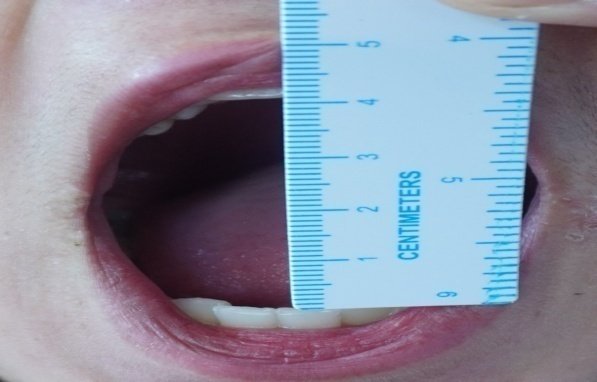
Extraoral examination showed asymmetry due to flatness with respect to right side of face and fullness with respect to left side (FIG.1). Mandibular movements were restricted and maximum interincisal opening was 18mm (FIG.2). Deviation was present towards right side while opening mouth.
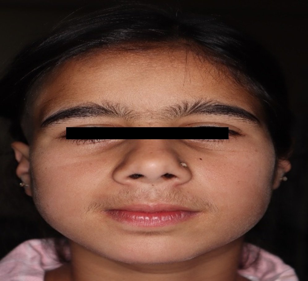
FIG.1
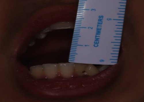
FIG .2
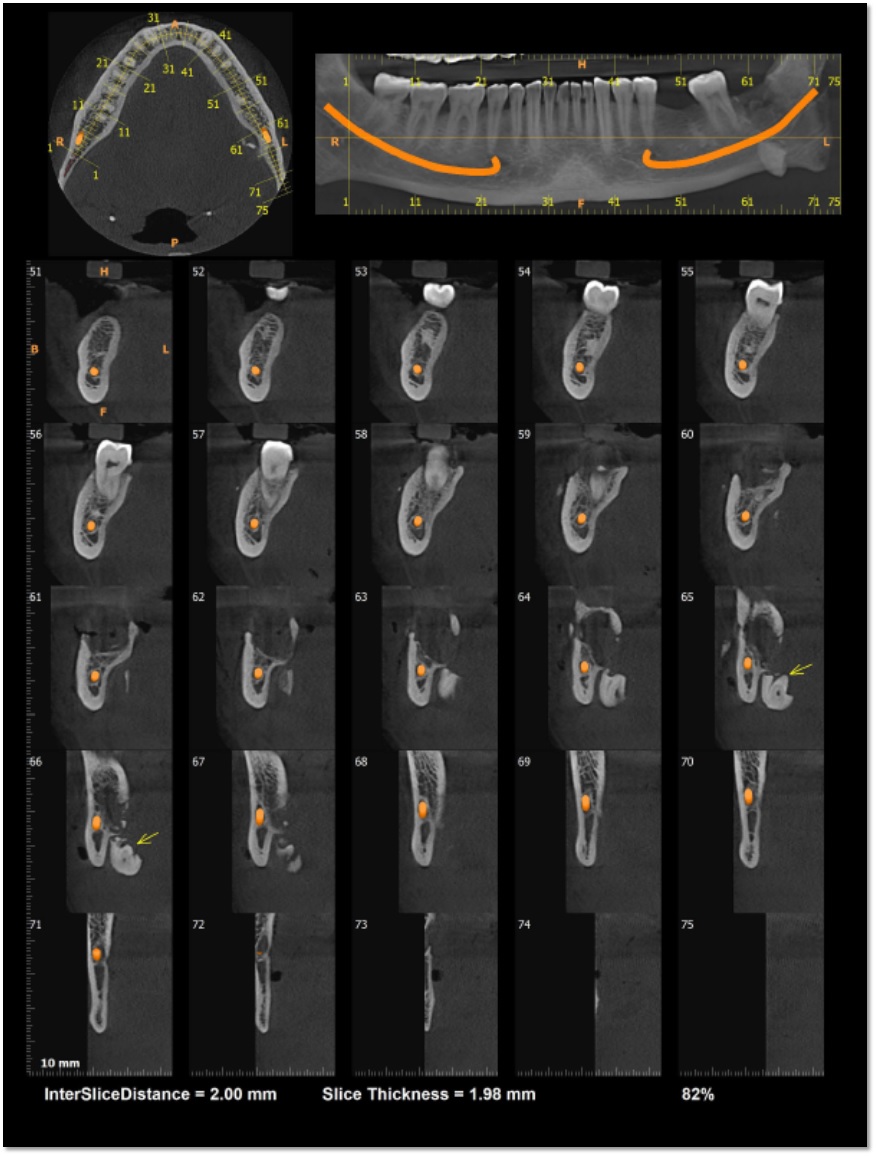
An orthopantomagram (OPG) showed a narrowed TMJ space and evidence of right TMJ ankylosis of lateral aspect of mandibular condyle with right zygomatic arch was detected (Figure 3), which was subsequently confirmed by 3Dcoronal section computed tomography scans (Figure 4&5). The child was eventually diagnosed with right TMJ ankylosis secondary to trauma, and a surgical intervention is achieved by selective lateral arthroplasty of the affected TMJ, by simply removing the lateral bony chunk and thus preserving the disc and condyle thereby allowing the mandibular condyle to grow in normal manner.
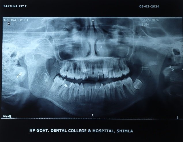
FIG .3

Fig 4
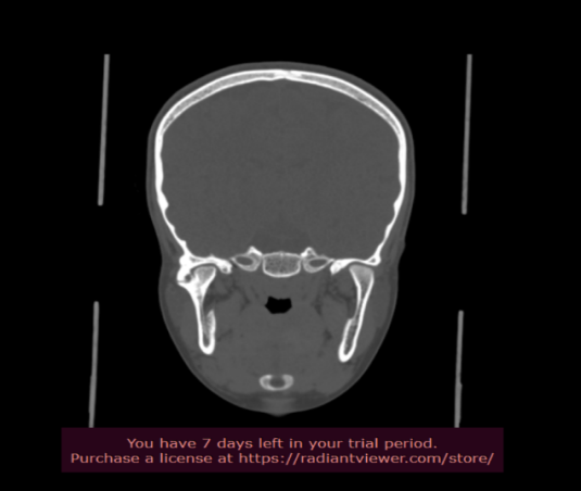
Fig 5
Alkayat Bramley incision was given with respect to right side and layer wise dissection was done to reach the affected joint space. A dense, hard sclerotic bony mass was discovered around right TMJ. Selective lateral arthroplasty of the affected TMJ done , by simply removing the lateral bony chunk and preserving the disc .Drain was placed and secured using 2-0 silk. Layerwise closure was done using 3-0 vicryl and 5-0 prolene.
MATERIAL AND METHODS: Lateral cephalometric tracing done both affected side preoperatively &postoperatively.
Relationship of mandible to rest of facial bone was analyzed .Measurements was made point A TO POG (lower anterior facial height),mandible plane angle (CO-GO-GN),occlusal plane angle (molar cusp to incisal edge),and( CO-GO )ramal height ,(CO–GN) measurement of total mandibular length,(GO-GN )length of the body of mandible.(FIG. 6)
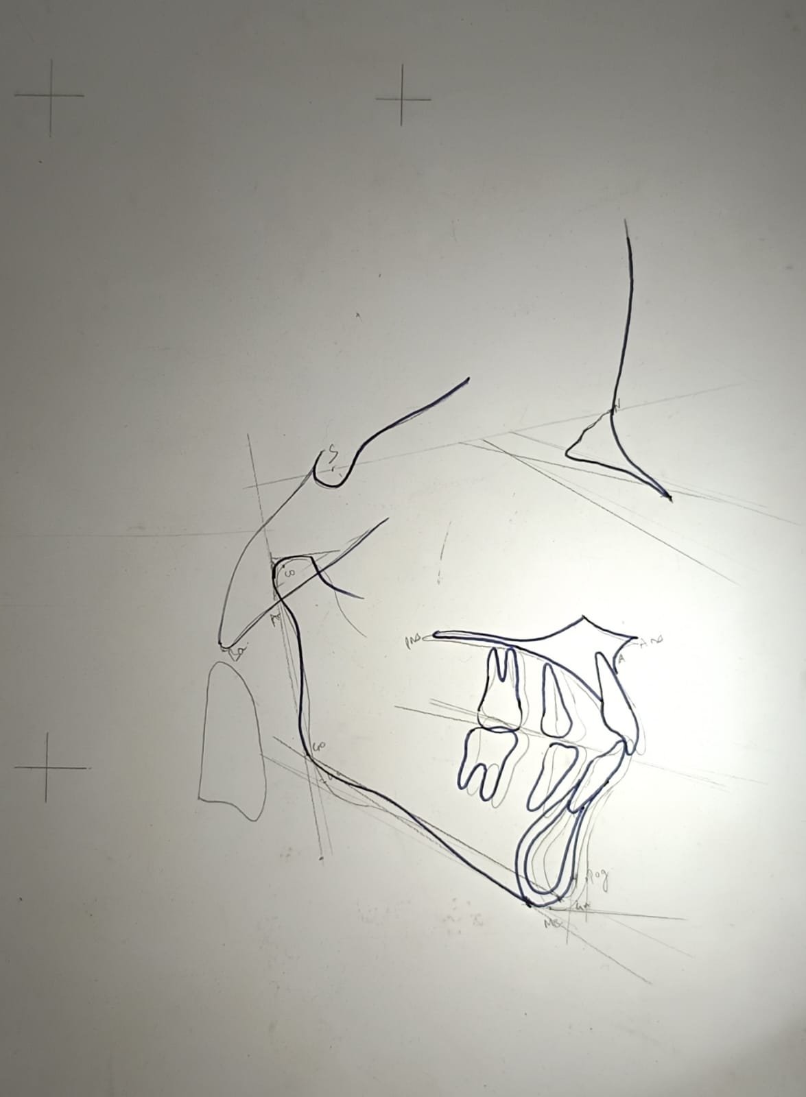
PREOPERATIVE:BLACK OUTLINE (FIG. 6)
POSTOPERATIVE:GRAY OUTLINE
RESULTS:
| PARAMETERS | PREOPERATIVE MEASUREMENT | POSTOPEARTIVE MEASUREMENTS |
|---|---|---|
| LOWER ANTERIORFACIAL HEIGHT | 69 MM | 57 MM |
| MANDIBULAR PLANE ANGLE | 41 DEGREE | 37 DEGREE |
| OCCLUSAL PLANE ANGLE | 24 DEGREE | 28 DEGREE |
| CO-GN(TOTAL MANDIBULAR LENGTH) | 94MM | 96 MM |
| CO-GO(RAMAL HEIGHT) | 42MM | 47 MM |
|
GO-GN(MANDIBULAR BODY LENGTH) |
62 MM | 60 MM |
Postoperative mouthopening:41 mm
DISCUSSION
The mandibular condyles in children are more prone to intra-capsular fractures due to trauma OR surgical manipulation because they have a broad head and a relatively narrow neck.2 Management of TMJ ankylosis in children is often challenging due to the unpredictability of the mandibular growth and the high rate of recurrence of ankylosis in children.3 Moreover, paediatric condyles have a high regenerative and remodeling capacity, which significantly increases the likelihood of ankylosis after trauma or re-ankylosis after surgery.11
TMJ ankylosis is a condition that has a significant impact on the patient's quality of life, with deteriorating effects on nutrition, speech, oral hygiene, growth, occlusion, facial aesthetics, and—in severe cases—can cause micrognathia, which could lead to obstructive sleep apnoea. In children, unilateral ankylosis of TMJ often results in facial asymmetry due to deviation of the chin towards the affected side, which often has a compromised mandibular growth.12
In this case we did selective lateral arthroplasty of the affected TMJ, by simply removing the lateral bony chunk and preserving the disc thus allowing the remaining mandibular condyle to grow in normal pattern. On cephalometrics analysis postoperatively 6 months follow up increase in ramal body height, increase in body length and mandibular length. Decrease in lower facial height clearly indicate mandibular growth forwardly and upwardly. It is important to note that early postoperative opening exercises, active postoperative physiotherapy, and strict follow-up are essential to prevent reankylosis or recurrence.13
REFERENCES
- N. Mittal, M. Goyal, D. Sardana, and J. S. Dua, Outcomes of surgical management of TMJ ankylosis: a systematic review and meta-analysis, Journal of Cranio-Maxillofacial Surgery, vol. 47, no. 7, pp. 1120–1133, 2019.
- E. P. Ramly, J. W. Yu, B. S. Eisemann et al., Temporomandibular joint ankylosis in pediatric patients with craniofacial differences: causes, recurrence and clinical outcomes, The Journal of Craniofacial Surgery, vol. 31, no. 5, pp. 1343–1347, 2020.
- C. Rozanski, K. Wood, P. Sanati-Mehrizy, H. Xu, and P. J. Taub, Ankylosis of the temporomandibular joint in pediatric patients, The Journal of Craniofacial Surgery, vol. 30, no. 4, pp. 1033–1038, 2019.
- N. D. Shashikiran, S. V. V. Reddy, R. Patil, and C. Yavagal, Management of temporo-mandibular joint ankylosis in growing children, Journal of the Indian Society of Pedodontics and Preventive Dentistry, vol. 23, no. 1, pp. 35–37, 2005.
- E. A. Al-Moraissi, T. M. El-Sharkawy, R. M. Mounair, and T. I. El-Ghareeb, A systematic review and meta-analysis of the clinical outcomes for various surgical modalities in the man agement of temporomandibular joint ankylosis, International Journal of Oral and Maxillofacial Surgery, vol. 44, no. 4, pp. 470–482, 2015.
- Management of temporomandibular joint ankylosis: 11years' clinical experience Zhi, Keqian et al.Oral Surgery, Oral Medicine, Oral Pathology, Oral Radiology and Endodontics, Volume 108, Issue 5, 687 – 692.
- Dean, Alicia M.D, Alamillos, Francisco M.D. Mandibular Distraction in Temporomandibular Joint Ankylosis,Plastic and Reconstructive Surgery 104(7):p 2021-2031, December 1999.
- Nitzan, D. W., Bar-Ziv, J., & Shteyer, A.(1998), Surgical management of temporomandibular joint ankylosis type III by retaining the displaced condyle and disc, Journal of Oral and Maxillofacial Surgery, 56(10), 1133–1138.
- V. Shetty, P. N. Kishore, A.Khanum,A.Yadav, and H. F. Sailer,Retrospective analysis of a TMJ ankylosis protocol with a 9 year follow up, Journal of Cranio-Maxillofacial Surgery, vol. 47, no. 12, pp. 1903–1912, 2019.
- N. De Roo, L. Van Doorne, A. Troch, H. Vermeersch, and N.Brusselaers, Quantifying the outcome of surgical treatment of temporomndibular joint ankylosis: a systematic review and meta-analysis, Journal of Cranio-Maxillofacial Surgery, vol. 44, no. 1, pp. 6–15, 2016.
- O. Güven and A. Keskin, Remodelling following condylar fractures in children, Journal of Cranio-Maxillofacial Surgery, vol. 29, no. 4, pp. 232–237, 2001.
- U. M. Das, R. Keerthi, D. P. Ashwin, R. VenkataSubramanian, D. Reddy, and N. Shiggaon,Ankylosis oftemporomandibular joint in children, Journal of the Indian Society of Pedodontics and Preventive Dentistry, vol. 27, no. 2, pp. 116–120, 2009.
- Y. Jiang, Y. Huang, B. Ye, Y. Li, and S. Zhu,Management of temporomandibular joint Ankylosis with dentofacial deformi ties in children, Journal of Craniofacial Surgery, vol. 29, no. 2, article e150, 2018.
Disclaimer from Guident
The views and opinions expressed in articles published in Guident are solely those of the authors and do not necessarily reflect the views of the editors, the editorial board or the publisher, Ivory India. Guident does not endorse any product, technique or treatment described. Clinical content is intended for qualified dental and medical professionals and is not a substitute for individual clinical judgement. Guident, its editors and the publisher accept no liability for any consequence arising from the use of information published in the journal.
How to cite this article
Dr. Monika Parmar, Dr. Priyanka, Dr. Yogita. Effect of Lateral Arthroplasty on Mandibular Growth: A Cephalometric Analysis Case Report. Guident. 2025 May;18(6). Available from: https://guident.in/articles/oral-surgery/effect-of-lateral-arthroplasty-on-mandibular-growth-a-cephalometric-analysis-case-report.html