
Treatment Options for Aesthetic Management of Fluorosed Teeth
Crowns and Roots Dental Solution, Mira Road, Mumbai, India
Abstract:
Tooth discolouration is a common problem and affects people of all ages. Apart from the conventional treatment modalities for the same, newer options are available today with better techniques and materials. This article describes the various options available for different grades of fluorosis along with cases performed using these techniques. The pros and cons of all treatment options are also carefully weighed. The execution of the planned treatment must yield good aesthetic and functional outcome.
Key words: Dental fluorosis, Ceramic veneers, Direct composite bonding, Teeth Whitening
Introduction:
Dental fluorosis is a developmental disturbance of the enamel which is caused due to repetitive or continuous exposure to high fluoride concentrations during tooth development. McKay and G. V. Black published an article explaining the effects that fluoride has on the enamel of teeth1. The harmful effects of fluorides can be attributed to their systemic absorption during tooth development, thus resulting in dental fluorosis2. Dental fluorosis is the result of chronic endogenic intake of fluorides in amounts exceeding the optimal daily dose of 1 ppm 3. Dental fluorosis features hypomineralization of enamel which occurs due to the effects of excessive fluoride on ameloblasts during amelogenesis. Fluorides disturb mineralization of the enamel by decreasing the concentration of free calcium ions in the mineralizing matrix, which interferes with the proteinases; thus degrading matrix proteins during enamel maturation. This causes degradation of enamel matrix proteins or an inhibition of the removal of degraded enamel matrix proteins4. Fluoride-induced retention of these proteins causes impaired and incomplete crystal growth. Poor interlocking of crystals accounts for the increased porosity of enamel and thus the subsequent optical and physical changes5.
The biggest concerns in dental fluorosis are the subsequent aesthetic changes and these occur primarily in children who are exposed to high fluoride concentrations between 1 to 4 years of age. The safe daily fluoride intake is 0.05 to 0.07 mg F/Kg/day. Exceeding this level, there is a risk of developing fluorosis due to chronic fluoride consumption6. The optimum level of fluoride in drinking water as given by Dean and McKay was 0.9-1.0 ppm7. The World Health Organization (WHO) has recommended the maximum limit of fluoride in drinking water to be 1.5 mg/l8. The Bureau of Indian Standards has laid down Indian standards at 1.0 mg/l as maximum permissible fluoride limit in drinking water9.
Clinical manifestations and classification:
Depending on the quantity of fluoride intake, dental changes of varying degrees are observed. This was used as a guideline for Dean’s classification for dental fluorosis10. The very mild form of changes shows small, opaque, paper white areas scattered irregularly over the tooth but not involving as much as 25 percent of the tooth surface. Frequently included in this classification are teeth showing no more than about 1-2 mm of white opacity at the tip of the summit of the cusps of the bicuspids or second molars. Mild fluorosis is characterized by a more extensive spread of white opaque areas in the enamel but not involving as much as 50 percent of the tooth. In the moderate form of dental fluorosis, all enamel surfaces of the teeth are affected, and the surfaces subject to attrition show wear; brown stain is frequently a disfiguring feature. In severe fluorosis, all enamel surfaces are affected and hypoplasia is so marked that the general form of the tooth may be affected; the major diagnostic sign of the severe form is discrete or confluent pitting, brown stains are widespread and teeth often present a corroded-like appearance.
Treatment options for fluorosis:
- Teeth whitening:
Teeth whitening or dental bleaching can be considered as a treatment modality for cases of fluorosis showing mild to moderate discolouration, which is generally browninh in colour. Teeth whitening can help to either lighten or completely eliminate fluorosis stains. Tooth surfaces showing pitting don’t give good results and require other modalities of treatment.
- Direct composite bonding:
This paper presents a case of successful aesthetic management of severe dental fluorosis in an Indian female patient by the use of porcelain laminate veneers and direct composite veneers.
Case report:
A 17-year-old girl was referred for aesthetic management of discoloured teeth. The patient primarily complained of an unpleasant smile due to stained teeth. Detailed clinical history was elicited. The patient was born at full term in Gangapur, Rajasthan, India after an uneventful pregnancy and had stayed in the same region ever since. The medical history of the patient was non contributory. She had an elder brother who also suffered from a similar condition.
Intraoral examination revealed generalized discolouration of her dentition. All teeth were affected with pitting and chalky white areas (figure 1a). The pits on the enamel were generalized and yellowish brown in colour. A diastema was present between the maxillary central incisors. Both the maxillary lateral incisors had Ellis class I fractures of their incisal edges and both maxillary canines had Ellis class I fractures of their cusp tips.
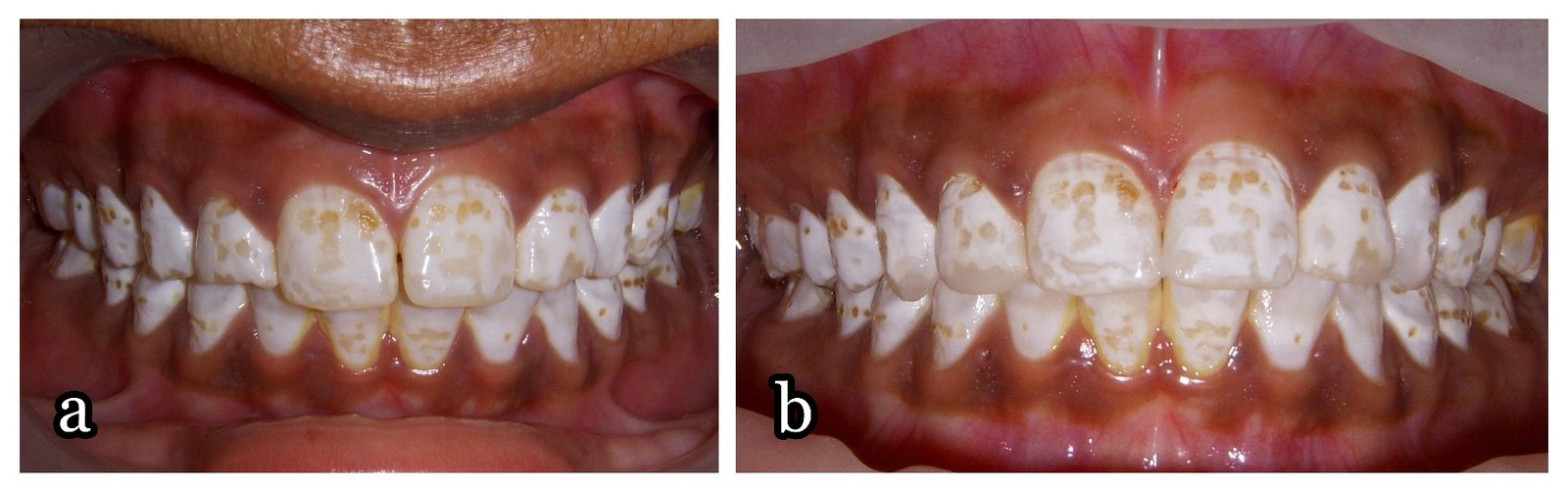
Figure 1 (a): Preoperative presentation showing generalized dental fluorosis with discolouration and pitting, (b): Composite mock-up on the fractured maxillary lateral incisors and anines and on the central incisors which had a diastema.
A diagnosis of severe form of dental fluorosis was made owing to the generalized and bilateral distribution of the discolouration and pitting, and due to the suggestive history that the patient belonged to a fluoride - rich region. The aetiology for the same was attributed to exposure of the patient to drinking water containing high concentrations of fluoride during enamel formation. As per a 2010 report11, the average fluoride level in the groundwater of Gangapur district was 2.5-2.9mg/l which is above the permissible limits. The critical period for fluoride overexposure is 1 to 4 years old, and the child would not be at risk around 8 years old12. Considering that the patient had spent this period of her life in Gangapur, the diagnosis can be further confirmed.
A problem list and treatment objectives were formulated and included: improvement of the shade of teeth, masking the pitting on the facial tooth surfaces, restoration of the fractured maxillary lateral incisors and canines and closure of the midline diastema.
The preparatory stage of the treatment started with smile analysis, preliminary shade selection, photographs and study models. The occlusion of the patient was determined as class I. The various treatment modalities for dental fluorosis including polishing, micro/macroabrasion, dental bleaching, composite veneering, porcelain laminate veneers and full veneer crowns were explained to the patient’s parents. After discussing the treatment options and considering the age of the patient and the severity of fluorosis, it was decided to place ceramic veneers on the maxillary incisors and canines. Direct composite veneers were planned for the maxillary premolars and mandibular incisors, canines and first premolars.
Before commencing the tooth preparation, direct composite mockup (figure 1b) was done to build up the fractured teeth and to close the midline diastema. The size and shape of the teeth were approved by the patient and her parents. A polyvinyl siloxane impression was made and was used as a preparation template in the further procedures. No anaesthesia was administered as the preparation was to be restricted to enamel. Tooth preparation was first started with a 0.5 mm depth cutting diamond point and the depth grooves were marked with non-water-soluble ink. The labial and incisal tooth reduction was completed (figure 2a) using the preparation template as a guide to gauge the depth of preparation. Proximal preparation was not carried out and the contacts were not involved. Only the mesial surfaces of the maxillary central incisors were included in the preparation.

Figure 2 (a): Showing completed tooth preparation of maxillary anterior teeth for ceramic veneers, (b): Cementation of ceramic veneers
After adequate gingival retraction with a retraction cord (Ultrapak, Ultradent, South Jordan, Utah, United States), a two step dual impression was made and sent to the laboratory for fabrication of IPS e.max veneers (Ivoclar Vivadent AG, Schaan/Liechtenstain, Germany).
The prepared teeth were rinsed and polished with pumice and water before cementation procedures. The fit of the ceramic veneers was verified onto the cast and on the prepared teeth individually and collectively and the luting sequence was determined. A water-soluble try-in paste (Variolink II, Ivoclar Vivadent AG, Schaan/Liechtenstain, Germany) was used to determine the shade of the luting cement. The veneers were rinsed thoroughly, etched with 5% hydrofluoric acid for 20 seconds (as per the manufacturer’s instructions), rinsed with water and silanized. The tooth was phosphoric acid-etched and adhesive was applied. Cementation of the veneers was carried out following the predetermined luting sequence. It was made certain that the veneers were exactly seated and not occupying any space meant for the adjacent veneer. The luting cement was initially light cured for 1-2 seconds and excess was gently removed using a no. 12 blade, followed by thorough curing of the luting agent.
Once the porcelain veneers were bonded (figure 2b), the labial surfaces of the maxillary premolars and the mandibular anteriors and first premolars were prepared. Minimal tooth preparation was required. 37% phosphoric acid etching was done followed by application of a bonding agent. Composites were layered according to the desired shades on all the teeth (figure 3a).
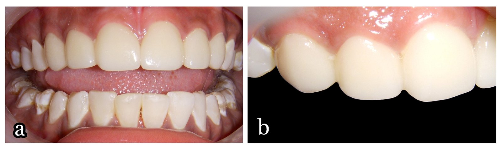
Figure 3 (a): Excellent aesthetics achieved after completion of direct composite bonding on the maxillary bicuspids and mandibular anteriors and first bicuspids, (b): Healthy gingival nd periodontal status and ideal emergence profile.
Final finishing of the composite restorations and the veneer luting margins was postponed until the next appointment (figure 4). The patient’s occlusion was evaluated and adjusted so as to avoid any veneer fracture. The patient was instructed about the precautions to be taken after veneer placement. Periodic follow-up was scheduled to evaluate the gingival health and patient comfort.
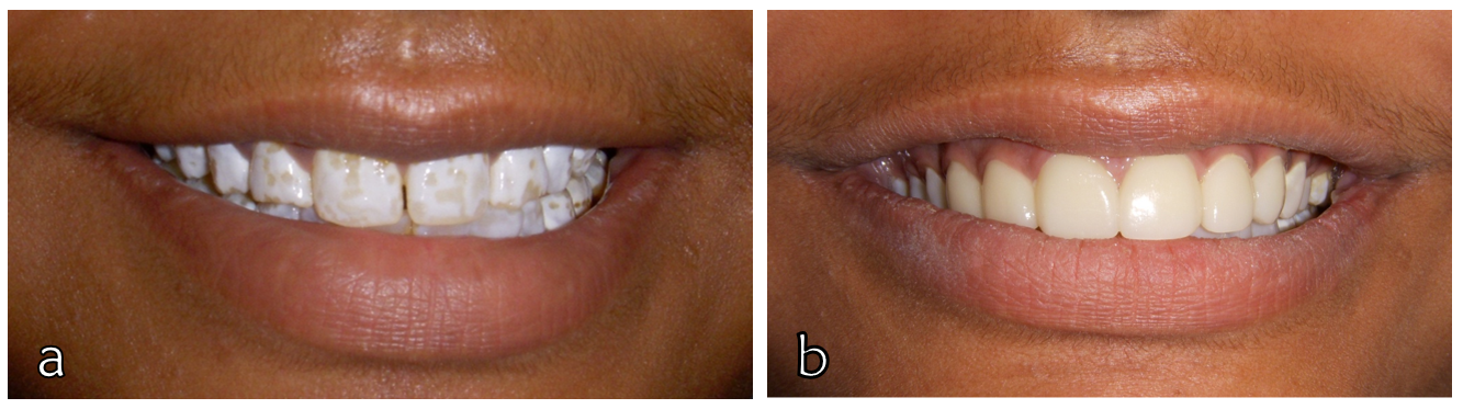
Figure 4: Comparision of pre-operative and post operative close-up smile pictures for showing treatment outcome.
Discussion:
Dental fluorosis, with all its adverse effects on dental tissues, is a debilitating condition for the patient’s smile. Due to the early age at which it affects the dentition and thus the appearance of the patient, it has adverse psychological effects which may in turn affect the overall development and confidence of the patient. This is especially true in early and late teens when the patient is most conscious about his/her appearance in public. It thus becomes imperative to provide prompt treatment to the patient.
Lesser invasive treatment modalities such as polishing, microabrasion or bleaching would not have sufficed to mask the pitting present on the facial surfaces. Composite layering on the entire facial surfaces was a viable treatment option and was therefore explained to the patient and considered in the treatment plan. However, concerns regarding discolouration and need for frequent repairs had to be taken into account considering the young age of the patient. The patient was thus consulted to opt for ceramic veneers on all the teeth which were revealed during smiling. Due to cost concerns however, the number of ceramic veneers had to be restricted and composite veneering had to be opted for. Thus after careful evaluation and discussion, it was decided to place ceramic veneers on the six maxillary anterior teeth which are the most prominent ones during smiling, and to layer composite veneers on the four maxillary premolars. In the mandibular arch, composite veneers were planned for teeth upto the first bicuspids on either side.
Porcelain laminate veneers were first introduced in around 193813 and have gained ever increasing popularity ever since. They are a useful treatment option for discolored teeth. Porcelain veneers have been said to have high survival rates and good clinical success14-24. They have excellent biocompatibility with gingival and periodontal tissues (figure 3b). However, to ensure these, pre-operative patient consultation and post-operative instructions are essential. Their main indications include stained or darkened teeth, hypocalcifications, diastema, chipped teeth, slightly rotated teeth, discrepancies in size and shape of teeth25, worn acrylic veneers, foreshortened teeth, slight deviation of midlines, stained restorations and lingually positioned teeth26. Labially placed teeth, excessive interdental spacing, poor oral hygiene and periodontal status, clenching or nail-biting habits, severe discolouration, weak non-vital teeth25 and extreme midline deviation are some of the cases wherein veneers are contraindicated26. These were all considered and the contraindications were individually ruled out before planning the treatment.
The smile was carefully analyzed to check the teeth visible during talking, smiling and laughing. Also, the occlusion was assessed in centric and eccentric relations, to ensure multiple tooth contacts during excursive movements. Accordingly, porcelain laminate veneers were planned for the patient. But keeping the economical constraints in mind, it became essential to use composite veneers on some teeth instead so that we could give the patient her very much desired smile. Thus, considering all the various factors the final treatment plan was devised.
Before preparing the teeth, the fractured teeth and the diastema were built up to their ideal shape using composite (figure 1b) without etching or bonding. This enabled the patient to understand the final shape of her teeth. Once satisfied, a putty index of this composite mock-up was made so that it could be used to guide us to gauge the amount of tooth preparation. The silicone indices were very helpful to guide in the adequacy of the tooth preparation so that the strength, translucency and the final contours of the veneers could be maintained. The patient and her parents were satisfied mainly because their expectations had been understood and the final outcome was demonstrated to them. It is essential to understand the patient’s requirements before suggesting any treatment.
The incisal finish line can either be placed at the incisal edge ending in a feather edge or short of it as in window preparations, or can be extended onto the lingual surface after preparing the incisal edge. An incisal overlap may be preferred in the preparation as the bulk of porcelain is stronger and provides a positive seat during cementation27. Accurate seating of the veneer occurs by the vertical stop that is provided by the incisal overlap28. Smales et al22 determined the clinical success rate of 110 ceramic veneers for seven years and stated a 96% success rate for incisal overlap design as opposed to the 86% success in veneers without incisal coverage. Accurate placement of all the veneers was essential in this case as multiple veneers had to be provided. Thus, incisal overlap design was preferred. The proximal contacts of none of the teeth were involved in the tooth preparation. Preparation had to be extended upto the lingual line angles on 11 and 21 to allow for closure of the diastema between them. This is in accordance with established literature29 which states that closing a diastema may require preparation of the interproximal area. This allows the technician greater freedom in alteration of the form or position30. Entire tooth preparation was restricted to the enamel to achieve reliable bonding. This ensures adequate bond strengths and lengthens the life of the veneers. As no dentin is involved, the incidences of intra-operative and post-operative sensitivity are also greatly reduced.
Written post-operative instructions must be given to the patient such as the use of a soft toothbrush, regular flossing as with natural teeth, and use of a mouthguard when involved in any contact sport and for parafunctional habits. They must also be instructed to not use alcohol and alcohol-containing mouthwashes during the first 48 hours and to avoid hard foods, chewing on ice, nail-biting etc.31 In spite of the good clinical success rates and life of porcelain veneers, certain failures may occur which may be due to a variety of reasons. Failures include partial/complete fracture or debond, inaccurate placement, colour mismatch, marginal discolouration, loss of marginal integrity, postoperative sensitivity etc.31 Regular dental check-up visits and proper care of the veneers may however, increase their life with no complications.
Conclusion:
Dental fluorosis is a debilitating dental condition affecting the aesthetics, psychology and confidence of the patients. In spite of the various treatment options that are available today, a single treatment modality may not suffice and there may be a need to opt for a multi-treatment approach. This case was successfully managed by the combined use of porcelain laminate veneers and direct composite bonding to give the young girl, her much deserved smile, back.
References:
- McKay FS, Black GV. Investigation of mottled teeth: An endemic imperfection of the enamel of teeth heretofore unknown in literature of dentistry. Dent Cosmos. 1916;58:129-56;477-84;627-34;781-92;894-904.
- Fejerskov O, Kidd AM. Clinical use of fluoride. In: Dental caries: the disease and its management. 1st ed. United Kingdom: Blackwell Munksgaard;2003.p. 189-202.
- Kidd E. Fluoride Supplementation. In: Kidd E. Essentials of Dental Caries, Oxford. 2005. p 109-125.
- Aoba T, Fejerskov O. Dental fluorosis: chemistry and biology. Crit Rev Oral Biol Med 2002;13:155-170.
- Robinson C, Connell S, Kirkham J, Brookes SJ, Shore RC, Smith AM. The effect of fluoride on the developing tooth. Caries Res 2004;38:268-276.
- Burt BA. The changing patterns of systemic fluoride intake. J Dent Res. 1992;71:1228-37.
- Dean HT, McKay FS. Production of mottled enamel halted by a change in common water supply. Am J Public Health. 1939;29:590-6.
- World Health Organization. Guidelines for drinking water quality. WHO: Geneva; 2004.
- Government of India. Prevention and control of fluorosis in India. New Delhi: Rajiv Gandhi National Drinking Water Mission; 1993. p. 25.
- Dean H.T. 1942. The Investigation of physiological effects by the epidemiological method. In: Moulton FR, Fluorine and dental health. Washington, DC: American Association for the Advancement of Science, Publication No. 19, p.23-31.
- Integrated Management Information System (IMIS) Ministry of Drinking Water and Sanitation. Available from: http://indiawater.gov.in
- Alvarez JA, Rezende KM, Marocho SMS, Alves FBT, Celiberti P, Ciamponi AL. Dental fluorosis: Exposure, prevention and management. J Clin Exp Dent. 2009;1:e14-18.
- Pincus CR. Building mouth personality. J Calif State Dent Assoc 1938;14:125-9.
- Dunne SM, Millar JA. A longitudinal study of the clinical performance of porcelain veneers. Br Dent J 1993;175:317-321.
- Walls AWG. The use of adhesively retained all-porcelain veneers during the management of fractured and worn anterior teeth. Part II. Clinical results after 5-years follow-up. Br Dent J 1995;178:337-339.
- Shaini FU, Shortall ACC, Marquis PM. Clinical performance of porcelain laminate veneers. A retrospective evaluation over a period of 6.5 years. J Oral Rehab 1997;24:553-559.
- Fradeani M. Six-year follow-up with Empress veneers. Int J Periodont Rest Dent 1998;18:216-225.
- Friedman MJ. A 15-year review of porcelain veneer failure - a clinician’s observations. Compend Contin Educ Dent 1998;19:625-38.
- Magne P, Perroud R, Hodges JS, Belser UC. Clinical performance of novel-design porcelain veneers for the recovery of coronal volume and length. Int J Periodont Rest Dent 2000;20:441-457.
- Dumfahrt H, Schaffer H. Porcelain laminate veneers. A retrospective evaluation after 1 to 10 years of service: Part II— clinical results. Int J Prosthodont 2000;13:9-18.
- Aristidis GA, Dimitra B. Five-year clinical performance of porcelain laminate veneers. Quintessence Int 2002;33:185-189.
- Smales RJ, Etemadi S. Long-term survival of porcelain laminate veneers using two preparation designs: a retrospective study. Int J Prosthodont 2004;17:323-6.
- Fradeani M, Redemagni M, Corrado M. Porcelain laminate veneers: 6- to 12-year clinical evaluation—a retrospective study. Int J Periodontics Restorative Dent 2005;25:9-17.
- Wiedhahn K, Kerschbaum T, Fasbinder DF. Clinical long-term results with 617 Cerec veneers: a nine-year report. Int J Comput Dent 2005;8:233-46.
- Newsome P, Owen S. Ceramic veneers in general dental practice. Part 1: Treatment planning. Int Dent SA. 2008;10:66-71.
- Freedman GA, McLaughlin GL. Porcelain Veneers: Indications and Contraindications. In: Color Atlas of Porcelain Laminate Veneers. 1990. Ishiyaku EuroAmerica Inc. Publishers, USA. p 37-62.
- Gürel G . Atlas of Porcelain Laminate Veneers. In: The Science and Art of Porcelain Laminate Veneers. 2003. Germany. Quintessence Publishing. p 231-344.
- Calamia JR. Etched porcelain veneers: The current state of the art. Quintessence Int 1985;16:5-12.
- Touati B, Miara P, Nathanson D. Ceramic Laminate Veneers. In: Esthetic Dentistry and Ceramic Restorations. 1999. New York: Martin Dunitz. p 161-213.
- Rouse JS. Full veneer versus traditional veneer preparation with a medium wrap: A discussion of interproximal extension. J Prosthet Dent 1997;78:545-549.
- Newsome P, Owen S. Ceramic veneers in general dental practice. Part five: After care and dealing with failure. Aesthetic Dentistry Today. 2008;2(4).
Disclaimer from Guident
The views and opinions expressed in articles published in Guident are solely those of the authors and do not necessarily reflect the views of the editors, the editorial board or the publisher, Ivory India. Guident does not endorse any product, technique or treatment described. Clinical content is intended for qualified dental and medical professionals and is not a substitute for individual clinical judgement. Guident, its editors and the publisher accept no liability for any consequence arising from the use of information published in the journal.
How to cite this article
Dr. Harsh Haren Shah, Dr. Jigna Sanghavi. Treatment Options for Aesthetic Management of Fluorosed Teeth. Guident. 2025 May;18(6). Available from: https://guident.in/articles/aesthetic-dentistry/treatment-options-for-aesthetic-management-of-fluorosed-teeth.html


