Revolutionizing Atrophic Maxilla Rehabilitation with the Pterygoid Implants: A Review
Department of Prosthodontics, Post Graduate Institute of Dental Sciences, Pt. B.D. Sharma University of Health Sciences, Rohtak, Haryana, India
ABSTRACT
Dental Implantologists have several anatomical and structural challenges when rehabilitating lost teeth in the posterior maxilla because of the sinus space and the fragility of the posterior maxillary bone. Numerous surgical methods have been practiced to solve these issues, including sinus lifts, bone augmentation, tilted implants, short implants, and zygomatic implants. Each of these strategies does, however, have certain drawbacks. A good substitute for successful rehabilitation of the atrophic posterior maxilla is the pterygomaxillary area, which can be effectively used for implant insertion without requiring further complicated surgical procedures. Implants can be placed strategically in the pterygoid plate region to overcome anatomical constraints.
Keywords- Petrygomaxillary region; posterior maxilla; the pterygoid process; pterygoid implant; pterygoid plate; posterior occlusion.
INTRODUCTION
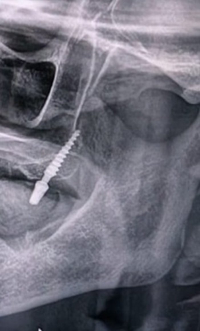
Figure 1: Panoramic radiograph demonstrating the pterygoid implant.
Due to several factors, rehabilitation of the posterior maxillary region is considerably more challenging than that of the maxillary anterior region.1 Issues with the posterior maxillary area include: 1) Alveolar bone resorption resulting in less residual bone height and/or maxillary sinus enlargement. 2) Type three and four low bone density, as defined by Lekholm & Zarb's classification method.2,3 The pterygoid implant is a revolutionary alternative to successful rehabilitation of the atrophic posterior maxilla. Paul Tessier gave this idea in 1989, and Tulsane JF later presented it in 1992.4 Implants inserted into the pterygomaxillary region's compact bone encourage osseointegration and offer improved stability and retention.5-8 Pterygoid implants are usually between 15 and 20 mm long. The benefits of pterygoid implants include improved axial loading, avoiding cantilever stresses, bicortical anchorage, enabling anchorage of atrophic maxilla in the posterior region, and avoiding the requirement for sinus lifts or bone augmentation.5-8 When positioned properly, pterygomaxillary implants are a good substitute because they take advantage of the area's thick bone. Other names for these implants are pterygomaxillary implants and tuberosity implants.4
Maxillary Bone Remodeling and Implant Difficulties
A continual process of bone remodeling occurs in the alveolar ridge after tooth extraction or periodontal bone loss. As a result of this three-dimensional remodeling, the residual ridge's height and width are decreased. Therefore, without augmentation treatments, there is not enough bone left for implant insertion.
There are fewer and fewer surgical alternatives available in situations of advanced maxillary bone resorption. Genetically low maxillary bone density can also affect physiological bone remodeling, causing a more rapid and substantial resorption than in the mandible. Furthermore, trauma-induced resorption, like that brought on by prolonged denture wear, can hasten bone loss and decrease the amount of bone accessible in the front and posterior maxilla, making it more difficult to insert implants without augmentation.9
Pterygoid region's anatomy

Figure 2: Human skull region demonstrating the pterygoid region’s anatomy and inferior relationship of tuberosity and pyramidal process of palatine bone and pterygoid process of sphenoid bone.
The maxillary tuberosity, the palatine bone's pyramidal process, and the sphenoid bone's pterygoid process are the main anatomical features that may offer support for implant insertion. The pyramidal process defines the medial and posterior boundaries of the maxillary tuberosity, which generates the posterior convexity of the alveolar ridge.4 The pyramidal process of the palatine bone attaches to the anterior aspect of the pterygoid process of the sphenoid bone, which is situated posteriorly and slightly medial to the tuberosity. This process forms a structural connection between the maxillary tuberosity and the inferior end of the pterygoid plates by integrating with their anterior surface. The pterygoid pillar, a narrow, dense bony column, forms at this intersection and provides a solid location for implant apical attachment. The maxillary tuberosity( Type 3 and Type 4 cancellous bone); the pyramidal process of the palatine bone; the pterygoid process of the sphenoid bone; and the pterygopalatine fossa (PPF), are some of the important structures that make up the pterygoid region's anatomy, an important region situated within the deep space. These structures must be carefully examined during imaging to guarantee precise assessment and planning. The center portion of the pterygoid process, between plates, has the thickest supporting bone. The implant placement is slightly angled medially to transect the thickest part of bone available in the pterygoid area, which is 3–4 mm medial to the alveolar ridge. In the oropharynx, the hamular process on the medial pterygoid plate is palpable. Laterally, this landmark is used for implant positioning. When full arch rehabilitation is required, pterygoid implants avoid the uneven distribution of functional stress, distal cantilevers, and posterior occlusion extension.
Reiser's classification10
Based on their anatomical position, Reiser's classification of pterygoid implants comprises the following types: maxillary tuberosity, maxillary tuberosity-pterygoid process, pyramidal process-pterygoid process, and tuberosity-pterygoid process-pyramidal process.
Both the pterygoid process and the more anterior pterygomaxillary process are suitable sites for pterygoid implants. There is no apparent distinction between these placements in the literature currently in publication, and there is no agreement on their nomenclature. Implant angulation and length, however, differ between these sites. To replicate the third molar's natural location, shorter implants are usually positioned in the pterygomaxillary region with an angulation of 10° to 20°. Contrarily, longer implants are fixed to the sphenoid bone's pterygoid plate.
Pterygoid Analytical Radiography Prediction, or PARP
|
PARP Stage |
Location |
Bone Residual |
Sinus Invasion |
PARP 1 |
Retromolar/pterygoid |
>13mm of bone |
Without sinus invasion |
PARP 2 |
Retromolar/pterygoid |
10-13 mm of residual bone |
With or without sinus invasion |
PARP 3 |
Pterygoid |
5-9.99mm of bone left |
Moderate sinus invasion |
PARP 4 |
Pterygoid |
<5mm of bone left |
Critical sinus invasion |
The Bedrossian Surgical Planning Classification
A structured guideline for choosing the surgical strategy for implant placement in the atrophic maxilla is provided by the Bedrossian classification. Using a panoramic radiograph, this classification is based on a methodical evaluation of the available bone. The Bedrossian Classification's Maxillary Zones-There are four separate zones within the maxilla:
|
Zone |
Area |
|
Zone I |
Canine to Canine |
|
Zone II |
Bicuspids |
|
Zone III |
Molars |
|
Zone IV |
Zygomatic area |
The right surgical strategy depends on whether bone is present in these areas or not. In order to identify implant placement options, this classification takes into account the relationship between the maxillary sinus, nasal floor, and alveolus.
Guidelines for Implant Placement:
|
Bone Level |
Treatment Plan |
|
Sufficient amount of bone in Zones I, II, and III |
Four to eight axial (non-angled) implants are placed |
|
Zone III has insufficient bone, but Zones I and II have enough |
Using the All-on-X technique, four to six implants are positioned axially or tilted to maximize arch spread and the A-P (anteroposterior) ratio. |
|
Only Zones I and IV have enough bone, while Zones II and III have insufficient bone |
Bilateral zygomatic implants are positioned in Zone IV, while two to four implants are positioned in Zone I.11 |
|
Zones I, III, and IV include bone |
To improve support, pterygoid implants might be positioned in Zone III in addition to implants in Zones I and IV.8 |
|
Zones I and II have insufficient bone (without bone augmentation) |
Dual bilateral (quad) zygomatic implants are used as treatment. Pterygoid implants may be used in Zone III to expand the A-P spread for a full-arch restoration if the spread of zygomatic implants is not enough for stability. |
In situations of maxillary atrophy, the Bedrossian categorization is an essential tool for planning implant placement, guaranteeing the best possible distribution and support for prosthetic rehabilitation.12
Procedure for placing pterygoid implants
The preoperative examination seeks to identify the best implant location, confirm that osseointegrated implants are appropriate, and address any problems that need to be fixed before implantation. The site's anatomical condition and structure, its relationship to neighboring structures, the location, amount, and quality of the bones, and the ridge's relationship to neighboring teeth are all important factors. Assessments of anterior esthetic elements, occlusal connections, and soft tissue measurements are also necessary.
The shape and properties of the bone must be thoroughly assessed in three dimensions. While CT imaging offers comprehensive information on bone amount, quality, and cortical plate lengths, conventional radiographs are inadequate for evaluating the quality of hard tissue. A radiopaque marker applied to a stent or temporary acrylic resin restoration at the edentulous ridge level facilitates accurate identification of the implant site.13
The TTPHIL (Tall tilted pin hole immediate loading) procedure is used to insert the pterygoid implant slanted. It is oriented toward the intersection of the maxillary surface, palatine process, and sphenoid’s posteroinferior projection in 2nd to 3rd molar edentulous region. The maxillary tuberosity height and floor determine the distal angle of implant, which ranges from 45 to 60 degrees.14,15 The single-drill method is used to prepare the implant site. A pilot drill is used first, followed by a tapered drill that is oriented superiorly, posteriorly, distally, and palatally. Implants usually have a diameter of 3.75 mm or 4.2 mm and a length of 18 to 25 mm. For initial loading, torque values greater than 40 Ncm are necessary to guarantee implant stability. Multiunit abutments with different angulations (30°, 40°, and 50°) and lengths (3–5 mm) are placed.
While drilling techniques help prepare the implant site, particularly in areas of dense cortical bone, the osteotome technique reduces surgical risks, saves bone, and improves tactile control. A 22 mm long pterygoid implant is anchored into the pterygoid plate of the sphenoid bone using the traditional method outlined by Tulasne. Depending on the position of the sinus floor and the tuberosity height, the implant is positioned through the palate and maxilla with an angulation of 35° to 55° distally. The procedure outlined by Valeron & Valeron for implant site preparation uses a combination of drills and straight osteotomes.15 First, a round bur is used to identify the entry spot. Next, the smallest osteotome is used to start the preparation process. 2. To determine the orientation of the implant axis, pilot drill. 3. Cylindrical osteotomes in succession and Pterygomaxillary implants are positioned parallel to the posterior sinus wall within the maxillary tuberosity. The use of curved osteotomes rather than straight ones is the main distinction between the procedure and pterygoid implant insertion. To replicate the third molar's normal placement, the implant angulation is usually between 10° and 20°.
Numerous studies highlight the benefits of osteotomes:
- To limit bleeding, lower surgical risks, and preserve bone, Valeron et al. suggested osteotomes. Anatomically adapted osteotomes were created by Nocini et al. to provide better access to the maxillary tuberosity.
- By combining burs and osteotomes, Penarrocha et al. capitalized on their respective advantages: Osteotomes for decreased surgical risks, tactile control, and bone preservation. In dense cortical bone, drills are used to construct the implant bed precisely.
A 90.7% success rate was reported by Candel et al.8 in their assessment of 1,053 maxillary implants in 676 individuals. According to their research, pterygoid implants are a dependable option for treating the atrophic posterior maxilla since they show success rates and bone loss levels that are similar to those of traditional implants. Pterygoid implants also have low complications and good patient acceptance, making them a dependable option for treating atrophic posterior maxilla. They are therefore a good substitute for people who have significant
MAXILLARY ATROPHY.
Drawbacks
Pterygomaxillary implants require a high level of surgical skill and technical accuracy. However, the intricate and little-documented anatomy of the implant site makes this technique difficult. A sufficient mouth aperture is necessary to fit the drill and handpiece. Additionally, because the region is hard to reach, prosthodontic restoration of pterygomaxillary implants is challenging. Careful component handling is essential to avoiding issues, such as aspiration hazards. Even with encouraging results, success depends on selecting cases carefully and fully comprehending procedural obstacles.
Recent Advances
To capitalize on the physiologic benefits of preparing the implant site with a piezosurgical device, piezosurgical tips may be used in place of conventional osteotomes.16-17 The implant site preparation with ultrasonics may accelerate early osseointegration by increasing bone morphogenetic proteins, regulating inflammation, and stimulating bone remodeling, indicating more active bone growth. This method also reduced proinflammatory cytokines while promoting an earlier spike in TGF-β2 and BMP-4 proteins.18
CONCLUSION
Pterygoid implants are a good substitute for bone augmentation, sinus lifts, and 3D maxillary reconstruction. According to studies, pterygoid implants had 90% to 100% success rates, few problems, and follow-up times of 1 to 12 years[19-22]. The main drawbacks of bone grafting are the possibility of morbidity and the scarcity of alternatives if the graft fails, which frequently calls for a graftless procedure, which includes zygomatic and pterygoid implants that use the existing bone.[23] Because of their exceptional stability and suitability for quick loading, these implants also aid in the elimination of prosthetic distal cantilevers.
REFERENCES
- Albrektsson T, Zarb G, Worthington P, Eriksson AR. The long-term efficacy of currently used dental implants: A review and proposed criteria of success. Int J Oral Maxillofac Implants 1986;1:11-25.
- Cucchi A, Vignudelli E, Franco S, Corinaldesi G. Minimally invasive approach based on pterygoid and short implants for rehabilitation of an extremely atrophic maxilla: A case report. Implant Dent 2017;26:639-44.
- Balaji VR, Lambodharan R, Manikandan D, Deenadayalan S. Pterygoid implant for the atrophic posterior maxilla. J Pharm Bioallied Sci 2017;9:S261-S263.
- Tulasne JF. Osseointegrated fixtures in the pterygoid region. In: Worthington P, Branemark PI, editors. Advanced Osseointegration Surgery: Applications in the Maxillofacial Region. Chicago, Ill: Quintessence Publishing; 1992. pp. 182–188.
- Bidra AS, Huynh-Ba G. Implants in the pterygoid region: A systematic review of the literature. Int J Oral Maxillofac Surg. 2011;40:773–781.
- Balshi TJ, Wolfinger GJ, Slauch RW. Brånemark system implant lengths in the pterygomaxillary region: A retrospective comparison. Implant Dent. 2013;22:610–612.
- Curi MM, Cardoso CL, Ribeiro Kde C. Retrospective study of pterygoid implants in the atrophic posterior maxilla: Implant and prosthesis survival rates up to 3 years. Int J Oral Maxillofac Implants. 2015;30:378–383.
- Candel E, Peñarrocha D, Peñarrocha M. Rehabilitation of the atrophic posterior maxilla with pterygoid implants: A review. J Oral Implantol. 2012;38: 461–466.
- Alexandre Amir Aalam et al. The severely atrophic maxilla: Decision making with zygomatic and pterygoid dental implants. Journal of Oral Biology and Craniofacial Research 13 (2023) 202–206.
- Reiser GM. Implant use in the tuberosity, pterygoid, and palatine region: Anatomic and surgical considerations. In: Nevins M, Mellonig JT, editors. Implant Therapy Clinical Approaches and Evidence of Success. Vol.2. Chicago: Quintessence;1998. P. 197.
- Penarrocha M, García B, Martí E. Rehabilitation of severely atrophic maxillae with fixed implant-supported prostheses using zygomatic implants placed using the sinus slot technique: Clinical report on a series of 21 patients. Int J Oral Maxillofac Implants. 2007;22:645–650.
- Bedrossian E. Systematic pretreatment evaluation of the edentulous maxilla. In: Bedrossian E, ed. Implant Treatment Planning for the Edentulous Patient: A Graftless Approach to Immediate Loading. Missouri: Mosby Elsevier; 2011:37–40.
- Bahat O. Osseointegrated implants in the maxillary tuberosity: Report on 45 consecutive patients. Int J Oral Maxillofac Implants. 1992;7:459–467.
- Graves SL. The pterygoid plate implant: A solution for restoring the posterior maxilla. Int J Periodontics Restorative Dent. 1994;14:512–523.
- Valeron JF, Valeron PF. Long-term results in placement of screw-type implants in the pterygomaxillary-pyramidal region. Int J Oral Maxillofac Implants. 2007;22: 195–200.
- Esteves JC, Marcantonio E Jr, de Souza Faloni AP. Dynamics of bone healing after osteotomy with Piezosurgery or conventional drilling histomorphometrical, immunohistochemical, and molecular analysis. J Transl Med. 2013; 11:221.
- Pereira CC, Gealh WC, Meorin- Nogueira L. Piezosurgery applied to implant dentistry: Clinical and biological aspects. J Oral Implantol. 2014;40: 401–408.
- Preti G, Martinasso G, Peirone B. Cytokines and growth factors involved in the osseointegration of oral titanium implants positioned using piezoelectric bone surgery versus a drilling technique: A pilot study in minipigs. J Periodontol. 2007;78:716–722.
- Khayat P, Nader N. The use of osseointegrated implants in the maxillary tuberosity. Pract Periodontics Aesthet Dent. 1994;6:53–61.
- Balshi SF, Wolfinger GJ, Balshi TJ. Analysis of 164 titanium oxide-surface implants in completely edentulous arches for fixed prosthesis anchorage using the pterygomaxillary region. Int J Oral Maxillofac Implants. 2005;20:946–952.
- Ridell A, Grondahl K, Sennerby L. Placement of Brånemark implants in the maxillary tuber region: Anatomical considerations, surgical technique and long-term results. Clin Oral Implants Res. 2009;20:94–98.
- Park YJ, Cho SA. Retrospective chart analysis on the survival rate of fixtures installed at the tuberosity bone for cases with missing unilateral upper molars: A study of 7 cases. J Oral Maxillofac Surg. 2010;68:1338–1344.
- Cooper LF, Thalji G, Al-Tarawneh S. Are nongrafting solutions viable for dental implant treatment in limited bone volume? Comp Cont Educ Dent. 2020 Jul/Aug;41 (7):368–376.
Disclaimer from Guident
The views and opinions expressed in articles published in Guident are solely those of the authors and do not necessarily reflect the views of the editors, the editorial board or the publisher, Ivory India. Guident does not endorse any product, technique or treatment described. Clinical content is intended for qualified dental and medical professionals and is not a substitute for individual clinical judgement. Guident, its editors and the publisher accept no liability for any consequence arising from the use of information published in the journal.
How to cite this article
Dr. Savita Narwal, Dr. Pankaj Ghalaut, Dr. Priya Nagar. Revolutionizing Atrophic Maxilla Rehabilitation with the Pterygoid Implants: A Review. Guident. 2025 May;18(6). Available from: https://guident.in/articles/implantology/revolutionizing-atrophic-maxilla-rehabilitation-with-the-pterygoid-implants-a-review.html