Endodontic Management of a Mandibular Canine with Two Roots and Two Root Canals:: A Case Report
Department of Conservative Dentistry and Endodontics, K. D. Dental College and Hospital, Mathura
ABSTRACT
Effective root canal therapy depends on accurate access cavity preparation, thorough cleaning, proper shaping, and complete obturation. The location of all existing canals in the tooth is crucial in the early stages of treatments. Although the majority of teeth exhibit typical morphology, it is important to acknowledge the presence of deviations. Typical mandibular canine possesses one root and one root canal. Mandibular canines have a lower probability of having two roots and two root canals. So far, there have been only a limited number of documented instances in scientific literature where mandibular canines have been found to contain two roots (along with two canals). This case presentation describes a 45-year-old male patient who was sent to our dental institution for endodontic treatment by a private practitioner. The patient's mandibular canine was found to have two distinct roots and two canals.
Keywords: Root Canal Treatment, Mandibular Canine, two roots.
INTRODUCTION
Various root and root canal morphologies can be found associated with any tooth with varying degrees of incidence. Failure to appreciate the anomalous root canal anatomy in identifying a canal in such cases is one of the main reasons for failure of endodontic treatment.1 Due to the knowledge of the general root canal anatomy associated with a particular tooth, many clinicians misdiagnose the tooth and its anatomy, which leads to miss the specific number of roots and/or canals, and thus, failure of the root canal treatment occurs. Mandibular canals usually present with a single root and a single canal (Vertucci Type I). The occurrence of a single root with two canals joining into a single canal apically (Vertucci Type II) and two roots in a mandibular canine is very rare, ranging from 1 to 5%Treatment failures might result from ignorance of or incorrect identification of root canal configurations, underscoring the need for thorough understanding and diagnostic imaging in order to manage these difficulties.
In order to provide the best possible patient care, this article will provide a case study of a patient with varying root canal anatomy of the mandibular canine, with left canine having two separate roots, each having a single canal. This case study will emphasize the significance of customized treatment plans in handling a variety of root canal morphologies.2,3
CASE STUDY
A 45-year-old male, referred from a private dental practitioner to the Department of Conservative Dentistry & Endodontics at K D Dental College & Hospital for needful treatment with respect to lower left canine and first premolar. Patient was complaining of ongoing pain that was restricted to his lower left quadrant, specifically around his lower left canine and first premolar, tooth 33 and 34. He complained of continuous pain in the affected teeth, which was especially worsen when he was chewing and at night with no relevant medical history. He first visited a private practitioner 1 week ago where treatment was started. Following clinical examination, mandibular canine and first premolar was restored with some kind of restorative material, and pain following percussion suggested a possible pulpal involvement. During radiographic assessment radio-opaque materials found in the pulp chamber and in the coronal portion.
In peri apical radiograph (Figure 1A) variations of root configurations suspected in the mandibular canine therefore CBCT is advised to patient. After removing the restorative material from the coronal portion patient is referred to department of Oral Medicine and Radiology Department for CBCT. CBCT data confirmed (Figure 1B, 1C) two separate roots with two root canals.

Figure 1: Pre-operative radiograph and CBCT images shows 2 separate roots with 2 separate root canals. (A) Periapical radiograph. (B) Sagittal View. (C) Coronal view.
Diagnosis of irreversible pulpitis with symptomatic apical periodontitis and develop a comprehensive treatment strategy, with the goal of saving the tooth and minimizing discomfort through root canal therapy with respect to both the teeth were made.
The patient was fully informed of the procedure's complexities, expected results, and possible complications prior to the start of treatment.
To ensure sufficient pulpal anaesthesia, a local anaesthetic solution containing a 2% lidocaine with 1:100,000 epinephrine was used. Careful rubber dam isolation was done to preserve aseptic conditions and make treatment easier.
A high-speed handpiece with an endo access bur number 2 (Dentsply Mallifier, Swtzerland) was used to remove the restorative material from both teeth, and water cooling was used at the same time to avoid thermal stress. After that access cavity was refined with Endo Z bur (Dentsply Mallifier, Swtzerland). Two separate canal orifices buccal and lingual were located using a DG 16 (Hufriedy, US) in 33 and one single canal in 34.
Using an electronic apex locator (Root ZX mini, J moriita) in conjunction with radiograph accurate estimation of working length was determined with 10 no. K files (Dentsply Mallifier, Swtzerland) (Figure 2A).
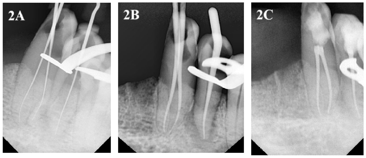
Figure 2: (A) Working length radiograph. (B) Master cone radiograph. (C) Post endo radiograph.
Glide path preparation done with number 10 K files. After glide path preparation canal is enlarged till number 15 K file. Then Protaper Universal rotary nickel instruments were used till F2 according to the manufracturer instruction. Recaputilation was done using 10 number k file in between every rotary file used to ensure the root canal system was carefully cleaned and shaped. Debris removal was aided by extensive irrigation with 5ml of sodium hypochlorite (5.25%) and 0.9% normal saline solution for each canal with double side vented 30G needle.
To lessen periapical inflammation and encourage disinfection, calcium hydroxide paste was used as an intracanal medicament for 14 days. A temporary seal was made using temporary restorative material (Cavit, 3M) and patient is recalled after 14 days.
After 14 days patient was asymptomatic so we went for obturation on the same appointment. F2 GP points were place inside the canals and tug back was achieved. A peri apical radiograph was taken to confirm the master cones (Figure 2B).
After that obturation was done using AH Plus (Dentsply, De-Trey Konstanz, Germany) sealer. Then, the access cavity was repaired to give structural support and stop coronal microleakage using composite resin (Figure 2C).
The patient was provided with thorough post-operative instructions that emphasized the importance of routine dental check-ups for maintenance and follow-up assessments, in addition to rigorous oral hygiene routines. To assure patient’s continuing oral health and well-being and to track the endodontic treatment's long-term success, a recall appointment was set up.
Patient did not report for follow up but on telephonic communication he confirmed he was asymptomatic with no complain or swelling.
DISCUSSION
The objective of root canal treatment is to clean any remaining pulp tissue, microorganism, bacteria, and its by-products from within the root canal space prior to obturation, so as to have an environment for the healing of periapical tissues. Diagnosis and identification of total number of roots, roots canals, and any possible permutation of the root canal morphology are of utmost importance for endodontic treatment. Failure to locate and disinfect an extra root canal is one of the most common cases for the failure of root canal treatment.3
Accurately diagnosing root canal configurations is crucial, but it can be difficult, particularly when there are anatomical differences. The straight angled radiograph should be enhanced with an angled x-ray; thus, any extra roots or canals might be noticeable. However, may not generally be satisfactory for evaluating morphological varieties in root anatomy. Advanced imaging techniques like cone-beam computed tomography (CBCT) may be required when traditional diagnostic procedures like periapical radiography fail to yield adequate information. The mandibular canine in this case study had two canals, which were discovered with the help of radiographic evaluation. This information helped determine the best course of therapy.
Effective root canal instrumentation is required for a thorough cleaning and contouring of the canal system. To guarantee appropriate canal debridement in the reported case, a combination of manual and rotational devices was used. Moreover, the ProTaper Universal system was chosen to perform instrumentation of the root canal system in this case due to one of its main characteristics: the rate of taper varies along the cutting flutes, which significantly enhances flexibility, allowing instrumentation of difficult areas with cutting efficiency and safety.4 Furthermore, irrigation with a sodium hypochlorite solution helps to remove debris and sanitize the area, facilitating periapical healing.
Post-instrumentation calcium hydroxide intracanal medicament performs several functions, including decreasing periapical irritation and sanitizing the canal system. Temporary sealing prevents bacterial entrance and protects the treatment space until the final obturation done. The combination of resin-based sealers and gutta-percha provides a hermetic seal, which is critical for long-term success.5
Post-operative care and follow-up appointments are essential elements of endodontic therapy. Tracking the progress of healing and the resolution of symptoms allows for prompt action in case any problems arise. Monitoring periapical healing and symptom resolution enables quick intervention if issues develop. Providing patients with full post-operative instructions and scheduling recall appointments supports continuous oral health maintenance and allows for long-term monitoring of treatment outcomes.
CONCLUSION
Understanding the fundamental structure of root canals and their deviations from the norm is essential for the effectiveness of non-surgical root canal therapy. Thoroughly analysing preoperative radiographs, together with improving access cavity and examining the pulpal floor, can greatly assist in effectively treating complex root canal anatomy during endodontic procedures.
REFERENCES
- Chugal NM, Clive JM, Spãngberg LS. A prognostic model for assessment of the outcome of endodontic treatment: effect of biologic and diagnostic variables. Oral Surg Oral Med Oral Pathol Oral Radiol Endod 2001 Mar;91(3):342-352.
- Victorino FR, Bernardes RA, Baldi JV, de Moraes IG, Bernardinelli N, Garcia RB, Bramante CM. Bilateral man dibular canines with two roots and two separate canals – case report. Braz Dent J 2009 Feb;20(1):84-86.
- Andrei OC, Margarit R, Daguci L. Treatment of a mandibular canine abutment with two canals. Rom J Morphol Embryol 2010 Jan;51(3):565-568.
- Grazziotin-Soares R, Baratto Filho F, Vanni J, Almeida S, Oliveira E, Barletta F, et al. Flexibility of K3 and ProTaper universal instruments. Brazilian dental journal. 2011 Jan; 22: 218–222.
- Ouellet R. Mandibular permanent cuspids with two roots. J Can Dent Assoc 1995 Feb;61(2):159-161.
Disclaimer from Guident
The views and opinions expressed in articles published in Guident are solely those of the authors and do not necessarily reflect the views of the editors, the editorial board or the publisher, Ivory India. Guident does not endorse any product, technique or treatment described. Clinical content is intended for qualified dental and medical professionals and is not a substitute for individual clinical judgement. Guident, its editors and the publisher accept no liability for any consequence arising from the use of information published in the journal.
How to cite this article
Dr. Arhana De, Dr. Ajay Kumar Nagpal, Dr. Mutiur Rahman. Endodontic Management of a Mandibular Canine with Two Roots and Two Root Canals:: A Case Report. Guident. 2025 May;18(6). Available from: https://guident.in/articles/endodontics/endodontic-management-of-a-mandibular-canine-with-two-roots-and-two-root-canals-a-case-report.html