A Case of Hyperdontia in a Non Syndromic Patient: A Case Report
Department of Oral and Maxillofacial Surgery, MES Dental College, Perinthalmanna, India
ABSTRACT
Multiple impacted supernumerary teeth are often rare and usually associated with uncommon syndromes like cleidocranial dysplasia; familial adenomatous polyposis; trichorhinophalangeal syndrome, type I; Rubinstein-Taybi syndrome; Nance-Horan syndrome etc. Supernumerary teeth are those that exceed the normal dental formula. They are more common in men, more common in the upper maxilla, and more prevalent in permanent dentition. Complications associated with supernumerary teeth include dental impaction, delayed eruption, ectopic eruption, overcrowding, spacing anomalies and the formation of follicular cysts. The treatment of supernumerary teeth depends on their type, position, and possible complications, detected clinically and radiographically. No clear consensus exists as to the best time to extract unerupted supernumerary teeth. This article reports a case of a 17 year old female with multiple impacted supernumerary teeth in the maxillary and mandibular arch.
INTRODUCTION
Hyperdontia, also called supernumerary teeth is a ‘number' dependent developmental dental anomaly, referring to any excess dental or odontological structure that is not a part of the normal dentition.1 Multiple impacted supernumerary teeth are rare in occurrence. Furthermore, most of these cases are associated with syndromes such as cleidocranial dysostosis, Gardner’s syndrome and cleft lip and palate.6,7,8 The prevalence of five or more supernumerary teeth was found to be 0.06%.9 It is often found as an incidental finding but can be seldom symptomatic. Apart from the symptomatic nature, impacted supernumerary teeth can also cause serious problems like formation of dentigerous cyst, complication due to its proximity with nasal floor and maxillary sinus, esthetic and functional complications such as crowding, midline diastema, root resorption, ectopic eruption, cystic lesions, displacement of the crowns of the neighboring teeth, intraoral infections, and delayed or failed eruption. In this report, we highlight about a case scenario of multiple impacted supernumerary teeth in the maxillary and mandibular arch managed under GA.
CASE REPORT
A 17 year old female patient was referred from Department of Orthodontics on the basis of incidental finding of multiple impacted supernumerary teeth in routine radiographic examination (OPG).
Her medical history is unremarkable and she has no relevant family history. On blood investigation her Hb was found to be slightly lower (10.4g/dl) and platelet count on a higher end 6.6 lakh/mm.3
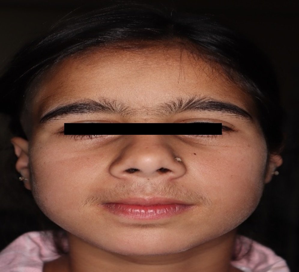
No gross asymmetry was noted on extraoral examination. Intraoral examination revealed a mild prominence in the palatal aspect of deciduous right maxillary canine as well as missing mandibular left first molar, right second molar & permanent maxillary canines bilaterally. Also presence of retained deciduous maxillary canine bilaterally and retained deciduous maxillary left first molar was noted.

FIGURE 1: MILD PALATAL SWELLING IN RELATION TO MAXILLARY RIGHT DECIDUOUS CANINE
Preoperative panoramic radiograph revealed supernumerary teeth in association with retained deciduous maxillary canine bilaterally, retained deciduous maxillary left first molar, mandibular permanent right & left second premolar. It also showed impacted maxillary left first premolar and maxillary permanent canines bilaterally.

FIGURE 2: PREOPERATIVE OPG
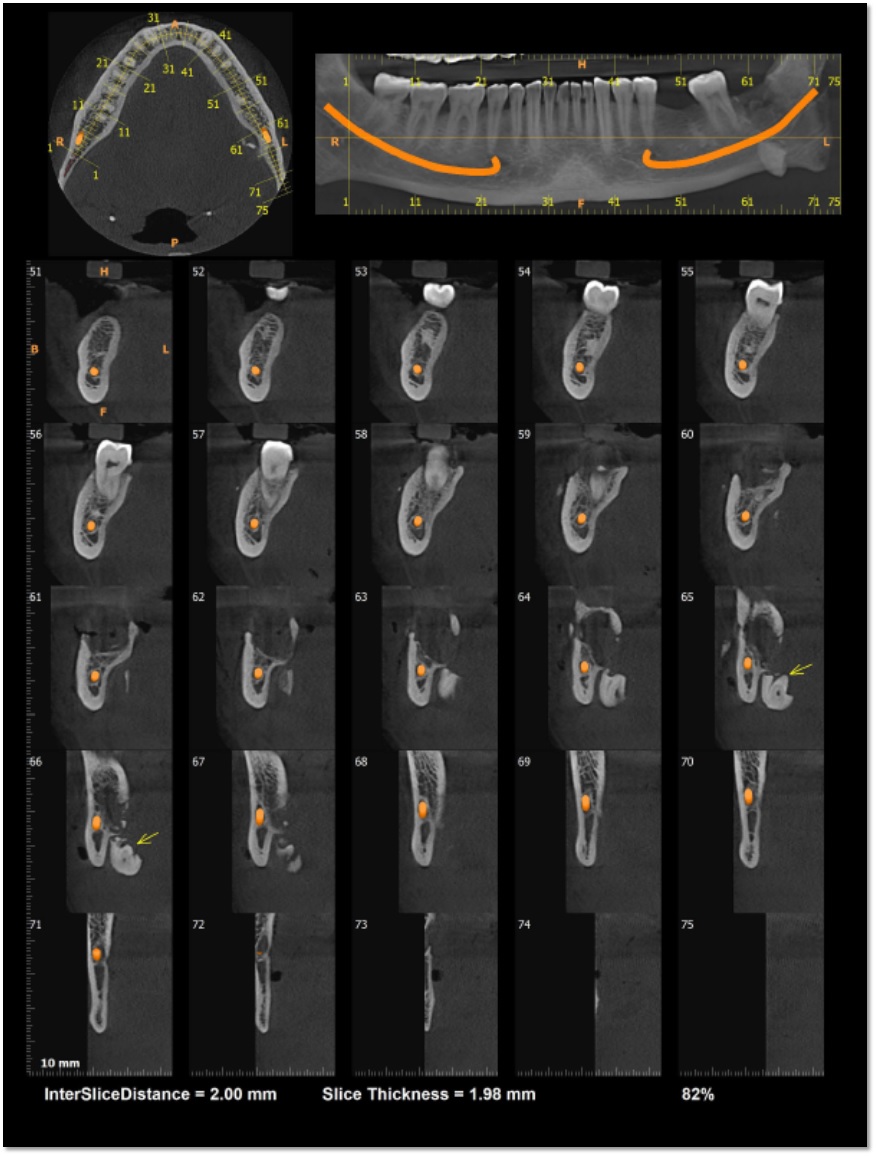
CBCT was taken to assess the nerve pathway and tooth morphology which reports the presence of an impacted supernumerary tooth with the crown resembling premolar located palatal & in contact with 53. Tooth is almost vertically impacted with the crown facing antero-inferiorly & root postero-superiorly.
Another impacted supernumerary tooth with the crown resembling premolar is located palatal & in contact with 14. Tooth is almost vertically impacted with the crown antero-inferiorly & root postero-superiorly. Root with open apex is also noticed.
Permanent right maxillary canine is impacted & located in such a way that; it is almost vertically impacted, in the antero- posterior plane. The crown of 13 is directed antero- medially and root is directed postero-laterally with no space in between adjacent tooth.
The crown of 13 is located palatal & superior to 11, in close approximation with the lateral wall of nasal cavity.
Middle part of root of 13 is seen palatal & superior to 12, in close approximation with the lateral wall of nasal cavity. Apical 3rd of root of 13 is located palatal & superior to 53, in close approximation with the lateral wall of nasal cavity.

FIGURE 3: CBCT IMAGE SHOWING 13 CLOSE TO LATERAL NASAL WALL (LINGUAL ASPECT)
Permanent maxillary left canine (23) is located palatal & in contact with 22 in close apposition with the lateral wall of nasal cavity. it is almost vertically impacted, in the anterior posterior plane. The crown of 13 is directed antero- medially and root is directed postero-laterally.
Another impacted supernumerary tooth with the crown resembling premolar is located palatal & in contact with 63. Tooth is almost vertically impacted with the crown antero-inferiorly & root postero-superiorly.
In addition, an impacted premolar 24 is located superior to 64. Tooth is vertically impacted. in close approximation with the floor of maxillary sinus.
And also impacted supernumerary teeth is located in the 35 and 45 region.
Findings were discussed and the patient was given an explanation of the indication to extract all impacted molars and all supernumerary teeth except 23 which was planned for orthodontic extrusion due to its favourable position.
SURGICAL TECHNIQUE
Patient was planned for surgical removal of impacted and supernumerary teeth under general anesthesia after getting fitness for the same. 1 g Cefixime intravenously was given preoperatively. Patient was aseptically painted and draped. 2% lignocaine with 1: 2,00,000 adrenaline infiltration given along the proposed site. In the mandibular arch, crevicular incision placed in the lingual aspect from 32 to 37 region & full thickness mucoperiosteal flap raised. A round bur was used to expose the crown region and bone guttering done with a straight fissure bur to expose the root of supernumerary tooth in relation to mandibular left second premolar tooth. Likewise, crevicular incision was placed from 42 to 46 region & full thickness mucoperiosteal flap raised. Bone guttering was done & supernumerary tooth in relation to mandibular right second premolar was exposed & removed.
In the maxillary arch, crevicular incision placed in the palatal aspect from 11 to 18 & full thickness mucoperiosteal flap raised. Palatal guttering done & vertically impacted maxillary right canine with root proximity to the lateral nasal wall was cautiously exposed & removed, once the impacted tooth was removed the supernumerary tooth in relation to 53 &14 behind it was visible and it was then elevated and taken out. Retained deciduous 53 also extracted.

FIGURE 4: INTRAOPERATIVE IMAGE POST REMOVAL OF SUPERNUMERARY TEETH IN RELATION TO 53 &14, IMPACTED 13 AND 53.
In order to approach the supernumerary teeth in the second quadrant, crevicular incision was placed from 21 to 25 in the buccal aspect, full thickness mucoperiosteal flap reflected. Buccal bone guttering done, supernumerary tooth in relation to 63 & 64 exposed, elevated & removed. The vertically impacted 24 which was close to the maxillary sinus was sectioned and taken out. Retained deciduous 63 & 64 also extracted. Wound closure done with 4-0 Vicryl. Patient was kept on IV antibiotics and analgesics. Postoperative follow up done for one month. Postoperative period was uneventful and healing was satisfactory.
DISCUSSION
There are limited publications of cases on numerous supernumerary teeth in healthy patients with no genetic involvement.8,10 Hence our case is of particular interest.
Multiple impacted teeth are a rare condition and are typically associated with certain syndromes. Lack of space due to crowding or early loss of deciduous teeth, prolonged retention of deciduous teeth, abnormal position of tooth bud, excessive fibrous tissue, ankylosis and cystic formation are the common etiology of impacted teeth.3,4
Delayed or arrested eruption is probably caused by diminished resorption of bone and of primary teeth and due to the presence of multiple supernumerary teeth.11 Conditions where we find retained teeth are: hemifacial atrophy, hypopitutarism, hypothyroidism, cherubism, gingival fibromatosis, cleft palate and preceding syndromes.12
Preoperative radiographic evaluation is the best method to identify the position and location of impacted supernumerary teeth. Since CBCT is a new technology that has transformed maxillofacial imaging, it was utilised as one of the diagnostic methods. It offers advantages of high contrast 3D images and image reconstruction for improved treatment planning.5
The presence of supernumerary tooth can cause a number of issues, including pathological alterations like chance of cyst formation, root resorption, delayed tooth eruption, and failure to erupt. A multidisciplinary approach may be appropriate choice since therapy involves issues with oral health, function, and aesthetics.
Treatment depends on the type and position of the supernumerary tooth and on its effect or potential effect on adjacent teeth. The management of a supernumerary tooth should form part of a comprehensive treatment plan and should not be considered in isolation.
Extraction is not always the treatment of choice for supernumerary teeth. They may be monitored without removal where:
- Satisfactory eruption of related teeth has occurred;
- no active orthodontic treatment is envisaged;
- there is no associated pathology;
- removal would prejudice the vitality of the related teeth.
However, surgical removal was advised due to the impacted teeth's pathological potential and undesirable position. It is very important to remove supernumerary teeth at a young age if it is damaging adjacent teeth or causing any other complication. Extraction of impacted teeth, including supernumerary teeth, is a commonly performed procedure associated with several complications and potentially substantial patient morbidity.13 With the assistance of a prosthodontist, pedodontist, and implantologist, it is necessary to plan space maintenance until the eruption of all permanent teeth and the patient's rehabilitation with fixed dentures. The aim of the therapy would be to achieve stable outcomes and improved aesthetics, dental health, and function.
CONCLUSION
The presence of multiple impacted supernumerary teeth is usually associated with syndromes like Gardner's syndrome and cleidocranial dysplasia. The simultaneous occurrence of multiple supernumerary and impaction of permanent teeth without association with complex syndromes is infrequent and is normally asymptomatic, usually diagnosed as a casual finding during routine panoramic X–ray studies. The close proximity to the lateral nasal wall & maxillary sinus makes the case more challenging. Managing such case scenario with the help of CBCT helped in better evaluation of the vital structures, nerve proximity, depth of tooth within the bone and also the orientation of tooth. Management of such condition also requires a multidisciplinary approach involving Dept of Oral Medicine & Radiology, Dept of Oral & Maxillofacial Surgey, Dept of Orthodontics and Dept of Prosthodontics to optimize patient specific superior results.
REFERENCES
- Tworkowski K., Gasowska E., Baryla D., Gabiec K. Supernumerary teeth – literature review. Journal of Pre-Clinical and Clinical Research . 2020;14(1):18–21. doi: 10.26444/jpccr/119037.
- Garvey MT, Barry HJ, Blake M. Supernumerary teeth overview of classification, diagnosis and management. J Canadian Dent Assoc. 1999;65(11):612–616.
- Sivakumar A, Valiathan A, Gandhi S, Mohandas AA. Idiopathic failure of eruption of multiple permanent teeth: report of 2 adults with a highlight on molecular biology. American Journal of Orthodontics and Dentofacial Orthopedics. 2007;132:687–92.
- Tanaka E, Kawazoe A, Nakamura S, Ito G, Hirose N, Tanne Y, Kawai N, Tanimoto K, Tanne K. An adolescent patient with multiple impacted teeth. The Angle Orthodontist. 2008 Nov;78(6):1110-8.
- Saluja KS, Singh B, Bhatia TK. An atypical case of nonsyndromic multiple impacted supernumerary teeth – a case report. International Journal of Contemporary Medical Research. 2016;3:1423–5.
- Golan I, Baumert U, Hrala B, Müssig D. Early craniofacial signs of cleidocranial dysplasia. Int J Paediatr Dent 2004;14(1):49–53.
- Atasu M, Dumlu A, Ozbayrak S. Multiple supernumerary teeth in association with cleidocranial dysplasia. J Clin Pediatr Dent 1996;21(1):85–91.
- Yusof WZ. Non-syndrome multiple supernumerary teeth: literature review. J Can Dent Assoc 1990;56(2):147–9.
- Ac?kgoz A, Ac?kgoz G, Tunga U, Otan. Characteristics and prevalence of nonsyndrome multiple supernumerary teeth: a retrospective study. Dentomaxillofac Radiol 2006;35(3):185–90.
- Pakdaman A, et al. Diagnosis and management of supernumerary (mesiodens): a review of the literature. Front Dent 2010;7(1):41–9.
- Babu V, Nagesh KS, Diwakar NR. A rare case of hereditary multiple impacted normal and supernumerary teeth. J Clin Pediatr Dent. 1998;23:59–62.
- Yalcin S, Gurbuzer B. Multiple impacted teeth in the maxilla. Oral Surg Oral Med Oral Pathol. 1993;76:130.
- Bouloux GF, Steed MB, Perciaccante VJ: Complications of third molar surgery. Oral Maxillofac Surg Clin North Am 19: 117, 2007
Disclaimer from Guident
The views and opinions expressed in articles published in Guident are solely those of the authors and do not necessarily reflect the views of the editors, the editorial board or the publisher, Ivory India. Guident does not endorse any product, technique or treatment described. Clinical content is intended for qualified dental and medical professionals and is not a substitute for individual clinical judgement. Guident, its editors and the publisher accept no liability for any consequence arising from the use of information published in the journal.
How to cite this article
Dr. Varsha Achuthan, Dr. Sooraj Soman, Dr. Roshni A. A Case of Hyperdontia in a Non Syndromic Patient: A Case Report. Guident. 2024 Oct;17(11). Available from: https://guident.in/articles/oral-surgery/a-case-of-hyperdontia-in-a-non-syndromic-patient-a-case-report.html