Contemporary Concept in Gingival Retraction for Implant Prosthesis
Department of Prosthodontics, Rajarajeshwari Dental College and Hospital, Bangalore
INTRODUCTION
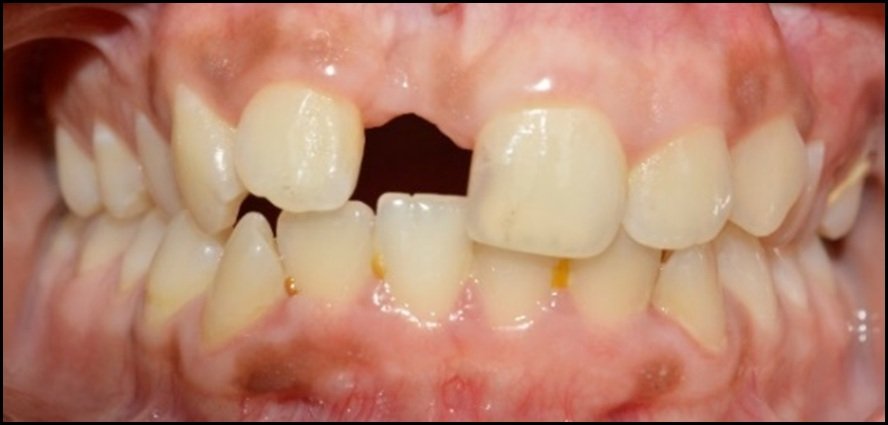
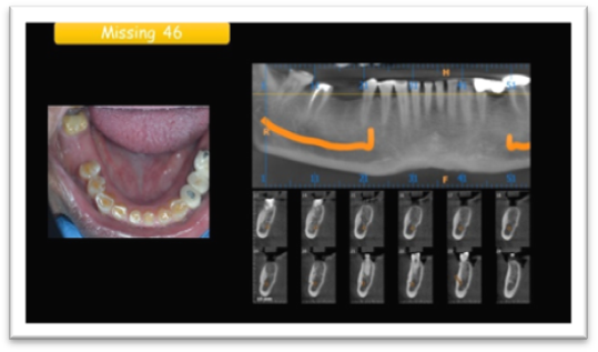
Osseointegrated implants have successfully provided alternative treatment to conventional prosthesis for the patients who have lost one, multiple, or all teeth who are seeking for a prosthetic substitute that can provide adequate masticatory, phonetic, and esthetic function.1 Today, the focus is more on the soft tissue than bony tissue in creating an esthetic restoration.2 In oral implants, fabrication of subgingivally placed abutment's margin is required, especially in esthetic regions and where minimal interarch space exists which necessitates reduced height of the abutment.3 The goal of soft tissue retraction in implants is to reversibly displace tissues and to expose margins of implant abutments before impression making.4 Inaccuracy in superstructure as a result of improper impression will lead to pathological biological cascades in peri-implant support tissues.This article reviews about present day concept involved in gingival retraction process in implant prosthetic replacement.
SOFT TISSUE HISTOPATHOLOGY MECHANISM IN IMPLANT TREATMENT
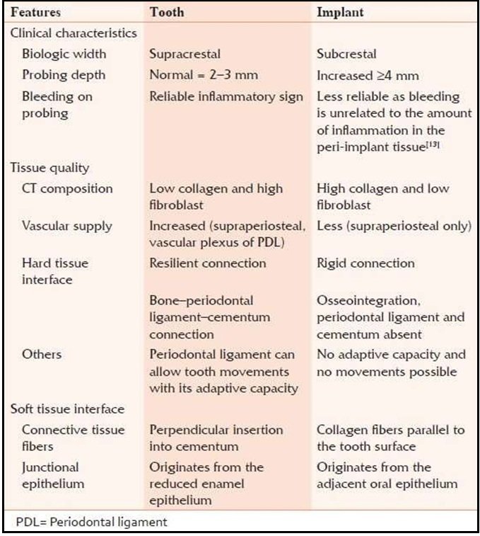
The junctional epithelium associated with natural teeth has a high rate of cell turnover, which occurs rapidly during the wound healing that takes place after recovering from infection or trauma.5 The rate of junctional epithelium cell turnover is twice that of oral gingival epithelium. At the base of the sulcus, the rate of exfoliation is as much as fifty times that of oral gingival epithelium that hinders bacterial colonization of the sulcus. When the junctional epithelium that surrounds implants is exposed to gingival retraction procedures, it is at greater risk of experiencing penetration damage than is the sulcus of natural teeth. Implant mucosa lacks keratinized epithelium at the base of the sulcus, which forms the junctional epithelium and has a hemidesmosomal attachment and internal basal lamina in the lower regions of the interface.6 It adheres poorly to implant surfaces, is more permeable and has a lower capacity for proliferation and regeneration than does the junctional epithelium around situations. Another consideration that has a bearing on the ability of epithelial tissues to withstand chemo mechanical manipulative procedures is the influence of the gingival biotype, categorized as thick or thin variety. Thick gingival biotype is more conducive for implant prosthesis.
In peridental tissue, the fiber-rich, highly organized periodontal complex surrounding natural teeth provides support for gingival tissues when they are retracted, mitigating the collapse of the tissues when the retraction agents are removed before making the impression. The periimplantfiber structure, however, does not provide the same level of support and is not able to prevent the collapse of retracted tissues to the same extent, which complicates attempts to successfully make impressions. This is particularly true in situations in which the depth of sulcus is greater than average, such as when an implant has been placed deeply.
GINGIVAL RETRACTION PROBLEMS IN IMPLANT PROSTHETIC PHASE
Several impression techniques are used in implant dentistry, and some require gingival displacement while making impressions. Others, such as the pickup impression technique, do not require any gingival retraction. For screw retained implant restorations, most systems use mechanical components impression copings that can be adapted accurately and directly to the fixture head on the abutment shoulder. With cement-retained prostheses that use customized abutments, the pickup impression technique cannot be used owing to the unique contour of the abutments. Therefore, clinicians must use another technique such as the conventional crown and bridge impression or optical impression. To ensure accuracy with polyvinyl siloxane impression materials, clinicians must maintain a minimum bulk of point two millimeter thickness in the sulcus area, which they can achieve by retracting the gingiva for at least four minutes before making the impression.7 Rapid reclosure of the sulcus requires that clinicians make the impression immediately after removing the retraction material.8
Placement of mechno chemical cords can cause inflammation and rebound hyperemia to the sulcular epithelium and underlying connective tissues. The filaments or fibers of conventional cords also may cause residual contamination of sulcular wounds, creating foreign body reactions and exacerbating inflammation. Healing of the sulcus can take few days time. Use of minimal force is necessary when packing cords to protect sharpe fibers. The use of metal instruments for cord placement in implant impression procedure might cause problem. Few lasers are contraindicated near implant surfaces, because they tend to absorb energy, which causes them to heat up and transmit the heat to bone, fragile subjunctional epithelium at the sulcus base around implants. Electro surgery is not recommended around implants because there is significant risk that the contacting electrode may arc by conducting electric current though the metal implant structure to the bone rather than via the more dispersive gingival tissue pathway. The concentrated electrical current at the tip of electrodes can generate heat, which may cause osseous or mucosal necrosis. Rotary curettage is inappropriate for use around implant restorations because of poor tactile control when cutting soft tissue, which could lead to bur contact damage to the implant surface and over instrumentation. The action of aluminum chloride is similar to that of aluminum sulfate, which is an astringent that causes precipitation of tissue proteins but less vasoconstriction than epinephrine.9 Aluminum chloride is the least irritating of the medicaments used for impregnating retraction cords, but it disturbs the setting of polyvinyl siloxane impression materials.10 Aluminum potassium sulphate and aluminum chloride medicated cords are more effective in keeping the sulcus open after clinicians remove the cord.
VIRTUAL GINGIVAL RETRACTION IN IMPLANT TREATMENT
Larger sulcus spaces than necessary for conventional crown and bridge impression techniques are needed when making digital computer aided design/computer-aided manufacturing impressions to ensure accurate recording of finishing lines. Direct optical impressions are limited to line of sight, which is facilitated by performing gingival retraction to expose finish lines. Artifacts caused by retraction cord fibers that remain in the sulcus may affect the accuracy of optical impressions. Fifteen percent aluminum chloride in an inject able kaolin matrix leaves a clean sulcus, reducing the influence of artifact-generated errors. However, the powders used when making optical impressions to reduce reflectivity and make tooth surfaces measurable can influence impression accuracy by increasing tooth surface thickness. Clinicians regard the indirect capture of digitized information as being potentially more accurate; however, the way in which clinicians can acquire data is influenced by the thickness of the impression material in the sulcus area. Significant errors can result from thin impression margins with a radius less than the contacting probe tip.
CONCLUSION
The use of fifteen percent aluminum chloride in an injectable kaolin matrix is effective. It also is safe, with the reports of no adverse effects. Gingival recession associated with an injection of aluminum chloride into the gingival sulcus is almost undetectable. The injectable matrix is hydrophilic and can be flushed away relatively easily from the gingival crevice. Gcuff which is newly introduced retraction system seems to yield predictable restoration for implant patients.
REFERENCES
- Goldstein, Ronald E. Esthetics in Dentistry. 2nd ed. Hamilton, On: B.C. Decker; 1998.
- Pad bury A Jr, Eber R, Wang HL. Interactions between the gingival and the margin of restorations. J Clin Periodontol 2003; 30:379-85.
- Misch CE. Cement retained implant prostheses: Implant protective occlusion. Dental Implant Prosthetics. St. Louis: Mosby; 2005.
- Bennani V, Schwass D, Chandler N. Gingival retraction techniques for implants versus teeth: Current status. J Am Dent Assoc 2008; 139:1354-63.
- Shimono M, Ishikawa T, Enokiya Y, et al. Biological characteristics of the junctional epithelium. J Electron Microsc (Tokyo) 2003;52(6):627-639.
- Glauser R, Schupbach P, Gottlow J, Hammerle CH. Periimplantsoft tissue barrier at experimental one-piece mini-implants with different surface topography in humans: a light-microscopic overview and histometric analysis.Clin Implant Dent Relat Res 2005;7(suppl 1):S44-S51
- Baharav H, Laufer BZ, Langer Y, Cardash HS. The effect of displacement time on gingival crevice width. Int J Prosthodont 1997; 10(3):248-253
- Laufer BZ, Baharav H, Langer Y, Cardash HS. The closure of the gingival crevice following gingival retraction for impression making. J Oral Rehabil 1997; 24(9):629-63
- Kellam SA, Smith JR, Scheffel SJ. Epinephrine absorption from commercial gingival retraction cords in clinical patients. J Prosthet Dent 1992; 68(5):761-765.
- Dental product spotlight: gingival retraction cord. JADA 2002; 133(5):652-653.
Disclaimer from Guident
The views and opinions expressed in articles published in Guident are solely those of the authors and do not necessarily reflect the views of the editors, the editorial board or the publisher, Ivory India. Guident does not endorse any product, technique or treatment described. Clinical content is intended for qualified dental and medical professionals and is not a substitute for individual clinical judgement. Guident, its editors and the publisher accept no liability for any consequence arising from the use of information published in the journal.
How to cite this article
Dr. Krishna Kumar U. Contemporary Concept in Gingival Retraction for Implant Prosthesis. Guident. 2021 Oct;14(11). Available from: https://guident.in/articles/implantology/contemporary-concept-in-gingival-retraction-for-implant-prosthesis.html