Oral Mucocele : A Case Report
Department of Periodontology and Oral Implantology, Akola
ABSTRACT
The mucocele is one of the most common benign tissue masses that occur in oral cavity. It is basically of two types : mucous retention and mucous extravasation type. Clinical observation gives the provisional diagnosis which is confirmed histopathologically. Treatment is surgical removal of the lesion.
INTRODUCTION
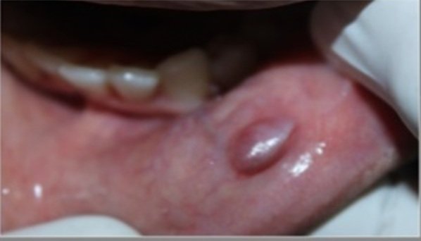
Mucocele is a benign, mucous containing cystic lesion of minor salivary gland.The term is derived from a Latin word, mucous and coele means cavity.1 It is caused as a result of accumulation of mucous and alteration of minor salivary glands which results in a limited swelling. It is the 17th most common salivary gland lesion in oral cavity.2

Oral Mucocele
Variants
There are two variants of mucocele : mucous retention cyst and mucous extravasation cyst. Mucous retention cyst is caused because of accumulation of mucous in minor salivary gland due to obstruction of ducts of these glands.3 Mucous extravasation cyst is caused due to rupture of duct of minor salivary gland and alteration of minor salivary gland which results in a limited swelling.3 It can also be: Superficial or deep. Superficial is located directly under the mucosa and deeper is located in the lower cornium.
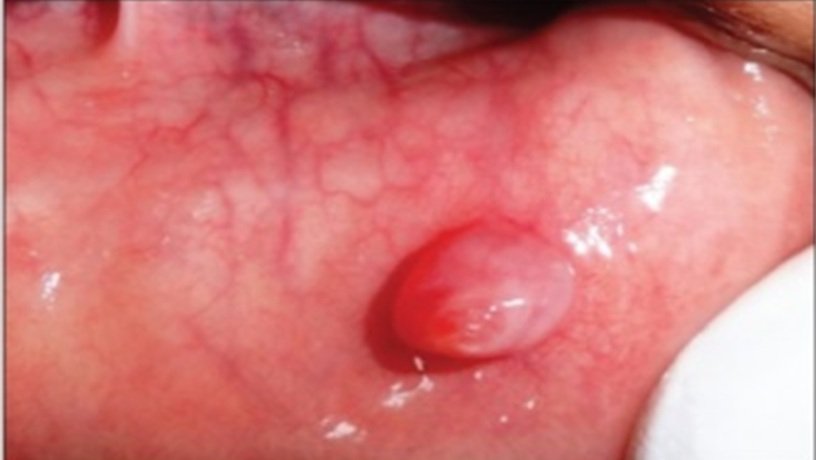
Superficial
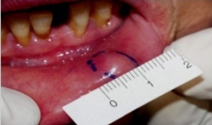
Deep
Clinical Features
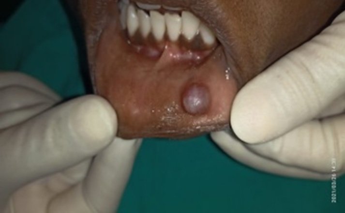
Mucocele is more prevalent in children and young adults. Its common location is on lower lip lateral to the midline. Clinically it is a raised vesicle usually of dome shaped approximately 1-2 mm to several cms in size. History of trauma to lower lip due to either lip biting or piercing repeatedly. Superficial lesion is bluish in colour and is transluscent, while deeper lesion is normal in colour same as that of surrounding mucosa due to the thickness of mucosa overlying the lesion.

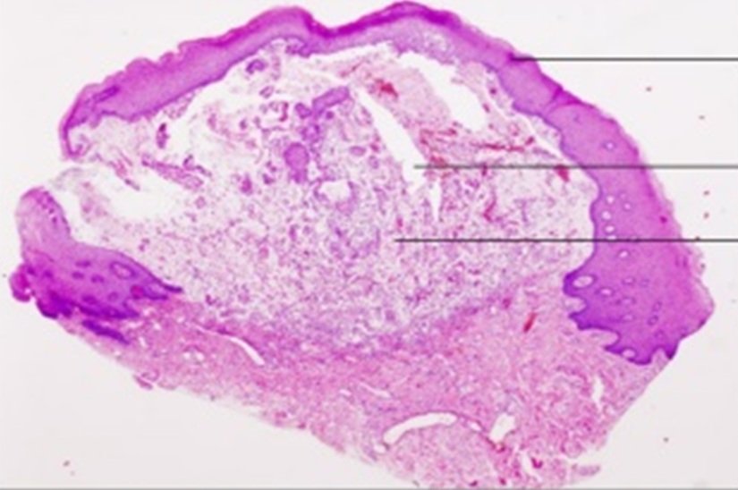
Normal Histologic Appearance
Diagnosis
Diagnosis is clinical. Fluctuation in the lesion helps to come to proper diagnosis. FNAC and Histopathology further confirms the diagnosis.
Treatment
Surgical excision of the cyst along with associated salivary acini. Excision can be done by following methods: Cryosurgery, Conventional surgical removal, Electrosurgery, Laser.4
Marsupialisation, micromarsupialization can also be done.
CASE REPORT
Clinical examination
A 30 year old female patient reported to the department of Periodontics, Dr R R Kambe Dental College, Akola with chief complaint of swelling in lower lip since a month and difficulty in chewing. The patient was apparently alright one month back when she noticed a small swelling in lower lip lateral to midline on left side which was initially small in size and has gradually increased to the present size. Patient
gave history of habit of lip biting. There was no pain associated with the swelling. Patient’s past medical and dental history were not significant and no significant family history was observed.
Extraoral Examination
Face appeared bilaterally symmetrical, Temperomndibular joint movements were synchronous and lips were competent. Lymph nodes were non palpable.
Intraoral examination
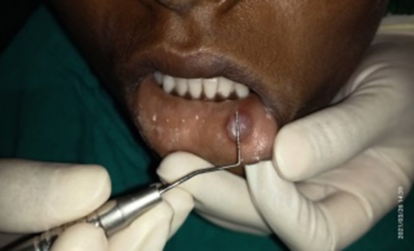
swelling present on lower lip lateral to midline on left side in relation with 34. Approximate size of the lesion is 10mm by 5mm. Shape is elliptical to ovoid. Colour is reddish blue.
Consistency is soft and is non tender, non reducible lesion, fixed to underlying tissue.
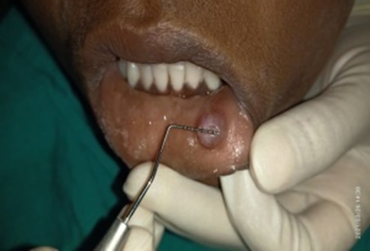
Investigations
The tentative diagnosis is made from clinical history, clinical presentation, and palpation. Routine hematological examinations were found to be normal including haemogram
& Bleeding time & Clotting time.
Histopathological examination to confirm the provisional diagnosis was done.
Provisional Diagnosis
Mucous extravasation cyst
Differential Diagnosis
Salivary gland tumour
Traumatic lesion (Traumatic fibroma)
Vesiculobullous lesion (mucous membrane pemphigoid)
Treatment
Phase I therapy included thorough scaling and root planing and oral hygiene instructions were given. Proper brushing technique was demonstrated. 0.2% chlorhexidine mouthwash was advised 10ml 1:1 dilution, twice daily for 15 days.
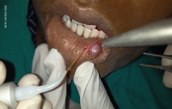
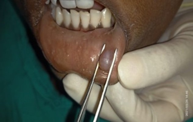
Phase II therapy included Surgical Excision using Laser. The lesion was removed along with associated glandular acini. The excised lesion was sent for histopathologic examination. Post operative instructions were given and patient was recalled after a week for follow up and to examine healing. Analgesics were prescribed.
The Lesion

Demarcation around lesion with laser
Demarcated lesion
Lesion held with holding forcep
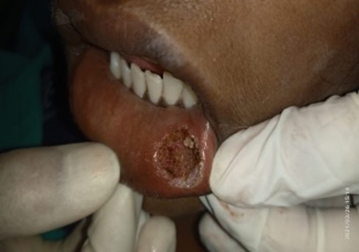
Excision of the Lesion
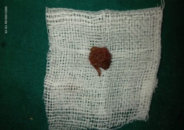
Excised tissue sent for histopathologic examination
DISCUSSION
Most common location of mucocele is in lower labial mucosa about 70% of total cases.
A rate of recurrence from 2.43% to 8.8% has been reported. Literature shows equal incidence in males and females but there are reports suggesting a slightly higher incidence in females 1.3:15 Lip biting is found to be common causative factor for mucocele to occur.6 If the vesicle is un roofed by suction pressure by any individual, they may report a chronic and recurrent history.7
The key point in avoiding recurrence is to eliminate adjacent surrounding glandular acini and to remove the lesion upto the muscle layer.
A localized and terminal paresthesia of labial mucosa close to surgical site in 10.58% of total patients 3 months after surgery.
Laser – total treatment within 3 to 5 min whereas scalpel technique is meticulous and need to perform suturing at end of operation.8
Advantages of CO2 laser versus cold scalpel
- Minimal damage to neighboring tissues
- Bloodless and highly decontaminated surgical bed.
- Lessened swelling and pain during post operative period.
- Appearance of fewer myofibroblasts resulting in comparatively lesser wound contraction.9
CO2 laser have excellent aesthetic outcome without any fibrosis or scarring while scalpel leaves small residual unesthetic defect after the healing period in 5 of 25 lower lip mucoceles treated with surgical treatment.
Healing of wound caused by CO2 laser results in:
Appearance of fibro osseous membrane after 72 hrs that replaces superficial necrotic layer of irradiated tissues.
Epithelial covering occurs after 2 weeks.
Thinner and parakeratotic epithelium is seen in comparision with epithelium that appears after surgical resection.
REFERENCES
- Baurmash HD. Mucoceles and Ranulas. J Oral Maxillofac surg 2003; 61:369-78
- Flaitz CM,Hicks JM. Mucocele and Ranula.eMedicine;2015
- J Ata-Ali, C Carrillo2 , C Bonet Oral Mucocele: review of literature J Clin Exp Dent. 2010;2(1):e18-21.
- Huang IY, Chen CM, Kao YH, Worthington P. Treatment of mucocele of lower lip with carbon dioxide laser. J Oral Maxillofac Surg 2007;65:855-8
- Rao PK, Hegde D, Shetty SR, Chatra L, Shenai P. Oral mucocele-diagnosis and management. J Dent Med Sci 2012;2:26-30.
- Jha M, Jogani V. Oral mucocele: Reviwe and case report. J Contemporary Dent 2012;2:119-24.
- Shafer’s Textbook of Oral Pathology. 2009. Sixth edition: 541-542
- Subramaniam Ramkumar, Lakshmi Ramkumar, Narasimha Malathi, Ramlingam Suganya. Case Report- Exision of Mucocele Using Diode Laser in Lower Lip, Case Report in Dentistry, 2016, Article ID 1746316
- Raffetto, Lasers for initial periodontal therapy, Dental Clin N Am 2004;48:923-936
Disclaimer from Guident
The views and opinions expressed in articles published in Guident are solely those of the authors and do not necessarily reflect the views of the editors, the editorial board or the publisher, Ivory India. Guident does not endorse any product, technique or treatment described. Clinical content is intended for qualified dental and medical professionals and is not a substitute for individual clinical judgement. Guident, its editors and the publisher accept no liability for any consequence arising from the use of information published in the journal.
How to cite this article
Dr. Aakanksha Tiwari, Dr. Reshma Avhad, Dr. Rajesh Ramdasji Kambe Dental College And Hospital. Oral Mucocele : A Case Report. Guident. 2021 Oct;14(11). Available from: https://guident.in/articles/periodontics/oral-mucocele-a-case-report.html