Separated Instrumental Retrieval "S.i.r." Call for a Protocol
Department of Pediatric and Preventive Dentistry, MNR DENTAL COLLEGE AND HOSPITAL, SANGAREEDY INDIA
ABSTRACT
AIM: To evaluate the knowledge about instrument separation and retrieval treatment options among postgraduate students and practising dentists
MATERIALS AND METHODS: A self-administered questionnaire was given to dentists and postgraduate students.
RESULTS: There exists difference in knowledge and practices among postgraduate students and practitioners.
CONCLUSION: Prevention is the best antidote for a separated file in the canal. Adhering to proven concepts with combining the best strategies and making safe techniques during preparation, will eliminate the separated instrument procedural mishaps.
KEYWORDS: Files, root canal treatment, separated endodontic instrument, causes, management, retrieval
INTRODUCTION
Endodontic instrument separation within the root canal is an untoward occurrence that will interfere with cleaning and shaping procedures affecting long-term prognosis of the tooth.1 It’s been reported that the prevalence of separated instrument ranges from 2 to 6% by Tronstad et al2 and 0.5 to 5% by Iqbal et al.3
MATERIALS & METHODS
A cross sectional study was conducted in 125 dental fraternity. Out of 125, a self-administered questionnaire (Table 1) was given to 67 postgraduate students and 58 dental practitioners.
Table 1: S.I.R. Questionnaire
|
S. No |
Questions |
|
1 |
What is your current professional status?
|
|
2 |
How many years have you been practicing dentistry?
|
|
3 |
If you break an instrument do you think it is required to inform your patient? a) Yes b) no |
|
4 |
Which types of files frequently break with you?
|
|
5 |
Which alloy of manufacture is frequently broken?
|
|
6 |
Which sizes are more broken?
|
|
7 |
In which part of root canal, instruments separate? a) Apical b) Middle c) Coronal |
|
8 |
In which stage of RCT, instruments separated?
|
|
9 |
Causes of breakage according to your preparation.
|
|
10 |
If file breakage is related to root canal anatomy, what is the most common cause?
|
|
11 |
In which teeth instruments break more?
|
|
12 |
If it is molars, in which root canal is more instrument breakage?
|
|
13 |
What you will do to deal with this separated instrument?
|
|
14 |
Which method will help you more in removing the separating file?
|
|
15 |
While attempting to bypass separated file, which procedure error result?
|
|
16 |
In follow up- what results do you find?
|
The collected data were entered into Microsoft Excel sheets and statistically analysed using SPSS version 22(IBM corporation, Washington DC, United States). The data was expressed as percentages.
RESULTS
Results of demographic data, current professional status and practising were presented in Table 2.
|
Professional status |
Postgraduate |
67 |
54% |
|
Dental Practitioner |
58 |
46% |
|
|
Experience |
Less than 3 years |
75 |
60% |
|
5-10 years |
46 |
37% |
|
|
More than 10 years |
4 |
3% |
62% of broken files were rotary files, 74% stainless steel files, and regarding size in 52% of cases smaller size files most separated in the canal. (Figure 1).
Concerning the site, higher percentage was separated in apical third (68%). With 72% of them broken during cleaning and shaping. [Fig-2]. With regard to different opinions concerning causes are as follows, 41% of breakage due to over usage of the file [Fig-3], most commonly were in molars by 95% [Fig-4] especially in mesiobuccal canal 32% followed by MB2 canal (26%), mesiolingual canal (21%), distobuccal canal (12%), palatal (5%) and distal canal (4%). [Fig 4].

FIGURE 1: Most frequent files, alloy of manufacture, file size separated in the canal

FIGURE 2: Which part and stage of root canal instrument commonly separate

FIGURE 3: Causes of breakage

FIGURE 4: Most common teeth and most common root canal in molars
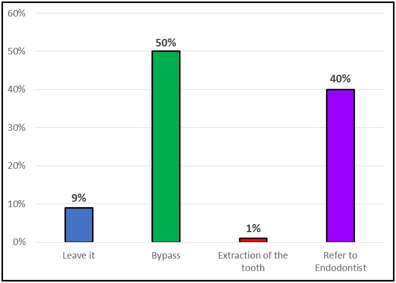
FIGURE 5: Management options

FIGURE 6: Methods used to remove separated file
Regarding dealing with the management, 50 % opted to bypass whereas 40 % preferred to refer to endodontist.
Concerning removal methods, 34% of dentists removed separated instrument using Masserann kit, while 28% used ultrasonics and conventional method and 10% IRS.
DISCUSSION
This is a cross-sectional study aimed to compare the incidence, factors contributing for separated endodontic files between dentists and postgraduates along with treatment options, management mishaps and methods used to retrieve endodontic files during endodontic treatment among dentists in Sangareddy.

Different methods for retrieval of instruments4 are: Conventional method; Tweezers, endodontic forceps, mosquito, dental pliers; Micro-tube, Injection needle, Tube like systems, Ultra sonics-Dental operating microscope, Laser, Electrolysis
Tube like systems: Aggressive, risk of root weakening, perforation; Cannot be applied to narrow and curved canals
- Endoextractor system
- Masserann kit
- Instrument removal system (IRS)
- Separated instrument removal system
- Canceliar instrument
- Mounce extractor
Ultrasonics-Dental operating microscope: Can be inserted in depth into the root canal, allows working under bright illumination and magnification, limits the risk of excessive removal of dentin and possible perforation and can be used without water increasing the visibility.
It causes increased temperature in outer surface of root and secondary separation of the fragment which can be prevented by air spray, low power and not using for longer period.
Facts from literature4:
- Rotary instrument separation frequency- 0% to 13%
- Manual instrument separation frequency- 0.25% to 6%
- Highest frequency of instrument separation in molars (more in mesiobuccal canal)- 77% to 89%
- Higher rate of separation is in apical third-41% to 82.7%
- Most common site separation site-2mm from tip of the instrument
- Most common sizes of instruments undergoing separation–NO 15-40(ISO); taper-4% and 9%.
- Clinical success rate for treatment of separated instrument- 53% to 95%
- Rate of successful fragment removal (44%-95%) is higher than instrument bypass (9%-47.7%)
- Stainless steel instruments are easily retrieved than NiTi
- Longer fragments are easily removed than shorter ones
Rotary files (62%) were more separated than hand files (38%) contrast to study by Samah et al5 where hand files (53%) were more separated than rotary (47%).
Stainless steel files (74%) were more separated than NiTi (26%) corresponds to study by Samah et al5 (68%).
Small size files are more separated (52%) that corresponds with Choksi et al6 who reported that they are more prone to distortion as a result of stressing on their small cross sections.
Concerning the site, higher percentage was separated in apical third (68%) corresponds to study by Samah et al5 (72%).
While 72% significantly broke during cleaning and shaping -corresponds to study by Samah et al 5(78%) and Madarati et al,2008.5
41% of causes of breakage were over usage of the file in contrast to this, root canal anatomy (45%) was the major cause as reported by Madarati et al7 where 28% of the cause of breakage is due to root canal anatomy.
95% of instruments separated in molars, 32% in mesiobuccal canal (26% MB2 canal) followed by 21% mesiolingual canal corresponding with Madarati et al.7
50% of the sample successfully bypassed which was in contrast to the study by Gandevivala et al8 where majority of them referred to the endodontist.
34% of dentists could successfully remove using masseran kit which was in contrast to the study by Gencoglu and Helvacioglu9 which confirmed that ultrasonics with the aid of an operating dental microscope is more successful in removing fractured instruments.
CONCLUSION
"SIR“ presents a call for a protocol to handle these endodontic mishaps as a fearless challenge.
Financial support: Nil
Conflict of interest: None
REFERENCES
- Sokhi R, Sumanthini MV, Shenoy V. Retrieval of Separated Instrument using Ultrasonics in a Permanent Mandibular Second Molar: A Case Report. J Contemp Dent. 2014;4(1).
- Kerekes K, Tronstad L. Long-term results of endodontic treatment performed with a standardized technique. J Endodon. 1979;5(3): 83-90.
- Iqbal MK, Kohli MR, Kim JS. A retrospective clinical study of incidence of root canal instrument separation in an endodontics graduate program: a PennEndo database study. J Endod. 2006;32: 1048-1052.
- Triantafyllia Vouzara, Maryam el Chares, Kleoniki Lyroudia. Separated Instrument in Endodontics: Frequency, Treatment and Prognosis. Balk J Dent Med. 2018;123-132.
- Samah et al. Evaluation of the Factors and Treatment Options of Separated Endodontic Files. Journal of Clinical and Diagnostic Research. 2016;10(3): ZC18-ZC23.
- ChoksiD,IdnaniB,KalariaD,Patel RN. Management of an Intracanal Separated Instrument: A CaseReport. IranEndodJ. 2013;8(4):205-7.
- Abmad A.Madarati,Mark J.Hunter,Paul M.H. Dummer. Management of Intracanal separated instruments, JOE. 2013;39(5).
- Gandevivala A, Parekh B, Poplai G, Sayed A. Surgical removal of fractured endodontic instrument in the periapex of mandibular first molar. J Int Oral Health. 2014;6(4):85-88.
- Nimet Gencoglua, Dilek Helvacioglub. Comparison of the Different Techniques to Remove Fractured Endodontic Instruments from Root Canal Systems. Eur J Dent. 2009;3:90-95.
Disclaimer from Guident
The views and opinions expressed in articles published in Guident are solely those of the authors and do not necessarily reflect the views of the editors, the editorial board or the publisher, Ivory India. Guident does not endorse any product, technique or treatment described. Clinical content is intended for qualified dental and medical professionals and is not a substitute for individual clinical judgement. Guident, its editors and the publisher accept no liability for any consequence arising from the use of information published in the journal.
How to cite this article
Dr. Uma Ramana Prakhya, Dr. Pranitha.V, Dr. Saumya.V. Separated Instrumental Retrieval "S.i.r." Call for a Protocol. Guident. 2021 Oct;14(11). Available from: https://guident.in/articles/research/separated-instrumental-retrieval-s-i-r-call-for-a-protocol.html