Bone Augmentation Using a Mandibular Symphysis Graft-a Case Report
Department of Prosthodontics and Crown and Bridge, Navi Mumbai India
ABSTRACT
Alveolar ridge resorption is a common phenomenon after tooth loss, which changes the size and shape of the bone available for placement of implants. In the age of prosthetic driven implant dentistry, the final prosthesis design and type dictates the size, number and the ideal implant position. Deficiency of bone volume is the primary reason for avoiding replacement treatment with implants. The solution lies in the re-establishment of the height of the alveolar ridge in conjunction with the prosthesis and with load-bearing lamellar bone for implant placement. The use of autogenous bone grafts is still the "gold standard" in implant site reconstructive surgery. The mandibular symphysis is a favorable donor site to increase alveolar bone volume both laterally and vertically to prepare the ridge for a correct placement of dental implants. The presented article is a case report of the clinical and radiological outcome of autologous onlay chin block grafts used to augment a unilateral defect due to loss of the upper central incisor for dental implant placement for the purpose of improving function and aesthetics.
INTRODUCTION
The upper central incisor region is a zone of high aesthetic value with a limited area available for a precise implant insertion.1 It may result in alveolar bone and buccal plate defects. The repair of acquired alveolar defects with autogenous bone grafts for implant placement is one of the most common surgical techniques in implant surgery. Once adequate quantity of bone has been augmented, implant placement plays a pivotal role in the rehabilitation of these patients.2
Bone reconstruction techniques have been improved in order to optimize the aesthetic and functional result.3,4 The extension of the bone defect determines whether bone augmentation procedures can be performed simultaneously with the placement of implants or as a separate procedure.5
Among the various bone augmentation materials available, only autologous bone combines osteoconductive, osteoinductive and osteogenic features when compared to any other bone substitute.6 Owing to its biological properties and absence of immunological reactions, autologous bone graft has been set as the “gold standard” for bone regenerative procedures.7
Intraoral harvesting may lead to donor site morbidity and possible complications that could occur during the bone harvesting procedure.8
Intraoral donor sites such as the mandibular symphysis and ramus show reduced resorption and higher revascularization rates than endochondral bone grafts, like the tibial plateau and iliac crest.9,10 Bone harvesting can be performed with local anesthesia without resorting to general anesthesia.
The mandibular symphysis provides a greater graft volume with a simpler surgical access. The corticocancellous composition of the bone harvested from the mandibular symphysis region aids in rapid vascularization and integration after it has been positioned at the recipient site. Better postoperative patient morbidity and low rate of wound dehiscence make the ramus a preferred donor site.11,12
The use of bone substitutes with low turnover rates to cover the graft reduces the resorption rate of the bone block.13 Some authors found that deproteinized bovine bone (DBB) particles stabilized by resorbable membranes covering onlay block grafts reduced resorption by almost 50% in comparison to noncovered grafts.1,14 Bone substitutes contribute to the creation of a smooth connection between the block graft and the recipient bone and can provide a scaffold promoting bone regeneration.13,14 Another advantage of using resorbable rather than nonresorbable membranes is the elimination of a second surgical phase.
Based on the above rationale, the main aim of the presented case report was to evaluate the placement of dental implants in resorbed alveolar ridges augmented with autogenous onlay bone grafts obtained from the mandibular symphysis.
CASE REPORT
A 17 year old female reported to the Department of Prosthodontics in Dr.D.Y.Patil University School of Dentistry with a chief complaint of missing tooth in the upper right region. The anatomy of the upper jaw was evaluated by panoramic radiography and clinical examination. A unilateral bone defect in correspondence with the upper right central incisor region was observed. The patient had a thin gingival biotype. (Figure 1)
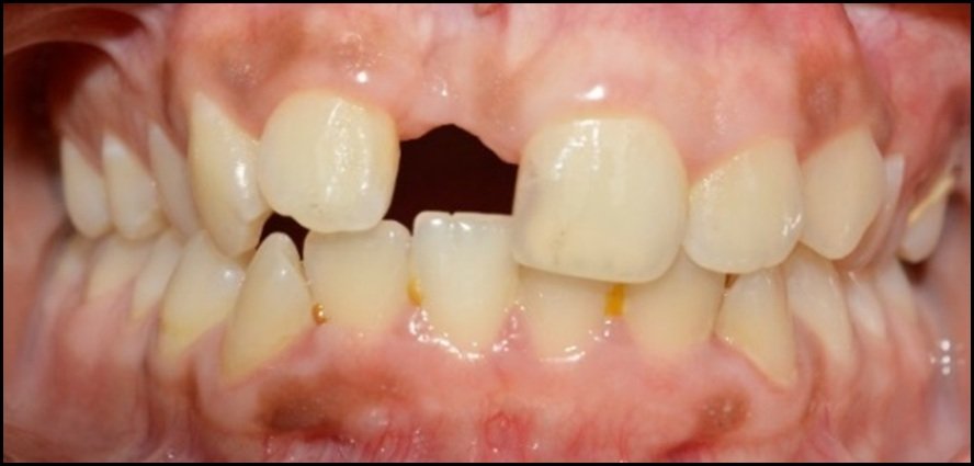
Figure 1: Intraoral pre-operative view
A bone augmentation procedure was planned using an intraoral autogenous bone harvested from the mandibular symphysis and delayed implant insertion. (Figure 2)
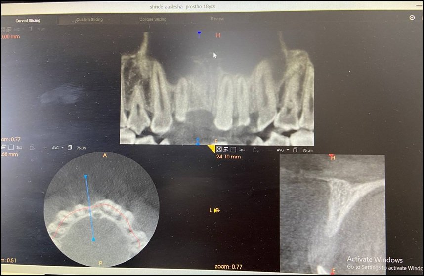
Figure 2: Bone augmentation planning using CBCT
Several injections were required as the anterior symphyseal region of the mandible is innervated by the mandibular branch of the fifth cranial nerve (V3) and cervical nerves from C-3 and C-4. Bilateral mandibular block with 2% lidocaine HCL (1:100,000 epinephrine) accomplish anesthesia for V3 innervation.15
Assessment of the periodontal status of lower anteriors, periodontal risk of root fenestration, amount of bone loss, subgingival margins of the restoration, amount of keratinized gingiva and local musculature should be done to determine the ideal incision design. Depending on the anatomy, surgical access to the symphysis region can be obtained via a crestal incision, mid-keratinized tissue incision, vestibular incision or sulcular approach.
Submarginal/attached gingiva incision is the horizontal or parasulcular, scalloped incision15,16,17 that should be given in the attached gingiva at least 1 mm above the mucogingival junction. If >3 mm of keratinized gingiva is present, beveled incision in apical direction is given. If >3 mm of keratinized gingiva is present, perpendicular incision to the underlying bone gives a butt joint for tissue adaptation during the suturing.17 The vertical incision is given either at the midline or between the canine and the first premolar, keeping a safe distance from a local neurovascular bundle. The midline vertical release can be given when the harvest is for 3 teeth or less and extends up to 5 mm of the inferior aspect of the mandible. (Figure 3)

Figure 3: Bone blocks obtained from the mandibular symphysis

Figure 4: Bone block stabilization using osteosynthesis screws

Figure 5: Sutures given
Misch in 1992, proposed the Rule of 5’s surgical technique to harvest a bone block graft from symphysis with bone cuts perpendicular to the cortex in a right angle to the vestibular plain of the symphysis. The superior cut should be atleast 5 mm below root apices to prevent any injury to tooth roots. The inferior cut should be 5 mm above the lower border of the mandible. Vertical cuts should be minimum 5 mm away from the mental foramen. To obtain a monocortical graft, depth of the cut should extend from the outer cortex to the opposite cortical plate. Lingual cortex should not be perforated.18

Figure 6: Implant planning using CBCT

Figure 7: Surgical guide for implant placement

Figure 8: Implant placed
Two rectangular monocortical block grafts were harvested from the mandibular symphysis region. Two osteosynthesis screws were used to fix them at the buccal aspect of the bone defects (Figure 3 and 4). Sutures were given to obtain a first-intention seal of the flaps (Figure 5). The sutures were removed after 14 days and an orthopantomograph was obtained. Six months post grafting, fixation screws were removed and an implant was placed with the aid of a surgical stent (Figure 6,7 and 8). The insertion torque was >35 Ncm. At this point, DBB particles (Bio-Oss®, Geistlich Biomaterials, Wolhusen, Switzerland) and a Type- I resorbable collagen membranes from bovine origin (Cologenesis Colo Gide GTR Membrane) were used to cover the block graft at each site. (Figure 9 and 10)
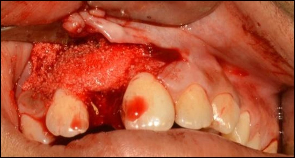
Figure 9: Bone graft placed buccally

Figure 10: Bone graft covered with Cologide GTR membrane
After five months, implants were uncovered to connect the healing abutments (Figure 11). After proper maturation of the soft tissues, impressions were taken to start the prosthetic phase (Figure 12). Temporary implant-supported acrylic resin prostheses were connected to the implants for soft tissue conditioning and initial load. Definitive implant-supported cemented-retained metal-ceramic prosthesis was delivered (Figure 13 and 14). The clinical and radiological assessments showed the presence of adequate marginal bone levels circumferentially around the dental implant.

Figure 11: Healing abutment placed

Figure 12: Closed tray impression taken

Figure 13: Abutment placed intraorally
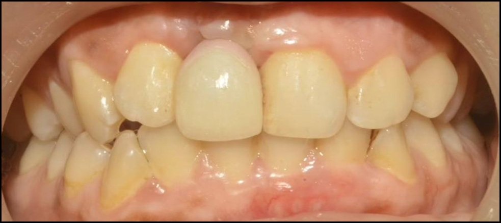
Figure 14: Intra-oral view of final prosthesis
DISCUSSION
The present case was reported to document clinically and radiographically the survival of dental implants placed in atrophic alveolar ridges augmented with mandibular symphysis autogenous onlay grafts. Implant rehabilitations in bone augmented with autologous mandibular symphysis block grafts are a reliable treatment modality in patients with bone defects.
In a recent retrospective study,19 it has been claimed that intraoral bone grafts harvested from the mandibular symphysis, mandibular ramus, and maxillary tuberosity provide a good treatment modality for ridge augmentation. Bone availability from these regions is adequate for anatomical bone defects spanning up to the width of three teeth.20 The surgical access to the mandibular symphysis is easier than that of the mandibular ramus.11 Both the procedures can be undertaken on an outpatient basis, while harvesting of bone from distant sites or extraorally requires inpatient care along with increased costs.21 Both the harvesting and grafting procedures are usually performed in the same surgical field.22 The use of autologous bone in the present case showed excellent survival and success rates. The aesthetic aspect of the treatment is a success when the gingival scalloping has been preserved.23,24 Bone augmentation resulted in stable lamellar-bone conditions along with a low risk of mucosal recession.
Loss of bone architecture and volume, can be solved using narrower sized implants and careful management of soft tissue. Soft tissue augmentation techniques have demonstrated good aesthetic results so that more invasive bone augmentation procedures may be avoided.25 At the time of surgery, the patient being only 17 years old, an alternative approach with an additional waiting period of three to four years was contemplated to prevent the intrusion of the peri-implant tissues that could occur due to premature implant insertion.26 The patient also refused presurgical orthodontic treatment aimed at optimizing the alignment of the anterior sector.
The hypothesis that bone substitutes could be effectively used for the replacement of autogenous bone with its osteoconductive, osteoinductive and osteogenic properties is still being investigated. Studies have also proved benefits and appropriateness of autogenous tissue for an ridge reconstructive procedures of atrophied ridges before implant surgery.27
Regarding surgical complications, the postoperative morbidity and flap dehiscence with or without exposure of the grafts or membrane.28 The peri-implant mucosa needs to be supported by an adequate three-dimensional volume of alveolar bone, including an intact buccal plate of sufficient height and thickness.4,29,30 Deficiency of the buccal bone anatomy has a negative impact on the aesthetic outcome and is therefore considered a critical causative factor for implant complications and failures.4,30,31
Postoperative morbidity after mandibular bone harvesting procedures was reported to be mainly related to temporary or permanent neural disturbances involving the inferior alveolar nerve and its branches.27 Although the mandibular incisal canal has defined anatomical limits, harvesting bone from the mandibular symphysis, may still lead to neurosensory alterations. Physiological changes in the course of the mandibular incisal canal may be the causative factors for such occurences. It is therefore advisable to subjectively evaluate the feasibility of the technique on a case-by-case basis through orthopantomographs and second-level investigations such as dental scan, computed tomography and stereolithographic prototypes. No complications related to neurosensory function were reported in this case.
Marginal bone loss measurement on the mesial and distal aspects may be a limitation to this case owing to 2D imaging. The loss of buccal and lingual bone can only be measured using 3D imaging modalities. However, panoramic radiographs are frequently used in clinical settings for the evaluation of bone peak stability.2,32
In the given case report, loss of the upper central incisor can be treated with bone grafting from the mandibular symphysis and delayed implant placement leading to an enhanced functional and aesthetic results. Rate of resorption of the graft may be reduced with correct management of the peri-implant soft tissues and the prosthesis.
CONCLUSION
The mandibular symphysis is a convenient intramembranous source that supplies a dense quality transplant. The thick cortical layer of the graft reduces the amount and rate of resorption, while the cancellous part fastens the regenerative process. This transplant does not cause any immune reactions and incorporates osteoclastic resorption with a shorter healing period compared with other methods of osseous repair. Precise case selection and a comprehensive surgical planning is the prerequisite for successfully harvesting a graft for bone augmentation procedures. Application of safety guidelines in mandibular symphysis harvesting could reduce the risk of altered post-operative neurosensations due to injury of the mandibular incisal nerve.
Conflict of Interests
The authors declare no conflict of interest.
REFERENCES
- C Maiorana. Histomorphometric evaluation of anorganic bovine bone coverage to reduce autogenous grafts resorption: preliminary results. The Open Dentistry Journal. 2011;5(1):71-8.
- C Maiorana, P Poli, A Mascellaro, S Ferrario, M Beretta. Dental implants placed in resorbed alveolar ridges reconstructed with iliac crest autogenous onlay grafts: a 26-year median follow-up retrospective study. Journal of Cranio-Maxillofacial Surgery. 2019;47(5):805-14.
- C Maiorana, P Poli, M Beretta. Guided bone regeneration and implant placement in association with a coronally positioned palatal sliding flap: a 17-year follow-up case report. The Journal of Oral Implantology. 2018;44(5):371–6.
- M Beretta, P Poli, S Pieriboni et al. Peri-implant soft tissue conditioning by means of customized healing abutment: a randomized controlled clinical trial. Materials. 2019;12(18):3041.
- D Buser, K Dula, H Hirt, R Schenk. Lateral ridge augmentation using autografts and barrier membranes: a clinical study with 40 partially edentulous patients. Journal of Oral and Maxillofacial Surgery. 1996;54(4):420–32.
- P Galindo-Moreno, G Ávila, J Fernández-Barbero, F Mesa, F O'Valle-Ravassa, H Wang. Clinical and histologic comparison of two different composite grafts for sinus augmentation: a pilot clinical trial. Clinical Oral Implants Research. 20018;19(8):755–9.
- M Chiapasco, M Zaniboni, L Rimondini. Autogenous onlay bone grafts vs alveolar distraction osteogenesis for the correction of vertically deficient edentulous ridges: a 2-4-year prospective study on humans. Clinical Oral Implants Research. 2007;18(4):432–40.
- A Jensen, S Jensen, N Worsaae. Complications related to bone augmentation procedures of localized defects in the alveolar ridge. A retrospective clinical study. Oral and Maxillofacial Surgery. 2016;20(2):115–22.
- J Smith, M Abramson. Membranous vs. endochondral bone autografts. Archives of Otolaryngology. 1974;99(3):203–5.
- A Esen, G Yıldırım, Ö Ä°nan, D Dolanmaz. The use of autogeneous mandibular bone block grafts for reconstruction of alveolar defects. Annals of Maxillofacial Surgery. 2015;5(1):71–6.
- C Misch. Comparison of intraoral donor sites for onlay grafting prior to implant placement. International Journal of Oral & Maxillofacial Implants. 1997;12(6):767–76.
- C Misch, C Misch, R Resnik, Y Ismail. Reconstruction of maxillary alveolar defects with mandibular symphysis grafts for dental implants: a preliminary procedural report. The International Journal of Oral & Maxillofacial Implants. 1992;7(3):360–6.
- T Arx, D Buser. Horizontal ridge augmentation using autogenous block grafts and the guided bone regeneration technique with collagen membranes: a clinical study with 42 patients. Clinical Oral Implants Research. 2006;17(4):359–66.
- C Maiorana, M Beretta, S Salina, F Santoro. Reduction of autogenous bone graft resorption by means of bio-oss coverage: a prospective study. International Journal of Periodontics & Restorative Dentistry. 2005;25(1):18–25.
- Misch CE. Mandibular donor block bone grafts: symphysis and ramus. Contemporary Implant Dentistry. Missouri: Elsevier. 2011;3:975-1012.
- Gapski R, Wang HL, Misch CE. Management of incision design in symphysis graft procedures: A review of the literature. J Oral Implantol. 2001;27:134-42.
- Schuler R, Verardi S. A new incision design for mandibular symphysis bone-grafting procedures. J Periodontol. 2005;76:845-9.
- Misch CM, Misch CE, Resnik RR, Ismail YH. Reconstruction of maxillary alveolar defects with mandibular symphysis grafts for dental implants: A preliminary procedural report. Int J Oral Maxillofac Implants. 1992;7:360-6.
- A Sakkas, F Wilde, M Heufelder, K Winter, A Schramm. Autogenous bone grafts in oral implantology—is it still a “gold standard? A consecutive review of 279 patients with 456 clinical procedures. International Journal of Implant Dentistry. 2017;3(1):23.
- V Moraschini, L Poubel, V Ferreira, E Barboza. Evaluation of survival and success rates of dental implants reported in longitudinal studies with a follow-up period of at least 10 years: a systematic review. International Journal of Oral and Maxillofacial Surgery. 2015;44(3):377–88.
- G Sàndor, I Nish, R Carmichael. Comparison of conventional surgery with motorized trephine in bone harvest from the anterior iliac crest. Oral Surgery, Oral Medicine, Oral Pathology, Oral Radiology, and Endodontology. 2003;95(2):150–5.
- E Nkenke, F Neukam. Autogenous bone harvesting and grafting in advanced jaw resorption: morbidity, resorption and implant survival. European Journal of Oral Implantology. 2014;7(2):S203–S217.
- D Buser, W Martin, U Belser. Optimizing esthetics for implant restorations in the anterior maxilla: anatomic and surgical considerations. International Journal of Oral & Maxillofacial Implants. 20014;19:43–61.
- V Chappuis, L Rahman, R Buser, S Janner, U Belser, D Buser. Effectiveness of contour augmentation with guided bone regeneration: 10-year results. Journal of Dental Research. 2017;97(3):266–74.
- T Testori, T Weinstein, F Scutellà, H Wang, G Zucchelli. Implant placement in the esthetic area: criteria for positioning single and multiple implants. Periodontol. 2000;77(1):176–96.
- L Bohner, M Hanisch, J Kleinheinz, S Jung. Dental implants in growing patients: a systematic review. The British Journal of Oral & Maxillofacial Surgery.2019;57(5):397–406.
- M Chiapasco, M Zaniboni, M Boisco. Augmentation procedures for the rehabilitation of deficient edentulous ridges with oral implants. Clinical Oral Implants Research. 2006;17(2):136–59.
- L Cordaro, F Torsello, S Morcavallo, V Torresanto. Effect of bovine bone and collagen membranes on healing of mandibular bone blocks: a prospective randomized controlled study. Clinical Oral Implants Research. 2011;22(10):1145–50.
- U Grunder, S Gracis, M Capelli. Influence of the 3-D bone-to-implant relationship on esthetics. International Journal of Periodontics & Restorative Dentistry. 2005;25(2):113–9.
- D Farronato, P Pasini, A Orsina, M Manfredini, L Azzi, M Farronato. Correlation between buccal bone thickness at implant placement in healed sites and buccal soft tissue maturation pattern: a prospective three-year study. Materials. 2020;13(3):511.
- S Chen. Clinical and esthetic outcomes of implants placed in postextraction sites. International Journal of Oral & Maxillofacial Implants. vol. 24, 2009.
- H Alkis, N Turker. Retrospective evaluation of marginal bone loss around implants in a mandibular locator-retained denture using panoramic radiographic images and finite element analysis: a pilot study. Clinical Implant Dentistry and Related Research. 2019;21(6):1199–205.
Disclaimer from Guident
The views and opinions expressed in articles published in Guident are solely those of the authors and do not necessarily reflect the views of the editors, the editorial board or the publisher, Ivory India. Guident does not endorse any product, technique or treatment described. Clinical content is intended for qualified dental and medical professionals and is not a substitute for individual clinical judgement. Guident, its editors and the publisher accept no liability for any consequence arising from the use of information published in the journal.
How to cite this article
Dr. Paloma Parulkar, Dr. Omkar Shetty, Dr. Gaurang Mistry, Dr. D.Y. Patil University School of Dentistry. Bone Augmentation Using a Mandibular Symphysis Graft-a Case Report. Guident. 2021 Dec;15(1-2). Available from: https://guident.in/articles/implantology/bone-augmentation-using-a-mandibular-symphysis-graft-a-case-report.html