Apexification Using MTA- a Case Series
Department of Conservative Dentistry and Endodontics, Seema Dental College And Hospital, Rishikesh
INTRODUCTION
Traumatic injuries to young permanent teeth affect 30% of children. These injuries often result in pulpal inflammation or necrosis and subsequent incomplete development of dentinal wall and root apices. The golden rule in the practice of endodontics is to debride and obturate the canals as efficiently and three dimensionally as possible in an amount of time and appointments that are reasonable to the patient.Before 1966 the clinical management of the “Blunder buss” canal usually required a surgical approach for the placement of an apical seal into the often fragile and flaring apex. Apicoectomy further reduces the root length resulting in a very unfavorable crown root ratio.The treatment of choice for necrotic young permanent teeth is apexification.
PURPOSE: The aim of this study is to investigate several cases with different root canal anatomies like in open apex with blunderbuss canals in maxillary central incisors and their management.
Material and methods: Four Cases of maxillary central incisors with immature open apex and blunderbuss canals w.r.t 11 & 21.
CASE SERIES
Apexification with Mineral Trioxide Aggregate
CASE REPORT 1
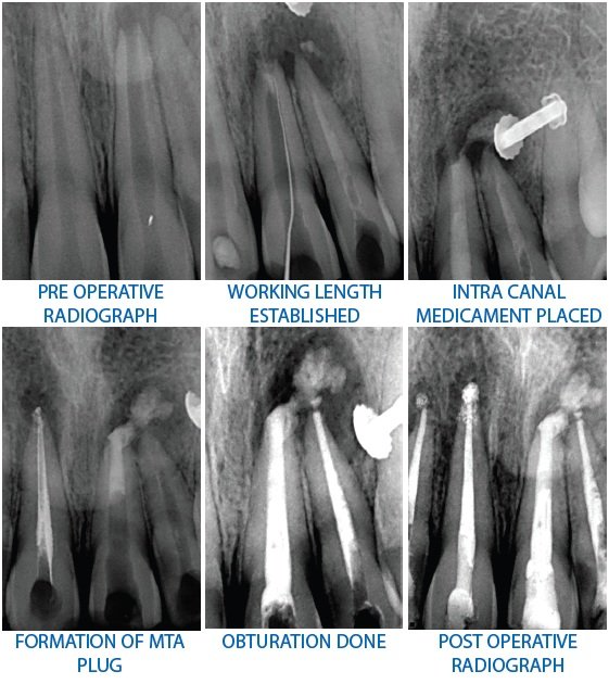
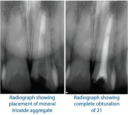
A 18-year-old female patient reported with a chief complaint of pain in her upper front tooth region for past two days. Patient gave history of dull throbbing pain for the past 6 months in the same region. Past medical and dental history was non-contributory. Clinical examination showed Ellis Davis Class IV Fracture in upper left central incisor (21). Sensibility test showed no response in 21.Radiographic examination showed blunderbuss canal and a periapical radiolucency in relation to 21. (Figure 1). Based on the clinical examination, sensibility test and radiographic findings, a provisional diagnosis of Ellis Davis Class IV fracture with open apex and periapical pathology was made. Apexification was planned with MTA (PROROOT Densply).A written consent was taken from the patient after explaining the treatment protocol. In the first visit, access opening was done under isolation using endo access bur #1.Canal patency was checked using a #10 K file and working length was determined using Ingle’s Radiographic method. Biomechanical preparation was done using K files using step back technique. Irrigation was done using 5.25 % NaOCl and saline). Chlorhexidine was used as a final irrigant. Calcium Hydroxide was placed using a Lentulo spiral and tooth was temporized. Patient was recalled after a week. In the second visit, access cavity was re-established, canal was irrigated copiously following the same protocol and dried with sterile paper points.MTA )was mixed according to the manufacturer’s protocol and and packed to a thickness of 5 mm in the apical third using a hand plugger (Figure 1).4 A sterilecotton pellet was placed in the canal and the tooth was temporized. Patient was recalled after two days.
In the subsequent visit, obturation was done using thermoplasticized guttapercha and Composite resin restoration was placed to seal the access cavity.
CASE REPORT 2
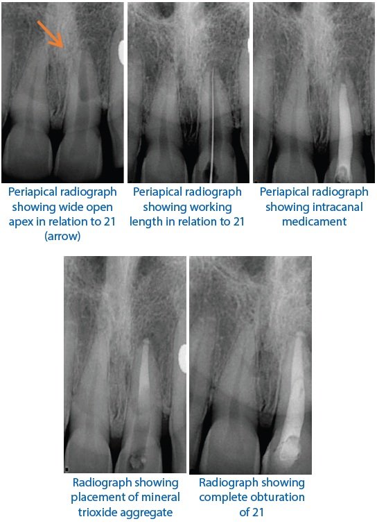
A 21-year-old female patient came with a chief complaint of pain in her upper front tooth region for past two days. Patient gave history of dull throbbing pain for the past 6 months in the same region. Past medical and dental history was non-contributory. Clinical examination showed Ellis Davis Class IV Fracture in upper left central incisor (21). Sensibility test showed no response in 21.Radiographic examination showed blunderbuss canal and a periapical radiolucency in relation to 21. Based on the clinical examination, sensibility test and radiographic findings, a provisional diagnosis of Ellis Davis Class IV fracture with open apex and periapical pathology was made. Apexification was planned with MTA (PROROOT Dentsply).A written consent was taken from the patient after explaining the treatment protocol. In the first visit, Access opening was done under isolation using endo access bur #1.

Canal patency was checked using a #10 K file and working length was determined using Ingle’s Radiographic method. Biomechanical preparation was done using K files usingstep back technique. Irrigation was done using 5.25 % NaOCl and saline). Chlorhexidine was used as a final irrigant. Calcium Hydroxide was placed using a Lentulo spiral) and tooth was temporized. Patient was recalled after a week. In the second visit, access cavity was re-established, canal was irrigated copiously following the same protocol and dried with sterile paper points. MTA was mixed according to the manufacturer’s protocol and and packed to a thickness of 5 mm in the apical third using a hand plugger. A sterilecotton pellet was placed in the canal andthe tooth was temporized. Patient was recalled after two days. In the subsequent visit, obturation was done using thermoplasticized guttapercha and Composite resin restoration was placed to seal the access cavity. Patient was recalled after a month for follow up.

CASE REPORT 3
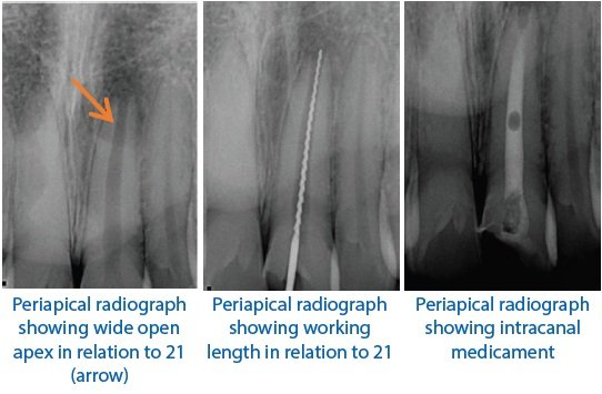
An 31year‑old female patient reported with a chief complaint of discolored left maxillary central incisor with a history of trauma 10 year back. The concerned tooth did not respond to both electric and heat test. The periapical radiograph revealed a large blunderbuss canal of the same tooth. On clinical examination, Ellis Class I fracture in permanent left maxillary central incisor was evident. Apexification with MTA was planned. Access opening was prepared and working length was determined ; irrigation was done with saline. Biomechanical preparation was carried out using 70 size k file with circumferential filing motion. Root canal debridement was done using alternative irrigation with 2.5% NaoCl and saline. Calcium hydroxide was placed in the root canal and patient recalled after 5 days. At subsequent appointment, canal was irrigated with 2.5% NaoCl and 2% chlorhexidine. The canal was dried with paper points and MTA placed with pluggers until thickness of 6 mm. A wet cotton pellet was placed in the canal and access cavity was sealed with temporary cement. In next appointment, root canal was obturated with using lateral condensation technique. Access cavity sealed with glass ionomer cement.
CASE REPORT 4
An 17 year‑old male patient reported with a chief complaint of discolored and broken left maxillary central incisor with a history of trauma 7 year back. The concerned tooth did not respond to both electric and heat test. The periapical radiograph revealed a large blunderbuss canal of the same tooth . On clinical examination, Ellis Class III fracture in permanent left maxillary central incisor was evident. Apexification with MTA was planned. Access opening was prepared and working length was determined ; irrigation was done with saline. Biomechanical preparation was carried out using 70 size k file with circumferential filing motion. Root canal debridement was done using alternative irrigation with 2.5% NaoCl and saline. Calcium hydroxide was placed in the root canal and patient recalled after 5 days. At subsequent appointment, canal was irrigated with 2.5% NaoCl and 2% chlorhexidine. The canal was dried with paper points and MTA placed with pluggers until thickness of 5 mm . A wet cotton pellet was placed in the canal and access cavity was sealed with temporary cement. In next appointment, root canal was obturated with using lateral condensation technique.
DISCUSSION
The goal of apexification is to obtain an apical barrier to prevent the passage of toxins and bacteria into periapical tissues from root canal. In the literature, many materials have been used for apexification, such as calcium hydroxide in combination with sterile water, saline, local anesthetic, CMCP, zinc oxide paste with cresol and iodoform, polyantibiotic paste and tricalcium phosphate. Calcium hydroxide is one of the most important medicaments used in treatments of pulp conditions and apical periodontitis.
MTA as an apexification material represents a primary monoblock. Appetite like interfacial deposits form during the maturation of MTA result in filling the gap induced during material shrinkage phase and improves the frictional resistance of MTA to root canal walls. MTA has superior biocompatibility and it is less cytotoxic due to its alkaline pH and presence of calcium and phosphate ions in its formulation results in capacity to attract blastic cells and promote favorable environment for cementum deposition. A total of 5 mm barrier is significantly stronger and shows less leakage than 2 mm barrier.In the present case, MTA was placed for around 6 mm in the apical region.
There is increasing popularity with one visit apexification technique using Mineral Trioxide Aggregate (MTA) as osteoconductive apical barrier. MTA is relatively non cytotoxic and stimulates cementogenesis. This Portland cement based material generates a highly alkaline aqueous environment by leaching of calcium and hydroxyl ions, rendering it bioactive by forming hydroxyappatite in presence of phosphate containing fluids. Unlike the extended use of Ca(OH)2 in immature roots, prolonged filling of these roots with MTA did not reduce their fracture resistance.3
Torabinejad reported the ingredients in MTA as tri calcium silicate, tricalcium aluminate, tricalcium oxide and silicate oxide with some other mineral oxides that were responsible for the chemical and physical properties of aggregate. The powder consists of fine hydrophilic particles that set in the presence of moisture. The hydration of the powder results in a colloidal gel with a pH of 12.5 that will set in approximately 3 hours. MTA has a compressive strength equal to intermediate restorative material and Super - EBA but less than that of amalgam. It is commercially available as ProRoot MTA ,and has been advocated for use in the immediate obturation of open root apex.
CONCLUSION
The apexification by Ca(OH)2 has been applied for many years back to the present as a valid therapy to perform an apexification treatment.
The MTA barrier as a treatment of apexification is a technique that is applied as a substitution to Ca(OH)2 apexification; this technique does not require several appointments, and the conformation of the barrier does not need an external factor to develop, as in the case of the apexification with Ca(OH)2, as well as in the regeneration of the pulp.
REFERENCES
- SeltzerS.Endodontology; Biologic Considerationsin Endodontic Procedures, 1988, 2nd edn. Philadelphia; Lea and Febiger.
- Lee SJ, Monset M, Torabinejad M. Sealing ability of a mineral trioxideaggregate for repair of lateral root perforations. J. Endod 1993; 19 : 541-4.
- Rebecca L, Martin BS, Francesca M et al.Sealing properties of mineral trioxide aggregate orthograde apical plugs and root fillings in an in vitro apexification model. J Endod 2007;33:272-275
- Torabinejad M, ChivianN.Clinical applications of mineral trioxide aggregate.JEndod 1999;25:197-205
- Shabahang S, TorabinejadM.Treatment of teeth with open apices using mineral trioxide aggregate. Pract Periodont Aesthet Dent 2000;12:315-20
Disclaimer from Guident
The views and opinions expressed in articles published in Guident are solely those of the authors and do not necessarily reflect the views of the editors, the editorial board or the publisher, Ivory India. Guident does not endorse any product, technique or treatment described. Clinical content is intended for qualified dental and medical professionals and is not a substitute for individual clinical judgement. Guident, its editors and the publisher accept no liability for any consequence arising from the use of information published in the journal.
How to cite this article
Dr. Rishika Luhach, Dr. Ayushi Bangari, Dr. Annil Dhingra. Apexification Using MTA- a Case Series. Guident. 2021 Oct;14(11). Available from: https://guident.in/articles/endodontics/apexification-using-mta-a-case-series.html