Management of Multiple Canals in Mandibular Incisors: A Case Series
Department of Conservative Dentistry and Endodontics, Seema Dental College And Hospital, Rishikesh
INTRODUCTION
Adequate knowledge and comprehensive understanding of the root canal system are an essential prerequisite for endodontic treatment. Clinicians must be aware of normal as well as complex root canal configurations encountered in daily practice to ensure complete chemomechanical instrumentation of all canals followed by three-dimensional filling of the root canal space; the presence of an untreated root canal is one of the main causes of root canal treatment failures. Generally, both maxillary and mandibular anterior teeth are described as single-rooted encasing single or double canals (in the mandibular anterior). Studies demonstrate a high prevalence of two canals in mandibular incisors, which varies according to ethnicity, gender and age.
Although the external morphology of permanent mandibular central and lateral incisors is differentiated mostly through the incisal aspect of the teeth, the internal morphology is interested to be investigated for both preclinical anatomies of the teeth and clinical prognostic indicator. Most prevalence proportion of internal morphology of permanent mandibular incisors is Vertucci’s Type Ι, one canal and one foramen configuration. The obvious differences can be found in second modes of Vertucci’s classification across the world
The main reason for the failure of endodontic therapy is incomplete knowledge about the anatomical variation of root canals. A canal is often left untreated because the dentist fails to recognize its presence either due to lack of knowledge of root canal morphology or due to lack of experience and skill to negotiate the canal. The dentist must have a thorough knowledge of root canal morphology before starting endodontic treatment.
CASE REPORT- I
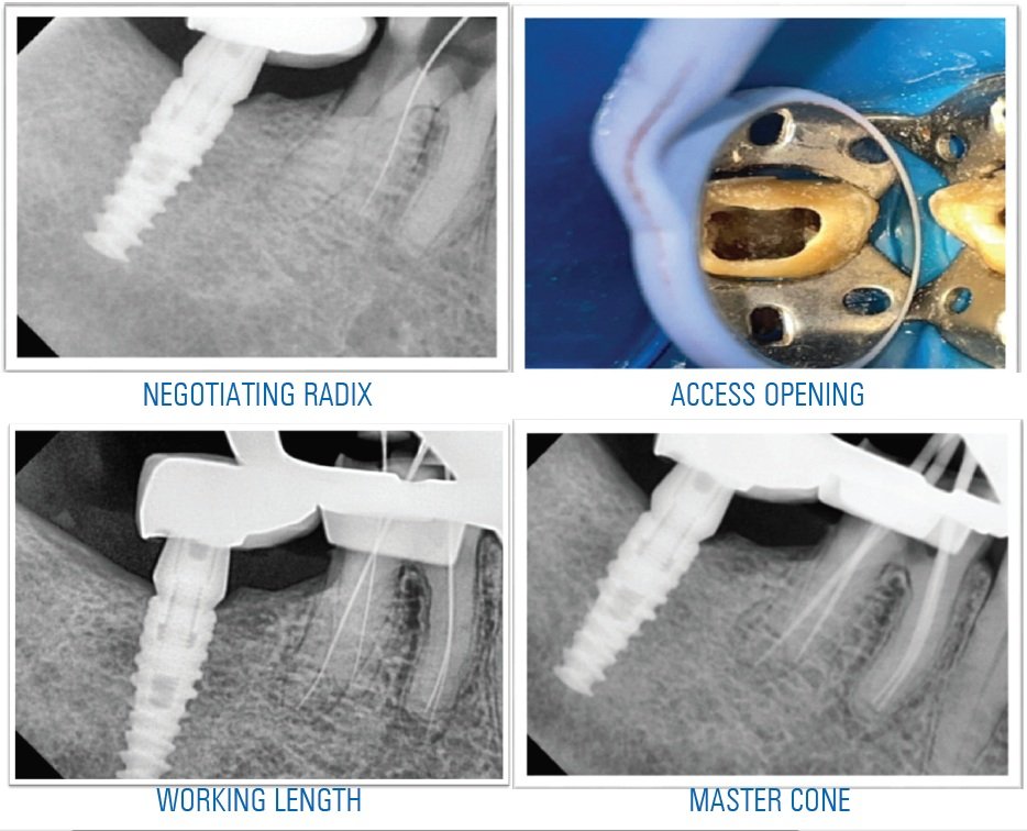
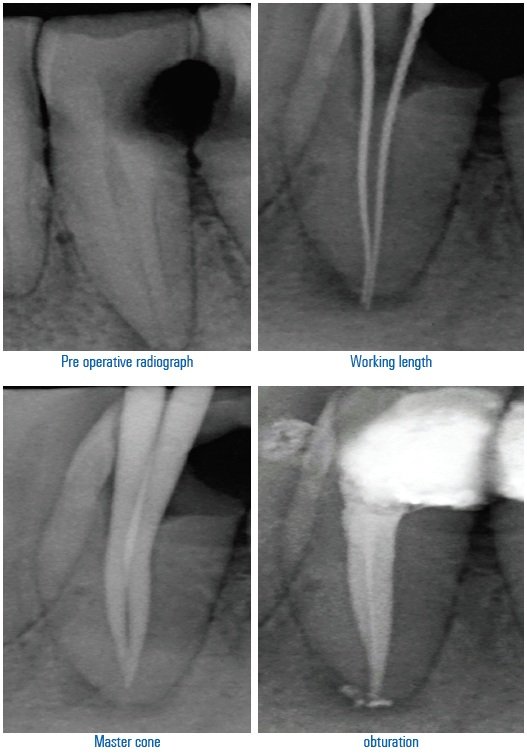
A patient named Ajay Pal Kandari 40 year old male came to the department of conservative dentistry and endodontics,seema dental college and hospital rishikesh with the chief complain of pain in lower right front tooth region for 4-5 days and gave the history of pain on having hot and cold bevereges. On examination it was seen deep class iv caries wrt 43, radiographically the radiolucency was involving the pulp. With the history of the pain the diagnosis was made of Acute Symptomatic irreversible pulpitis .
As the tooth was tilted so under rubber dam isolation straight line access opening was done, working length was establised using 8no and 10 no. K Files and was confirmed radiographically.
Shaping & cleaning was done till 25 no. K file and then rotatry Hyflex EDM file system was used for further shaping and cleaning ,irrigation was done using normal saline and Sodium Hypochlorite and then final master cone was selected . The obturation was done using Apexit plus sealer and finally post endo restoration was done with composite resin restorative material.

CASE REPORT- II
A 29-year-old patient reported to the department with a chief complaint of pain in the lower front tooth region for 1 week.
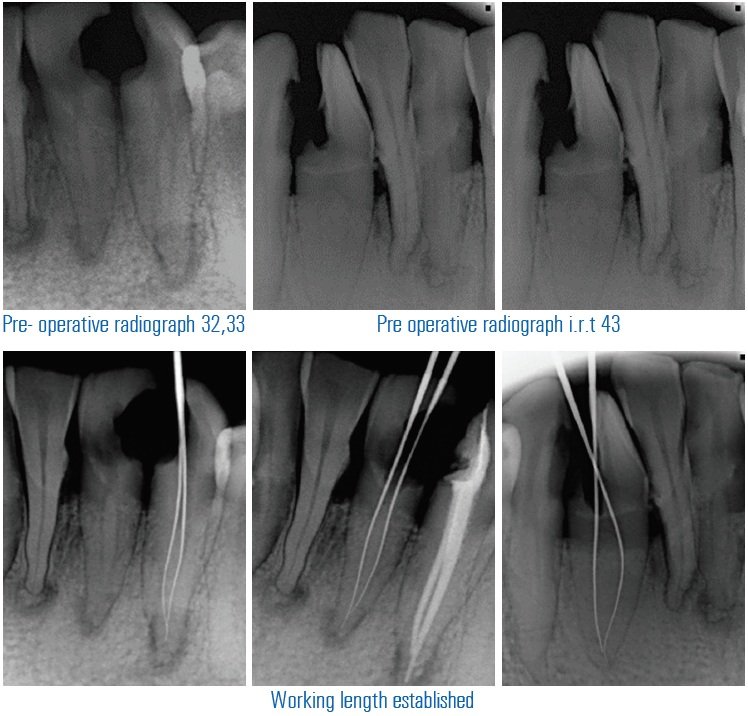
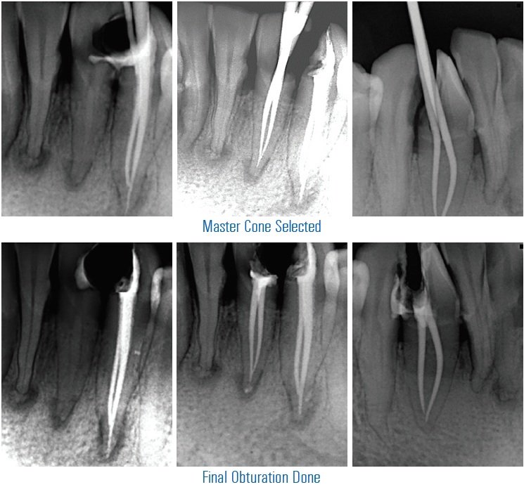
On clinical examination there was Deep class 2 caries i.r.t 32,33, 43 and radiographic examination revealed radiolucency involving enamel, dentin and pulp i.r.t 32,33,43. Cold test was negative i.r.t 32,33 and lingering response i.r.t 43 Teeth presented a delayed response to electric pulp testing i.r.t 43,32 and no response i.r.t 33. The treatment planned was non-surgical root canal treatment i.r.t 32,33,43. It was explained to the patient and consent was obtained. After the access opening of 11,21 was done the working length was determined using k file and thorough shaping and cleaning was done. The teeth were then obturated using warm vertical condensation.
CASE REPORT- III
A 50 year old patient came to the department of conservative dentistry and endodontics with the chief complain of pain in lower anterior tooth region. Pain aggravated on taking cold food and lingered on for 15 to 20 minutes. Clinical examination showed deep proximal caries i.r.t 31. Cold test gave positive response i.r.t 31 . radograph showed periapical radiolucency i.r.rt 31. The treatment plan of non surgical root canal treatment i.r.t 31explained to the patient and consent was obtained after the access opening of 31was done the working length was determined using k file and thorough shaping and cleaning was done. The teeth were then obturated using warm vertical condensation.
DISCUSSION
A well-designed access preparation is essential for a good endodontic result. Without adequate access, instruments and materials become difficult to handle properly in the highly complex and variable root canal system. Proper access cavity preparation provides straight or direct line access to the apical foramina or at least to the initial curvature of canal to locate all root canal orifices and it also conserve sound tooth structure. Thus, careful interpretation of the radiographic feature taken from different angles should be done before starting endodontic treatment. One must be careful while access opening, and initial buccolingual widening of mandibular incisors and gingival extension beneath the cingulum must be made to search for a possible second canal lingually.
CONCLUSION
The main reason for failure of endodontic treatment of mandibular incisor can be due to the inability to detect and treat second root canal, mostly lingual canal. Thus, careful interpretation of radiographs taken from different angulations is essential. Practice of extension of access cavity buccolingually and gingivally beneath cingulum will help to detect additional lingual canal if present in each mandibular incisor.
Disclaimer from Guident
The views and opinions expressed in articles published in Guident are solely those of the authors and do not necessarily reflect the views of the editors, the editorial board or the publisher, Ivory India. Guident does not endorse any product, technique or treatment described. Clinical content is intended for qualified dental and medical professionals and is not a substitute for individual clinical judgement. Guident, its editors and the publisher accept no liability for any consequence arising from the use of information published in the journal.
How to cite this article
Dr. Rishika Luhach, Dr. Advaitha Anand, Dr. Annil Dhingra. Management of Multiple Canals in Mandibular Incisors: A Case Series. Guident. 2021 Sep;14(10). Available from: https://guident.in/articles/endodontics/management-of-multiple-canals-in-mandibular-incisors-a-case-series.html