Endodontic Management of Radix Entomolaris in Mandibular First Molar- a Case Report
Department of Conservative Dentistry & Endodontics, K.D. Dental College & Hospital, Mathura
ABSTRACT
Usually mandibular first molar have three roots typically that is mesiolingual, mesiobuccal and distal canal. But sometimes the clinician encounters a fourth canal in the same buccolingual plane as the distobuccal root and superimposition of both roots can appear on the preoperative radiograph, resulting in an inaccurate diagnosis. In this case report we did the endodontic management of radix entomolaris of a 58 year old male which came with the chief complaint of spontaneous pain. Understanding such variant of root canal anatomy helps in the crucial approach toward working with such cases.
INTRODUCTION
The main purpose of endodontic therapy is the elimination of bacteria from the infected root canals and prevention of risk of further reinfection which is mainly achieved by thorough cleaning and shaping of root canals followed by three dimensional obturation with a coronal fluid tight seal. For this the clinicians must have a thorough knowledge of the diversity of morphology of root canal systems and its variations that may complicate the procedure. The majority of mandibular first molars have two roots, mesial and distal with two mesial and one distal canal. Many variations in root canal systems have been described. Fabra-Campos reported the presence of three mesial canals while Stroner reported three distal canals.1-4
A major but rare variant is the presence of three roots in mandibular first molar, first mentioned in the literature by Carabelli in 1844; known as radix entomolaris located in distolingual position.5 When located on mesiobuccal surface, the anomaly is known as radix paramolaris. The external morphology of this anomaly having additional lingual or buccal root, are described by Carlsen and Alexandersen. The Latin term Radix Entomolaris (RE) was coined by Mihaly Lenhossek in 1922.6
The main etiological factor behind this variant can be attributed to racial, genetic and external factors during odontogenesis7 or penetrance of an atavistic gene or polygenetic system (appearance of a trait belonging to a distant ancestor that has been dormant in recent generations).
The occurrence of third root varies in different ethnic groups, with the incidence of less than 5 percent. The maximum frequency occurs in African populations and is 3% .It appears to be 3.4-4.2% in Europeans, less than 5% in Eurasians and Indians , 5% to 40% in Mongoloid traits, such as Chinese, Eskimo and American Indians and 8.2% in Malaysian Borneos.8
Radiographic diagnosis plays an unparalleled role in successful endodontic treatment of almost every case, and when taken at different angulations gives information about extra canals or roots and aids in the better understanding of the anatomy of the root canal system and the treatment approach with sufficient `knowledge and absolute clinical thoroughness for successful root canal treatment.
This case report is about the detection and management of radix entomolaris (RE) in a mandibular first molar during its root canal treatment in a patient who came to the Department of Conservative Dentistry and Endodontics with a chief complaint of pain in right lower back molar region which aggravated during mastication and was spontaneous in nature indicating signs of irreversible pulpitis.
CASE REPORT
A 58 year old male patient was referred to Department of Conservative dentistry & Endodontics, K.D. Dental College Mathura, with spontaneous pain in his mandibular right first molar for the past four days. On clinical examination of the patient his mandibular right first molar had a deep carious lesion. Tooth was moderately tender on percussion. Intra Oral Periapical (IOPA) Radiograph of tooth showed a coronal radiolucent area on the distal aspect of the tooth which approached the pulp. Radiograph also revealed the presence of an extra distal root with accompanying apical radiolucency. From patient history and clinical examination a diagnosis of acute apical periodontitis was established and endodontic treatment was initiated after the informed consent from the patient. Buccal object rule (SLOB technique) confirmed the additional root as distolingual root [RADIX ENTOMOLARIS].
CLINICAL MANAGEMENT
Radiographic evaluation of the involved teeth revealed two completely formed roots with no indication of any variation in the root canal anatomy in 46. The teeth were anesthetized using lignocaine. After rubber dam isolation, caries excavation and access cavity was done on the tooth. On inspection with a DG‑16 endodontic explorer initially, the pulp chamber floor revealed three canals – mesiobuccal, mesiolingual, and the distal. A search for the second distal canal was made by further exploration of the pulpal floor with a DG‑16 endodontic explorer. A catch disto lingually unveiled the second distal orifice, and the access cavity was modified from a triangular form to a trapezoidal shape to include the distolingual canal. Negotiation of the canals was carried out with ISO size 6, 8, and 10 K files. Working length was determined using the Woodpecker apex locator with 15 No. K files and was verified using periapical radiograph. The working length radiograph taken with different horizontal angulations revealed the presence of a third root located distolingually. The additional root was narrow, short and tapers towards the apex. Thus, care should be taken to avoid overpreparation and any inadvertent perforation of the root during root canal treatment. A mild perforation site was restored using Glass Ionomer Cement at the chamber floor. The coronal access was defined with Gates Glidden burs (Dentsply, Maillefer) and canals were shaped with Hyflex CM files under copious irrigation with 2.5% sodium hypochlorite and lubrication with RCPrep. All canals were enlarged to 25/4 by crown down preparation to the working length to minimise the risk of strip perforation and also because of the age factor of patient that may have calcified the canal a bit. Two calcium hydroxide medicament dressings were placed at a week interval to aid in the healing process. At the fourth appointment, the patient was completely asymptomatic and canals were dry. The root canal system was then obturated with Gutta percha using single cone technique (25/4) and Zinc Oxide Eugenol sealer.
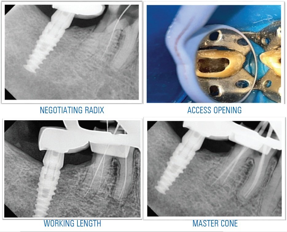
Patient was recalled after a week for check up, he was completely asymptomatic and then he was scheduled for permanent restoration with a PFM crown.

DISCUSSION
The presence of RE or a radix paramolaris has clinical implications in endodontics, oral surgery and orthodontics and an accurate diagnosis of these supernumerary roots can avoid complications or a “missed canal” during root canal treatment. RE is mostly situated in the same buccolingual plane as the distobuccal root, a superimposition of both roots can appear on the preoperative radiograph, resulting in an inaccurate diagnosis. Some particular marks or characteristics, such as an unclear view or outline of the distal root contour or the root canal, can indicate the presence of a “hidden” RE. To reveal the RE, a second radiograph should be taken from a more mesial or distal angle (30°).9,10
Classification: Carlsen & Alexandersen (1990)
Classified radix entomolaris (RE) into four different types based on the location of its cervical part11:
- Type A: the RE is located lingually to the distal root complex which has two cone-shaped macrostructures.
- Type B: the RE is located lingually to the distal root complex which has one cone-shaped macrostructures.
- Type C: the RE is located lingually to the mesial root complex.
- Type AC: the RE is located lingually between the mesial and distal root complexes.
De Moor et al. (2004) classified radix entomolaris based on the curvature of the root or root canal12:
- Type 1: a straight root or root canal.
- Type 2: a curved coronal third which becomes straighter in the middle and apical third.
- Type 3: an initial curve in the coronal third with a second buccally oriented curve which begins in the middle or apical third.
Song JS et al. (2010) further added two more newly defined variants of RE13:
1. Small type: length shorter than half of the length of the distobuccal root.
2. Conical type: smaller than the small type and having no root canal within it.
Morphology: The radix entomolaris is located distolingually ranging from short, conical extension to a normal mature root length with its coronal third partially or completely fixed to distal root.14 Externally, the distal furcation is slightly lower (1 mm.) than the furcation between mesial and distal roots.15 Clinically, tooth with additional distolingual root may present a more bulbous crown outline, an additional cusp, a prominent distolingual lobe or cervical prominence. These features can indicate the presence of additional root. Radiographically, third root is visible in 90% of cases.16,17 In addition to this, magnifying loupes, dental microscope or intraoral camera may also be useful. Recently, cone-beam computed tomography (CBCT) has emerged as a useful tool to aid in the diagnosis of teeth with complex root anatomies. However, cost and accessibility are the main limiting factors till now.18,19
CLINICAL IMPLICATIONS
Oral surgical procedures: Radix entomolaris may pose great difficulty during extraction and orthodontic procedure. Tooth should be thoroughly luxated during extraction, as distolingual root might fracture as it is smaller than mesial and distobuccal roots.
Orthodontic procedures: During orthodontic procedure, presence of distolingual root and its curvature makes tooth movement difficult.
CONCLUSION
Dental practitioners should be aware of this variation in anatomy of permanent mandibular first molars. The morphological variations of the RE in terms of root inclination and root canal curvature demand a careful and focused clinical approach to avoid or overcome procedural errors during endodontic therapy.
REFERENCES
- Vertucci FJ (1984) Root canal anatomy of the human permanent teeth. Oral Surg Oral Med Oral Pathol 58: 589-599.
- Barker BC, Parsons KC, Mills PR, Williams GL (1974) Anatomy of root canals. III. Permanent mandibular molars. Aust Dent J 19: 408-413.
- Fabra-Campos H (1989) Three canals in the mesial root of mandibular first permanent molars: a clinical study. Int Endod J 22: 39-43.
- Stroner WF, Remeikis NA, Carr GB (1984) Mandibular first molar with three distal canals. Oral Surg Oral Med Oral Pathol 57: 554-557.
- Carabelli G (Editor). Systematisches Handbuch der Zahn heilkunde (2nd edn.). Vienna: Braumuller and Seidel. 1844: 114.
- I. Stamfelj. Who coined the term radix entomolaris? International Endodontic Journal. 2014; 47: 810–811.
- Sperber GH, Moreau JL. Study of the number of roots and canals in Senegalese first permanent mandibular molars. Int Endod J. 1998; 31(2):117–22.
- Shekhar Bhatia S , Kohli S, Parolia A , Lim YN , Tung L , Hean T Prevalence of Radix Molar in Mandibular Permanent Molars: An Observational Study in Malaysian Population. Oral Health and Dental Management · January 2015
- Reichart PA, Metah D (1981) Three-rooted permanent mandibular first molars in the Thai. Community Dent Oral Epidemiol 9: 191-192. 23.
- Gu Y1, Zhou P, Ding Y, Wang P, Ni L (2011) Root canal morphology of permanent three-rooted mandibular first molars: Part III--An odontometric analysis. J Endod 37: 485-490.
- Carlsen O, Alexandersen V (1990) Radix entomolaris: identification and morphology. Scand J Dent Res 98: 363-373.
- De Moor RJ, Deroose CA, Calberson FL (2004) The radix entomolaris in mandibular first molars: an endodontic challenge. Int Endod J 37: 789-799.
- Song JS, Choi HJ, Jung IY, Jung HS, Kim SO (2010) The prevalence and morphologic classification of distolingual roots in the mandibular molars in a Korean population. J Endod 36: 653-657.
- Reichart PA, Metah D (1981) Three-rooted permanent mandibular first molars in the Thai. Community Dent Oral Epidemiol 9: 191-192.
- Gu Y1, Zhou P, Ding Y, Wang P, Ni L (2011) Root canal morphology of permanent three-rooted mandibular first molars: Part III--An odontometric analysis. J Endod 37: 485-490.
- Walker RT, Quackenbush LE (1985) Three-rooted lower first permanent molars in Hong Kong Chinese. Br Dent J 159: 298-299. 25..
- Ingle JI, Heithersay, GS, Hatwell GR (2002) Endodontic Diagnostic Procedures. BC Decker, London, UK.
- Gopikrishna V, Reuben, J., Kandaswamy, D (2008) Endodontic management of a maxillary first molar with two palatal roots and a single fused buccal root diagnosed with spiral computed tomography-a case report. Oral Surgery Oral Medicine, Oral Pathology, Oral Radiology and Endodontology 105: e74-e8.
- Aggarwal V, Singla M, Logani A, Shah N (2009) Endodontic management of a maxillary first molar with two palatal canals with the aid of spiral computed tomography: a case report. J Endod 35: 137-139
Disclaimer from Guident
The views and opinions expressed in articles published in Guident are solely those of the authors and do not necessarily reflect the views of the editors, the editorial board or the publisher, Ivory India. Guident does not endorse any product, technique or treatment described. Clinical content is intended for qualified dental and medical professionals and is not a substitute for individual clinical judgement. Guident, its editors and the publisher accept no liability for any consequence arising from the use of information published in the journal.
How to cite this article
Dr. Prakriti Kaul, Dr. Ajay Kumar Nagpal, Dr. Sunil Kumar. Endodontic Management of Radix Entomolaris in Mandibular First Molar- a Case Report. Guident. 2021 Oct;14(11). Available from: https://guident.in/articles/endodontics/endodontic-management-of-radix-entomolaris-in-mandibular-first-molar-a-case-report.html