Forced Eruption: A Boon for the Bone
Yenepoya Dental College, Yenepoya University, Mangalore (Kar) India, 2, 3K.D Dental College & Hospital, Mathura, (U.P) India, Department of Prosthodontics
Abstract
FORCED ERUPTION is defined as an orthodontic process whereby a tooth is intentionally moved in a coronal direction through the application of a small, continuous force in order to effect change in the soft tissue and bone.
INTRODUCTION
Forced eruption is performed in cases of structurally/ periodontally compromised and a challenged dentition where deficiencies are likely to occur post extraction. Therefore, to trigger valuable alterations in both the hard and soft tissues and to achieve harmony between teeth and the surrounding tissues forced eruption is carried out. It is an adjunct prosthodontic therapy for certain cases and can also be performed in combination with periodontal procedures for various situations. Alveolar ridge height is enhanced to precisely overcome various inadequacies of the tissues andwhere vertical ridge augmentation at the site of extraction might not yield favourable results.
HISTORICAL BACKGROUND
A lot has been written about Forced Eruption in the dental literature since 1940s, thenreferred as the “Artificial elongation of the teeth.” The clinical evidences in favour of forced eruption also made the treatment unanimous among the people.
In 1973, Heithersay introduced tooth traction. In 1974, Ingberexplained the method of treating a one and two wall infrabony osseous defects. He outlined the procedure of conveying tension stimulus to deposit bone through the attachment apparatus to the alveolar crest by the eruption of tooth orthodontically. According to Wolff’s Law, for maintenance of an ideal balance between strength and weight, bone is deposited and resorbed simultaneously; there is alignment of the trabeculaein the cancellous bone along the direction of principal stresses; a self regulating mechanism that react to the mechanical forces acting upon bone tissues is responsible for the occurrence of these events.
In 1976, Ingber described the method of treating a non restorable tooth with the help of forced eruption along with periodontal surgery to attain optimal length of the crown.
BIOLOGY OF FORCED ERUPTION
When gentle orthodontic force is applied in the periodontal ligament area during extrusion of the tooth, tension is generated in the gingival fibres which results in elongation of the periodontal fibres. This tension leads to the osteoblast stimulation and in turn deposition of new bone at the crest of alveolar bone at the level of periodontal attachment. There is increase in soft tissue volume as the gingiva moves coronally during extrusion leading to increased amount of attached gingiva and the mucogingival junction remains stable.
IMPORTANCE OF BUCCAL BONE
The success of forced eruption to enhance the pontic site without additional tissue augmentation is principally dependent upon the presence of the buccal plate.
The main causes for the buccal bone deficiency are disease involving the periodontium, resorption due to prolonged edentulism, extractions leading to fracture of buccal bone and abnormal topography of the bone. Adverse orthodontic teeth movement leading to the dehiscence of the alveolar can be an infrequent contributing cause for loss of alveolar bone.
The bone height can be accomplished even after loss of buccal plate but it is difficult to achieve adequate bone width, which would compromise the esthetic effect of the pontic emerging from the gingiva. The soft tissue can only mask small fenestrations but the involved tooth might shed off in case of large dehiscence of the bone after esthetic and functional compromises in the region of tooth.
FACTORS RESPONSIBLE FOR SUCCESSFUL TREATMENT OF FORCED ERUPTION
- Active periodontal disease
- Initial condition of the tooth or teeth
- Magnitude or speed of force
- Direction of force
- Bone height of adjacent tooth or teeth
- Number of teeth
- Retention time
Active periodontal disease
There is loss of bone height due to the destruction caused by prevalent periodontal disease. Before starting the procedure of forced eruption, the effective periodontal disease must be checked and treated as the pockets may be still prevalent (in case of non active disease). At every orthodontic appointment deep scaling is performed at the region of treatment to cause regeneration at the osseous level and gingival tissues.

Initial Condition of the Tooth
The periapical lesions should be treated before starting forced eruption even if the tooth is indicated for extraction. To prevent discomfort to the patient and to prevent the exposure of the pulpal tissues, the canals should be sealed with calcium hydroxide before starting the movement. To avoid dislodgement from extrusion, the full coverage restorations and the movement. To avoid dislodgement from extrusion, the full coverage restorations and the permanent cements should be replaced with quality provisional restoration.
The tooth should be grinded progressively to avoid interference with the opposing dentition during the course of movement. Thus the provisional must be replaced when they become short overtime.

Fig1. Shows periapical lesion that should be treated before starting the movement.
Magnitude of force
The treatment procedure mainly determines the magnitude of force applied to achieve particular results. Forces vary for every procedure; forces applied are rapid in case of crown lengthening whereas slow forces of 0.25N to 0.75N at a rate of 1mm per month are applied in case of pontic site development and at sites of implant placement. Force is also dependant on the length of the root in bone as this will influence the rate of movement.
Direction of force
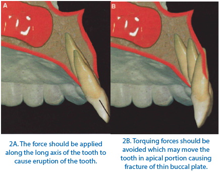
The force should be applied in the direction of eruption of teeth and along the long axis of the tooth. Torquing forces must be avoided as they can damage the thin cortical plate of bone by relocation of the apical portion of the root in the labial or buccal direction. This can lead to poor end results as the bone might not be deposited to the level expected. Orthodontic brackets or elastic threads can be used to apply the force along the long axis of tooth.
Bone height ofadjacent tooth or teeth
The interproximal bone height of the adjacent teeth should be evaluated before extrusion of single tooth. Tarnow and colleagues explained that papilla will be only stable when the distance from the underlying bone is not more than 5mm. The planned site should have equal amount of bone and tissue level on both sides of implants. Forced eruption is carried to the point when the involved tooth is moved to create an excess of bone and soft tissue along with the adjacent tooth or teeth to correct the interproximal attachment level.
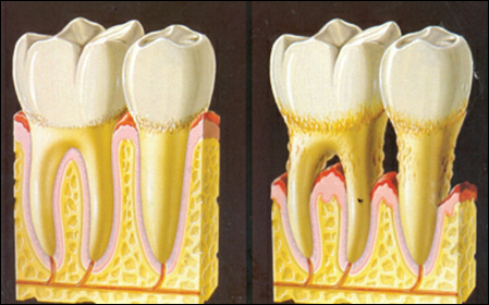
Fig 3: Interproximal bone height should be evaluated before extrusion of the tooth. A. normal bone height and contour. B. deficient bone height and contour.
Number of teeth
At times, to correct the patients’ alveolar bone loss single, multiple or a full dentition may be necessary
Retention time
Retention is very important after all the orthodontic treatments. The tooth or teeth should be retained for 2 to 3 months after extrusion so that bone deposition can take place at the apical portion. If the retention phase is absent then the tooth might intrude soon after removal of brackets.
APPLICATIONS OF FORCED ERUPTION
- Enhancement of Pontic Site
- Augmentation of Implant Site
- Crown Lengthening
- Treatment of Subgingival Fractures
- Treatment of One Or Two Walled Osseous Defects
- In Endodontic Perforations
- In Lateral Root Resorption
ENHANCEMENT OF PONTIC SITE AND PLACEMENT OF FIXED PARTIAL DENTURE
Forced eruption is performed if the buccal plate is present, for successful ridge augmentation and pontic site enhancement. Decoronation of the indicated tooth can also be performed by keeping the roots submerged if buccal plate is not present. Resorption can occur if multiple teeth are moved orthodontically and extracted as the interproximal bone would become unstable after extraction. The orthodontic forces applied are at a slow rate of 1-2mm per month after management of any periapical lesion. Both gingival and bone architecture are improved by this procedure, patient’s own bone is stimulated to produce more bone, and does not require additional surgical site. The only disadvantage is that the patient has to undergo endodontic therapy followed by orthodontic therapy for tooth extrusion.
Procedure
- Endodontic treatment of the indicated tooth or teeth should be performed at first.
- After completion of endodontic therapy, the brackets are placed on the indicated tooth 1.5mm cervically and 1mm cervically on the adjacent tooth or teeth and ligation is done with the help of 0.016inch nickel titanium round levelling wire to cause root extrusion.
- The protocol of 1mm per month should be carried out and the patient should be monitored every month for 6months to assess the extrusion.
- The incisal or occlusal surfaces should be grinded progressively for proper occlusal harmony.
- Meanwhile the exposed root surface can be restored with composite resin for proper esthetics until the treatment is complete.
- The extrusion process is completed at the end of 5months.
- The brackets should be placed for additional 6months to stabilize the tooth or teeth and after this, the indicated tooth or teeth are extracted atraumatically.
- The adjacent teeth are prepared and an interim restoration is given for 6 months for proper maturation of the bone at the edentulous site.
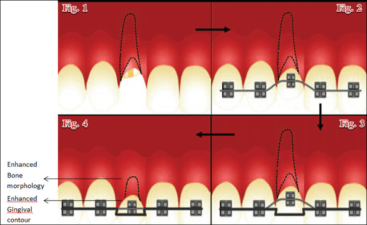
Fig: 4.1 Shows tooth to be extruded with poor bone morphology is treated endodontically before movement; 4.2. The orthodontic brackets are placed on the indicated tooth 1.5mm cervically and ligated with 0.016inch nickel titanium wire; 4.3. The tooth is moved coronally 1mm every month and the provisional is trimmed progressively; 4.4. After 5 months, the bone morphology and the gingival contours are enhanced and the tooth will be extracted after retention of another 6months.
AUGMENTATION AT IMPLANT PLACEMENT SITE
Ridge deficiencies are caused immediately after tooth extraction that progress in palatal and apical directions and are more severe during first 3-4 months. These defects are generally having both horizontal and vertical components. Distraction osteogenesis, guided bone regeneration and graft procedures are used to augment the alveolar ridge but due to the disadvantages like bleeding, soft-tissue injury, block fracture and mandibularfracture, trismus, pain, swelling, bruising, infection, bone resorption, neurosensory defects dehiscence, and graft failure, Forced eruption is preferredover these techniques to enhance the implant placement site. Forced eruption is a non surgical treatment and bone development at the future implant site is facilitated by hard and soft tissue remodelling.
Procedure
- The orthodontic therapy should be initiated after the endodontic therapy of the indicated tooth.
- The brackets are placed more apically to cause extrusionand ligation is done with the help of 0.016inch nickel titanium round levelling wire to cause root.
- The patient should be monitored every three weeks for selective grinding of the tooth interfering in the proper occlusion.
- 12 weeks of stabilization was done following the 6 months of extrusion phase.
- The radiographic assessments should be done to monitor the bone height regularly.
- After the complete maturation of the bone, the extraction should be performed atraumatically.
- The implant should be placed after 2weeks of extractionwith full-thickness flap technique without verticalreleasing incisions according to the implant placement protocol.
- Sutures should be removed 2weeks post- operatively.
- Punch technique is used to uncover the implant and healing abutments are connected after 2-3 months of soft tissue healing.
- Supported restoration should be placed with temporary cements.
- At follow up appointments radiographic and clinical evaluation should be performed every 1year.
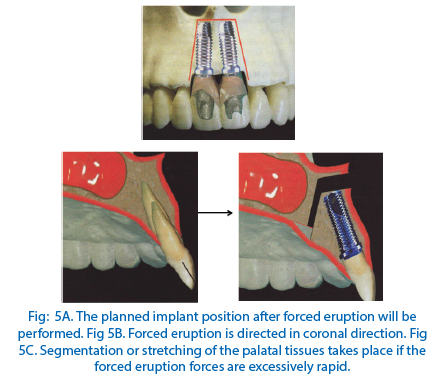
CROWN LENGTHENING
Crown lengthening of the tooth can be performed by applying rapid eruptive forces. Along with the tooth, the periodontal apparatus and alveolar bone also moves and follows the tooth. After the elimination of pseudopockets the procedure should be carried out. Regular fibrotomy should not be combined with forced orthodontic eruption to avoid coronal positioning of the apparatus.
The orthodontic therapy should be started after endodontic therapy and for extrusion, a0.036” SS wirehook is placed on the occlusal surface with double or four anchorages on the either side of the teeth. After the extrusion is done, stabilization is done for 4-6months and bonded retainers should be given for long term retention.

TREATMENT OF SUBGINGIVAL FRACTURES
The dental injury causes the tooth to intrude into the socket is called ‘Traumatic intrusion.’ This causes disruption of the marginal gingival seal, alveolar bone, periodontal fibres, cementum, and neuromuscular supply to the pulp, pulp necrosis, pulpobliteration, external root resorption and loss ofmarginal bone support. Forced eruption with fibrotomy is done of such teeth to avoid extraction of such hopeless tooth. Endondontic treatment of the tooth before starting the orthodontic therapy is necessary.Orthodontic treatment should be carried out for three weeks and on yielding favourable results, the extrusion should be carried out for another 2 weeks. After the therapy, stabilization should be done for 3weeks with stainless steel wire. After 3months, when the tooth has become firm, gingival supracrestalfibrotomy should be performed followed by fabrication of porcelain fused to metal crown.
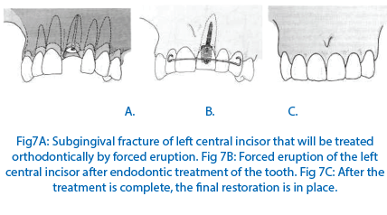
TREATMENT OF ONE OR TWO WALLED INFRABONY OSSEOUS DEFECT
Forced eruption can be used to enhance the gingival health by maintaining the biologic width of gingiva. The hemiseptal defects are more commonly found in mesially tipped teeth or supraerupted teeth. Some teeth might require endodontic treatment before the procedure. The periodontal inflammation is reduced before starting the orthodontic forced eruption procedure by debridement of the area. The teeth should be stabilized for atleast 6months after the orthodontic forced eruption and re- evaluated periodically to assess the periodontal tissues. The regular monitoring of the teeth should be executed as the defects have great sulcular depth initially and are more difficult to cleanse. Therefore patient should be recalled every 2-3 weeks during the extrusion procedure to regularly decrease the periodontal inflammation in the interproximal region mainly.
CONCLUSION
Performing forced eruption along with other procedures can enhance the outcomes of the procedures as it is less invasive, economic and conservative procedure. The cooperation of the patient is very important for the successful treatment and to reduce the chances of relapse of the procedure. It aids in augmentation of the bone and gingival contours before final cementation of prosthesis. Other techniques can be used but they are more invasive and discomfortable to the patient. This movement can be performed in single, multiple or full arched dentition. These movements can cause enhancement but not give rise to new attachments. It is used to analyse the various remodelling procedures and an extension or adjunct to various prosthodontics, endodontics and periodontics procedures. It causes deposition of bone by inducing the bone of the patient itself which is an added advantage over all other procedures that are performed surgically with artificial bone grafts.
REFERENCES
- AlperComut et al. Use of forced eruption to enhance a pontic site in the anterior maxilla. J of Prosthet Dent 2012; 108: 273-278.
- Kenneth F. Hinds. Alveolar ridge development with forced eruptionand distraction of retained natural dentition. Oral Maxillofacial SurgClin N Am 16 (2004) 75-89.
- Avinash S. Bidra et al. Treatment Planning Challenges in the Maxillary Anterior Region Consequent to Severe Loss of Buccal bone. J of Esthet and Rest Dent 2011, 23( 6) 354–360.
- Shyammohan A. Forced eruption: An Adjunct to Prosthodontic treatment Planning. Indian J Stomatol 2011; 2(4): 260-2.
- Luis Antonio Felippe et al. Reestabilishing biologic width with forced eruption.Quintessence Int 2003.34:733-738.
- Timorthy M. Durham et al. Rapid forced eruption: A case report and reviewof forced eruption techniques. Dent Article Review and Testing 2004,3-4; 167-174.
- Frank Celenza. The development of forced eruption as a modality for Implant Site Enhancement. Alpha Omegan 1997, 90(2); 40-43.
- Osbjorn M. Pearson et al.The Aging of Wolff’s “Law”:Ontogeny and Responses to Mechanical Loading in Cortical Bone.Yearbook Of Physical Anthropology 47:63–99 (2004).
- AnupamaSwarnkar et al.A multidisciplinary approach to treat a subgingivally fractured and intruded incisor - a case report. J Endodontology 19-22.
- Herbert T. Shillingburg. Fundamentals of Fixed Prosthodontics. 3rd Edition: Quintessence Publishing Co Inc 1997.
- Guy Huynh-Ba et al. Surgical lengthening of the clinical crown: a periodontal concept for reconstructive dentistry. QuintessenzPerio 2007; 4(3):193-201.
- Vincent G. Kokich. Carranza’s Clinical Periodontology. 9th Edition: W.B. Saunder’s Company, 2002.
Disclaimer from Guident
The views and opinions expressed in articles published in Guident are solely those of the authors and do not necessarily reflect the views of the editors, the editorial board or the publisher, Ivory India. Guident does not endorse any product, technique or treatment described. Clinical content is intended for qualified dental and medical professionals and is not a substitute for individual clinical judgement. Guident, its editors and the publisher accept no liability for any consequence arising from the use of information published in the journal.
How to cite this article
Dr. Hasan Sarfaraz, Dr. Manesh Lahori, Dr. Manpreet Kaur. Forced Eruption: A Boon for the Bone. Guident. 2014 Dec;8(1). Available from: https://guident.in/articles/general/forced-eruption-a-boon-for-the-bone.html