Optical Coherence Tomography
ABSTRACT
Dental prostheses are very complex systems, heterogenous in structure, made up from various materials, with different physical properties. They have to satisfy high stress requirements as well as aesthetic challenges. The failures of dental prostheses lead to functional, aesthetic and phonetic disturbances which finally render the prosthetic treatment inefficient. OCT is a tomography imaging technology capable of producing high-resolution cross-sectional images of the internal architecture of materials and tissues. This includes not only the images of normal and abnormal dental hard tissue structures but also teeth after several treatment methods. It can be used for non-invasive investigations for both in vivo and in vitro structural imaging within the oral cavity.
Key words: Optical Coherence Tomography, non-invasive investigation, dentistry.
INTRODUCTION
During the last 20 years, optical coherence tomography (OCT) has evolved into a powerful technique for imaging of transparent and translucent structures1 . OCT is an attractive noninvasive, nontouch
imaging technique for obtaining high-resolution images. The first application in the biomedical optics field was for the measurement of the eye length. Optical Coherence Tomography (OCT) is an optical imaging method with medium penetration and high resolution primarily used in medicine. It has the potential to create live cross-sectional images even of non-transparent tissue at depths of up to 3 millimeters at a resolution of little more than a micrometer2 .
PRINCPLES
One of the light characteristics which is used in the OCT is the phenomenon of interference. Two electromagnetic waves can interfere, add up or subtract each other and create a new one. Interferences could be fully constructive interference or completely destructive, but are usually partially destructive or constructive. (Fig.1)
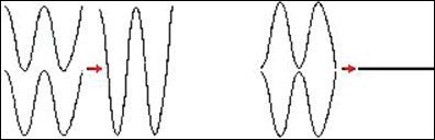
Fig 1: Constructive (left) and Destructive Interferences (right).
The light source has low-coherence like an SLD. Light is driven through the fibre to the beam splitter which divide by two (50/50) the beam. The first 50% are driven in direction of the sample (sample arm) to be reflected or backscattered by the different sample’s tissues. The second 50% go through the reference arm ended by a movable mirror. Both are reflected back to the detection arm which has a detector to convert light intensity into electrical current. That current is converted to a digital signal and analyzed by a computer which generates an image of the sample.3 (Fig.2)
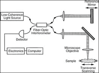
Fig 2: Interference Principle in a Michelson Interferometer.
USES IN DENTISTRY
OCT is useful widely in almost all the fields of dentistry as it is capable of imaging both the hard and soft tissues and also can be used both in vitro and in vivo. The various applications are:
- To detect the presence or absence of material defects and microleakage at the prosthetic interfaces.4 The investigations upon fixed partial prosthesis presented many defects that can lead to their deterioration. These defects are usually located inside the material and cannot be depicted visually or by other conventional imagistic method.
- OCT imaging offers the exciting potential to detect periimplantitis before significant osseous destruction occurs. OCT images of soft tissue surrounding failing implants are characterized by linear signal deficits, low-intensity collagen signals, and pronounced increases in vascular elements.5 The quality of the implant insertion could be investigated by implant bone interface analysis.
- Used to view the integrity of temporo-mandibular joint disc.
- OCT, as a relatively new imaging technique for detection and analysis of early caries, correlated well with an established method of quantifying demineralization. The reflectivity loss in tooth tissue following demineralization, measured using OCT, could be significantly related to the amount of mineral loss during the demineralization process.6
- OCT is a promising nondestructive imaging method for the diagnosis of vertical root fractures.7
- Optical coherence tomography clearly proves its utility in investigating orthodontic bonding. With this non-invasive method the material defects trapped inside the matrix resin can be localized and 3D reconstructed. Adding confocal microscopy to the OCT investigation leads to a much better evaluation of the communications of these gaps in order to forecast the strength of the orthodontic brackets’ bonding.8
- Malignant lesions of oral mucosa: Accounting for 96% of all oral cancers, squamous cell carcinoma (SCC) is usually preceded by dysplasia presenting as white epithelial lesions on the oral mucosa (leucoplakia). This high-resolution optical technique permits minimally invasive imaging of near-surface abnormalities in complex tissues, having a penetration depth of 1-2 mm.9,10 This permits in vivo noninvasive imaging of the macroscopic characteristics of epithelial and subepithelial structures, including: depth and thickness, histopathological appearance and peripheral margins.
- In the Todea C. et al. study.11 the quality of endodontic treatments and root canal fillings were investigated with eFOCT/CM technology. Areas of apical microleakage were detected between the gutta-percha cones and the root canal walls and the filling material of the root canal space.
- OCT is particularly well-suited for periodontal diagnosis, generating ultrahigh resolution crosssectional images of dental tissues. OCT provides rapid, consistent, and reproducible images of the surface topography, pocket morphology, and attachment level that are digitally recorded. These images pinpoint with great accuracy sites of disease progression. OCT also provides quantitative information regarding the thickness and character of the gingiva, root surface irregularities, and the distribution of subgingival calculus.5
CONCLUSION
OCT represents a valuable method for investigation and assessment of the health status of soft oral tissues and of hard dental structures. OCT can be used for evaluation of dental treatments reducing their failure
rate and saving time and resources, by eliminating incorrect restorations before their insertion in the oral cavity.
REFERENCES
- Carmen Todea1, Meda Lavinia Negrutiu1, Cosmin Balabuc1, Cosmin Sinescu1, Florin Ionel Topala1, Corina Marcauteanu1, Silvana Canjau1, Gianfranco Semez2, Adrian Gh. Podoleanu3. OPTICAL COHERENCE TOMOGRAPHY APPLICATIONS IN DENTISTRY. TMJ 2010, Vol. 60, No. 1: 5-17.
- Lorenz Diener. Optical Coherence Tomography.
- Optical Coherence Tomography: 3-D Dental Scanning Imaging Probe. Dubosson Fabrice
- Cosmin Sinescu, Meda Lavinia Negrutiu, Carmen Todea et al, Quality assessment of dental treatments using en-face optical coherence tomography, Journal of Biomedical Optics, 2008;13(5).
- Drexler W., Fujimoto J.G., Optical Coherence Tomography, Springer- Verlag Berlin Heidelberg, 2008;1166-1168.
- B. T. Amaechi, A. Gh. Podoleanu, G. N. Komarov, J. A. Rogers, S. M. Higham, and D. A. Jackson. Application of Optical Coherence Tomography for Imaging and Assessment of Early Dental Caries Lesions. Laser Physics, Vol. 13, No. 5, 2003, pp. 703–710.
- Hagay Shemesh, DMD,* Gijs van Soest, PhD,† Min-Kai Wu, PhD,* and Paul R. Wesselink, PhD. Diagnosis of Vertical Root Fractures with Optical Coherence Tomography. JOE — Volume 34, Number 6, June 2008
- R. O. Rominu, C. Sinescu, D.M. Pop, M. Hughes, A. Bradu, M. Rominu, and A. Gh. Podoleanu. En-Face Optical Coherence Tomography and Fluorescence in Evaluation of Orthodontic Interfaces. World Academy of Science, Engineering and Technology 32 2009.
- B. W. Colston, Jr., U. S. Sathyam, L. B. DaSilva et al, Dental OCT, Optic Express, 1998;3(6):230-238;
- Pedra Wilder-Smith, Kenneth Lee, Shuguang Guo et al, In Vivo Diagnosis of Oral Dysplasia and Malignacy Using Coherence Tomography: Preliminary Studies in 50 Patients. Lasers in Surgery and Medicine; 2009;41:353-357;
- Carmen Todea, Cosmin Balabuc, Cosmin Sinescu et al, En face optical coherence tomography investigation of apical microleakage after laser-assisted endodontic treatment, Lasers Med Sci 2009.
Disclaimer from Guident
The views and opinions expressed in articles published in Guident are solely those of the authors and do not necessarily reflect the views of the editors, the editorial board or the publisher, Ivory India. Guident does not endorse any product, technique or treatment described. Clinical content is intended for qualified dental and medical professionals and is not a substitute for individual clinical judgement. Guident, its editors and the publisher accept no liability for any consequence arising from the use of information published in the journal.
How to cite this article
Dr. Gunjan Gupta, Dr. Varun Goyal Dr. Alpa Gupta. Optical Coherence Tomography. Guident. 2014 Oct;7(11). Available from: https://guident.in/articles/general/optical-coherence-tomography.html