Single Step Apexification with MTA
Department of Conservative Dentistry and Endodontics Manipal college of dental sciences, Manipal, India
Abstract
Immature teeth with necrotic pulp and periapical lesion are difficult to treat via conventional endodontic therapy. Numerous procedures and materials have been utilized to induce root-end barrier formation. Traditionally, Calcium hydroxide has been the material of choice for the apexification of immature permanent teeth; however, Mineral Trioxide Aggregate has several advantages such as single step technique and ability to set in the presence of moisture. This case report describes the successful healing and apexification of an incisor using MTA as an apical barrier. After preparing the access cavity of 11, the working length was determined. The root canals were irrigated with 2.5% sodium hypochlorite and disinfected with calcium-hydroxide for a week. MTA was then placed in the apical 5 millimetres of the root canal. The remaining part of the root canal was filled with gutta-percha and the coronal restoration was finished with composite resin. So Mineral trioxide aggregate holds significant promise as an alternative to multiple treatments with Calcium hydroxide.
INTRODUCTION
Dental injuries to the young permanent incisors are the frequent finding following orofacial trauma. Prevalence of traumatized permanent anterior teeth varied from 2.6% to 30%.1The majority of these incidents occur before root formation is complete and may result in multiple complications like pulp necrosis, pulp canal obliteration, root resorption and open apex. The causes of open apices may be due to pulp necrosis as a result of caries or trauma, before root growth and development are complete or extensive apical resorption because of periapicalpathosis, trauma or orthodontic treatment. Root canal treatment at this time is a significant challenge, because of the size of the canal, the thin and fragile dentin walls and large open apex. In the past, techniques for management of the open apex in non-vital teeth were confined to custom fitting the filling material, paste fills and apical surgery.2 However, the limited success by these procedures resulted in significant interest in the phenomenon of continued apical development, or establishment of an apical barrier referred to as apexification.3Traditionally, calcium hydroxide (Ca(OH)2) has been the material of choice for the apexification of immature permanent teeth.4Whilst the advantages of calcium hydroxide lie in the fact that it has been widely studied and has shown success,5,6 the disadvantages are its prolonged treatment time, the need for multiple visits and increased susceptibility to cervical fracture.7,8 For these reasons, one visit apexification has been suggested. Mineral trioxide aggregate (MTA) has been proposed as a material suitable for one visit apexification, as it combines biocompatibility, bacteriostatic action, with favourable sealing ability when used to repair root/pulp chamber perforations or as root-end filling material.9,10,11 This case report presents the management of an immature tooth (with an open apex) with a single step apical barrier placement using MTA
CASE REPORT
A 19-year old female reported to the department of conservative dentistry &endodontics, Manipalcollege of dental sciences, Manipal with a complaint of fractured & discolored upper right front tooth. (Fig 1) The patient had a history of trauma that occurred 11 years back and no treatment was availed. Intraoral examination revealed fracture and discoloration in tooth no. 11 and Ellis class II fracture in 21. A negative response was obtained upon pulp sensibility testing of tooth no 11. On radiographic examination, tooth no. 11 presented with blunting of root apex and apparent loss of apical constriction leading to open apex.( Fig 2) On the basis of clinical and radiographic findings, the tooth was diagnosed as external inflammatory resorption with open apex. The patient and parents were informed about an endodontic treatment plan involving MTA apical plug. Upon approval of the treatment plan by the patient and obtaining of informed consent, root canal treatment was initiated at the same appointment.
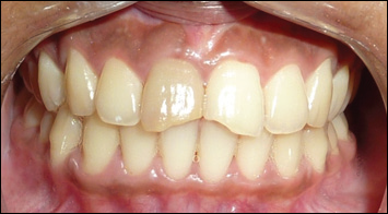
Fig: 1

Fig : 2
After the application of the rubber dam and access cavity preparation, the working length was obtained in tooth no 11.The canal was instrumented lightly with K files (Mani, Japan) with the aim of cleaning the root canal walls of debris. The canal was thoroughly debrided with a copious irrigation of 2.5% sodium hypochlorite (NaOCl) (Medilise chemicals, Kannur, Kerala, India) and 0.9% saline (Claris Ostuka Limited, Ahmedabad, India) to ensure complete removal of the necrotic pulp tissue. The canal was dried with sterile paper points and Ca(OH)2 was placed as an intracanal medicament and the access cavity was temporized with Cavit G (3M ESPE; Germany).
After 1 week, Ca(OH)2 was removed after repeated rinsing with NaOCl followed by rinsing with sterile water. The canal was then dried with paper points. MTA (White Pro-Root MTA; DentsplyMaillefer, Ballaigues, Switzerland) was prepared according to the manufacturer’s instructions,and a portion of the material was deposited in the canal using a small sterile amalgam carrier. The MTA was then gently condensed with an endodontic plugger to the working length. MTA plug were placed to the apex with a thickness of 5 mm. Correct placement of the MTA plug was verified with a periapical radiograph.(Fig 3)
A moistened cotton pellet was placed in the pulp chamber, and the access cavity was closed with a temporary filling material (Cavit; ESPE,Germany). One week later, the temporary filling was removed and the remaining root canal was obturated using cold lateral compaction technique using guttapercha with AH Plus sealer (Dentsply, Konstanz,Germany) (Fig 4)

The access cavity was restored with composite (3M ESPE,USA) and the tooth was rehabilitated with all ceramic crown 2 months post operatively.(Fig 5)
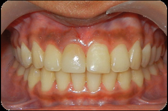
Fig: 5
DISCUSSION
Calcium hydroxide pastes have been considered as the material of choice to induce the formation of a hard tissue apical barrier.12Its efficiency has been demonstrated by many authors, even in the presence of an apical lesion. Despite its popularity for the apexification procedure, calcium hydroxide therapy has some inherent disadvantages that include unpredictability of apical closure, difficulty in patient follow up and delayed treatment. In addition, the canal is susceptible to reinfection because it is covered by a temporary seal.13 One alternative to calcium hydroxide apexification is a one-step technique using an artificial apical barrier with MTA. With the MTA apical plug technique, a one-step obturation after short canal disinfection with calcium hydroxide could be performed.MTA has an ability to facilitate normal architecture by inducing hard tissue barrier.14 Its excellent biocompatibity and ablility to set in presence of blood ,high predictability of apical closure, along with shorter treatment time and less dependence on patient compliance, makes it even more advantageous. MTA thus can be considered to be a very effective material to support regeneration of apical tissue and to promote apical obturation in traumatized, infected immature teeth with open apices.
REFERENCES
- Andreasen JO, Andreasen F, Andersson L. Textbook andColor Atlas of Traumatic Injuries to the Teeth, 4th Edn.Oxford: Blackwell Munksgaard, 2007.
- Rafter M. Apexification: A Review. Dent Traumatol. 2005;21:1–8
- Frank AL. Therapy for the divergent pulpless tooth by continued apical formation. J Am Dent Assoc. 1966;72:87–93
- Al Ansary MA, Day PF, Duggal MS, Brunton PA. Interventions for treating traumatized necrotic immature permanent anterior teeth: inducing a calcific barrier & root strengthening.DentTraumatol 2009; 25:367–79.
- Yates JA. Barrier formation time in non-vital teeth with open apices. IntEndod J 1988; 21:313–9.
- Ghose LJ, BaghdadyVS, Hikmat YM. Apexification of immature apices of pulpless permanent anterior teeth with calcium hydroxide. J Endod 1987; 13:285–90.
- Andreasen JO, Farik B, Munksgaard EC. Long-term calcium hydroxide as a root canal dressing may increase risk of root fracture. Dent Traumatol 2002; 18:134–7.
- Sahebi S, Moazami F, Abbott P. The effects of short-termcalcium hydroxide application on the strength of dentine. Dent Traumatol 2010; 26:43–6.
- Torabinejad M, Chiavian N. Clinical application of mineral trioxide aggregate. J Endod 1993; 25, 197–205.
- Mitchell PJ, Pitt Ford TR, Torabinejad M, McDonald F.Osteoblast biocompatibility of mineral trioxide aggregate.Biomaterials 1999;20:167–73.
- Al-Kahtani A, Shostad S, Schifferle R, Bhambhani S. In-vitro evaluation of microleakage of an orthograde apical plug of mineral trioxide aggregate in permanent teeth with simulatedimmature apices. J Endod 2005; 31:117–9.
- Felippe MCS, Felippe WT, Marques MM, Antoniazzi JH .The effect of renewal of calcium hydroxide paste on theapexification and periapical healing of teeth with incomplete root formation. IntEndod J 2005; 38,436–42.
- Magura ME, Kafrawy AH, Brown CE, Jr, Newton C. Human saliva coronal microleakage in obturated root canals: An in vitro study. J Endod. 1991; 17:324–31.
- Tittle K, Farley J, Linkhart T, TorabinejadM.Apical closure induction using bone growth factors and mineral trioxide aggregate. J.Endod 1996: 22, 198.
Disclaimer from Guident
The views and opinions expressed in articles published in Guident are solely those of the authors and do not necessarily reflect the views of the editors, the editorial board or the publisher, Ivory India. Guident does not endorse any product, technique or treatment described. Clinical content is intended for qualified dental and medical professionals and is not a substitute for individual clinical judgement. Guident, its editors and the publisher accept no liability for any consequence arising from the use of information published in the journal.
How to cite this article
Dr.Tina Puthen Purayil. Single Step Apexification with MTA. Guident. 2014 Oct;7(11). Available from: https://guident.in/articles/endodontics/single-step-apexification-with-mta-501.html