Fish: An Additional Diagnostic Tool for Head and Neck Cancer
Department of Oral Pathology, Santosh Dental College & Hospital, Ghaziabad, India, Department of Pedodontics, Sharma Dental Clinic, Kamla Nagar, Delhi
ABSTRACT
Fluorescence in situ hybridization (FISH) is a powerful and versatile tool for the detection and localisation of nucleic acids in fixed materials such as cells, metaphase chromosome spreads and tissue sections. This technique can be used to identify chromosomes, detect chromosomal abnormalities or determine the chromosomal location of specific sequences. FISH plays an increasingly important role in a variety of research areas, especially in tumor biology, gene amplification and gene mapping. Thus, the present paper emphasizes on the role of FISH as an important diagnostic and prognostic tool for head and neck cancer.
INTRODUCTION
Cytogenetics is the study of structure and function of cell, especially the chromosomes. Every human being has 46 chromosomes which play an important role in controlling the activities of a living cell. They are vital for the cell division process and are responsible for the division, replication as well as creation of daughter cells, which contain accurate sequences of DNA and proteins. Defects in these chromosome leads to various diseases.
FISH (Fluorescence In Situ Hybridization) technique is the principle of Cytogenetics which involves the study of human chromosomes in health and disease, any alterations in chromosomes number / structure is looked upon. It is the ability of a single stranded DNA molecule to find and bind to its complementary strand is pretty amazing. Any DNA of interest can be used as a probe for FISH studies e.g. whole human DNA, whole chromosomes, centromeric regions, any unique sequence of DNA. In FISH it is exploited to the utmost. It refers to using fluorescently labelled probe to hybridize to cytogenetic cell preparations. Probes are labelled either directly, by incorporation of fluorescent nucleotides, or indirectly, by incorporation of reporter molecules that are subsequently detected by fluorescent antibodies or other affinity molecules. Probes and targets are finally visualized in situ by microscopy analysis. FISH-based diagnostic assays have been developed within different fields of investigation, including clinical genetics, neuroscience, reproductive medicine, toxicology, microbial ecology, evolutionary biology, comparative genomics, cellular genomics, and chromosome biology.
The introduction of FISH in the late 1980s, as a technique that can readily detect trisomies and translocations in metaphase spreads and interphase nuclei using entire chromosome-specific DNA libraries, was heralded as a further revolution in cytogenetic analysis1, 2. The high sensitivity and specificity of FISH and the speed with which the assays can be performed have made FISH a powerful technique with numerous applications, and it has gained general acceptance as a clinical laboratory tool3.
FISH is essentially based upon the same principle as a Southern blot analysis, a cytogenetic equivalent that exploits the ability of single-stranded DNA to anneal to complementary DNA. In the case of FISH, the target is the nuclear DNA of either interphase cells or of metaphase chromosomes affixed to a microscope slide, although FISH can also be performed using bone marrow or peripheral blood smears, or fixed and sectioned tissue3. Once fixed to a microscope slide, the desired cells are hybridized to a nucleic acid probe. This anneals to its complementary sequence in the specimen DNA and is labelled with a reporter molecule which is either an attached fluorochrome, enabling direct detection of the probe via a coloured signal at the hybridization site visualized by fluorescence microscopy, or a hapten that can be detected indirectly3,4. The following figure describes it. (Fig 1)

Figure 1 . The principles of fluorescence in situ hybridization. (A) The basic elements are a DNA probe and a target sequence. (B) Before hybridization, the DNA probe is labelled indirectly with a hapten (left panel) or directly labelled via the incorporation of a fluorophore (right panel). (C) The labelled probe and the target DNA are denatured to yield single-stranded DNA. (D) They are then combined, which allows the annealing of complementary DNA sequences. (E) If the probe has been labelled indirectly, an extra step is required for visualization of the non-fluorescent hapten that uses an enzymatic or immunological detection system. Finally, the signals are evaluated by fluorescence microscopy.
Chromosomal abnormalities are very frequently found in head & neck tumors. With more than 70% of tumors are being aneuploid . Loss of 3q arm & 9p in over 80% of tumors are found. Tumor proliferation is known to result from activation of growth promoting pathways & inhibition of growth down of regulation. Mutations can occur in the genomes of all dividing cells as a result of misincorporation during DNA replication or through exposure to exogenous mutagens such as ionizing radiation or endogenous mutagens.5 (Fig. 3). Cellular responses to DNA damage are outlined in Fig.2

Figure 2 . Theoretical association of the signal transduction of checkpoint responses. DNA damages are recognized by sensor proteins. The signals are transmitted to tranducers (mainly kinases) via mediators and the regulated transducer molecules suppress effector kinases, such as Cdks and Cdc7, thereby arresting the cell cycle at the specific phases. Adapted from Niida and Nakanishi by permission of the UK Environmental Mutagen Society.9
Cancer genomes carry two biological classes of somatic mutation arising from these various processes. ‘Driver’ mutations emerge as a result of selective pressure during tumourgenesis. They confer growth advantage on the cell in which they occur, are directly implicated in cancer development and therefore are positively selected (passed on to the cancer progeny). Conversely, ‘passenger’ mutations are incidental, possible products of genome instability or the large number of cell divisions that lead from a single transformed cell to a clinically detectable cancer. They are present in the cell that was the progenitor of the final clonal expansion of the cancer, are biologically neutral and do not confer growth advantage.6,7 While certain types of chromosomal aberrations arelethal, others may lead to oncogenic transformation by inactivation of a tumour suppressor gene or activation of an oncogene via generating novel fusion proteins capable of initiating carcinogenesis. Increased frequencies of chromosomal aberrations are correlated with elevated risks of cancer and certain human neoplasias are associated with defined chromosomal aberrations branding chromosomal aberrations a hallmark of all tumour cells.8
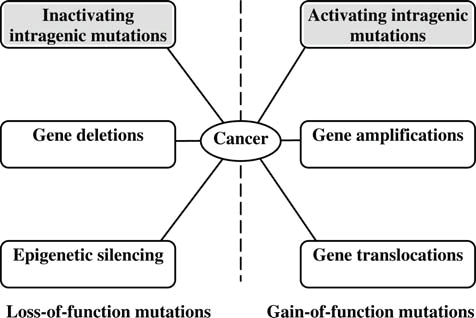
Figure 3. The diversity of mutations that can initiate human cancer. Intragenetic mutations—highlighted are small nucleotide changes that may be inherited by the cancer progeny from the progenitor cell, resulting in either a loss-of-function of genes or a gain-of-function of genes. Epigenetic silencing involves heritable modifications of nucleotides and histones in regulatory regions of genes, leading to suppression of gene expression in the absence of DNA mutations. Translocations lead to the fusion of DNA fragments from different chromosomal regions, either creating an abnormal fusion protein or leading to aberrant expression of a normal gene. These so-called ‘driver’ mutations contribute towards oncogenesis.9
ADVANTAGES
FISH has greatly expanded the capabilities of cytogenetics and pathology laboratories through its high sensitivity, specificity and rapid turnover with a high efficiency of hybridization and detection. Material for FISH can be processed in 4–24 h, and the analysis of 1000–2000 cells accomplished in 15–45 min, enabling the information on the cytogenetic pattern of tumour cells to be achieved within a sufficient time frame for use in treatment strategies.3,10
A particular advantage of FISH techniques is the possibility to also study chromosomal aberrations in nondividing cells, which is useful for the visualization of chromosomal aberrations directly in cytological preparations and tissue sections. This has led to chromosomal mapping of commonly deleted or amplified regions, providing starting points for the searchfor a gene (or genes) involved in growth control in certain tumour types.11 Amplification of oncogenes is an important factor in carcinogenesis and is often correlated to progressive tumour growth and poor prognosis; therefore, identification and characterization of genes subject to amplification can provide valuable molecular tools for evaluation of prognosis in human cancers.12
USES
Chromosomal studies are an important laboratory diagnostic procedure, such as in :
- Prenatal diagnosis
- Certain patients with mental retardation and multiple birth defects
- Abnormal sexual development
- Infertility or multiple miscarriages
- Malignancies & hematologic disorders
METHOD
Steps
- Cell Culture
- Harvesting
- Slide Preparation
- Banding: pre-treatment, denaturing and hybridization
FISH BASIC METHOD
- Each probe is specific to one region of a chromosome (pair), and is labeled with fluorescent molecules throughout its length.
- Each microscope slide contains many metaphases.
- Each metaphase consists of the complete set of chromosomes, one small segment of which each probe will seek out and bind itself to.
- The first step is to break apart (denature) the double strands of DNA in both the probe DNA and the chromosome DNA so they can bind to each other.
- This is done by heating the DNA in a solution of formamide at a high temperature (70 °C)
- Next, the probe is placed on the slide and a coverslip is placed on top. To prevent evaporation, the edges of the coverslip are sealed with rubber cement.
- The slide is then placed in a 37 °C incubator overnight for the probe to hybridize with the target chromosome.
- Overnight, the probe DNA seeks out its target sequence on the specific chromosome and binds to it. The strands slowly rejoin (reanneal).
- The next day, the coverslip is removed, and the slide is washed in a salt/detergent solution to remove any of the probe that did not bind to chromosomes.
- A differently colored fluorescent dye is added to the slide to stain all of the chromosomes so that they may then be viewed using a fluorescent light microscope.(Figure3)
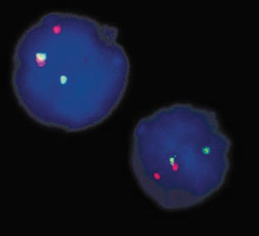
Figure3. Interphase FISH on bone marrow nuclei containing the translocation t(11;19)(q23;p13) using a dual-colour split-apart probe. Green-red fusion (yellow) signals indicate a normal cell. Separate green and red signals indicate the presence of translocations1
RESULTS
If the DNA sequence corresponding to the probe used is present in the target tissue a fluorescent signal will be seen. If the DNA sequence corresponding to the probe used is absent from the target tissue no signal will be seen.
Three Color FISH
Two (or more) different probes labeled with different fluorescent tags can be mixed and used at the same time. The chromosomes are then stained with a third color for contrast. This gives a metaphase or interphase cell with three colors which can be used to detect two different chromosomes at the same time, or to provide a "control probe" in case one of the other target sequences are deleted and a probe cannot bind to the chromosome.
Uses for three color FISH
1 )Example of chromosome enumeration:

Here, an interphase cell shows three pink signals and two green signals. This is the case in the detection of trisomy 21, where the chromosome 21 probe would be labeled pink and a control probe is labeled green.
2) Example of detection of deletion using control probe:

In this case, the left-hand chromosome is intact, but the other is deleted in the region where the probe was supposed to bind. The probe for the region of interest is labeled green, and another probe (control) which binds to the same chromosome is labeled pink.
CONCLUSION
The power of ability FISH to identify specific genetic aberrations has propelled FISH-based techniques to the forefront of screening procedures for prenatal, paediatric and adult cases in a wide variety of cell types making FISH analysis data a useful tool in the decision of therapy to combat cancer. This is supported by a recently conducted survey by Wordsworth et al. who reported that the most common techniques used for the testing of somatic mutations in laboratories were IHC and FISH.13
The ultimate goal of FISH utilization would be an arraybased screen using the complete oncogenic repertoire to diagnose any prenatal or postnatal aberration(s). Any mutations could be rectified accordingly using gene therapy as a method of cancer prevention.The death rate associated with head & neck cancer is particularly high not because it is hard to discover or diagnose, but due to the cancer being routinely discovered late in its development.This information may be used to enhance earlier detection & more effective preventive/treatment strategies to minimize mortality & morbidity.
REFERENCES
- Kearney L (2001) Molecular cytogenetics. Best Pract. Res. Clin. Haematol. 14:645–668.
- Pinkel D, Landegent J, Collins C et al. (1988) Fluorescence in situ hybridization with human chromosome-specific libraries: detection of trisomy 21 and translocations of chromosome 4. Proc Natl Acad Sci USA 85: 9138–9142.
- Gozzetti A, Le Beau MM (2000) Fluorescence in situ hybridization: uses and limitations. Semin Hematol 37: 320–333.
- Lichter P, Joos S (1996) Non-isotopic In situ Hybridization to Metaphase Chromosomes and Interphase Nuclei. EMBO Practical Course at the German Cancer Research Center, Heidelberg.
- Greenman C, Stephens P, Smith R et al. (2007) Patterns of somatic mutation in human cancer genomes. Nature 446: 153–158.
- Greenman C, Stephens P, Smith R et al. (2007) Patterns of somatic mutation in human cancer genomes. Nature 446: 153–158.
- Haber DA, Settleman J (2007) Drivers and Passengers. Nature 446: 145–146.
- Pfeiffer P, Goedecke W, Obe G (2000) Mechanisms of DNA double-strand break repair and their potential to induce chromosomal aberrations. Mutagenesis 15: 289–302.
- Niida H, Nakanishi M (2006) DNA damage checkpoints in mammals. Mutagenesis 21: 3–9.
- Jamil A, Theil KS, Kahwash S et al. (2000) TEL/AML-1 fusion gene: its frequency and prognostic significance in childhood acute lymphoblastic leukemia. Cancer Genet Cytogenet 122: 73–78.
- Ried T, Liyanage M, du Manoir S et al. (1997) Tumor cytogenetics: comparative genomic hybridization and spectral karyotyping. J Mol Med 75: 801–814.
- Samuelson E, Levan K, Adamovic T et al. (2008) Recurrent gene amplifications in human type I endometrial adenocarcinoma detected by fluorescence in situ hybridization. Cancer Genet Cytogenet 181: 25–30.
- Wordsworth S, Papanicolas I, Buchanan J et al. (2008) Molecular testing for somatic cancer mutations: a survey of current and future testing in UK laboratories. J Clin Pathol 61: 373–376.
- Ryan Bishop (2010)Applications of fluorescence in situ hybridization (FISH) in detecting genetic aberrations of medical significance biohorizons 3(1) : 85-95.
- Emanuela V. Volpi et al (2008) FISH glossary: an overview of the fluorescence in situ hybridization technique. BioTechniques 45:385-409.
Disclaimer from Guident
The views and opinions expressed in articles published in Guident are solely those of the authors and do not necessarily reflect the views of the editors, the editorial board or the publisher, Ivory India. Guident does not endorse any product, technique or treatment described. Clinical content is intended for qualified dental and medical professionals and is not a substitute for individual clinical judgement. Guident, its editors and the publisher accept no liability for any consequence arising from the use of information published in the journal.
How to cite this article
Dr. Geeta Sharma, Dr. Natasha Gambhir, Dr. Shourya Sharma. Fish: An Additional Diagnostic Tool for Head and Neck Cancer. Guident. 2014 Sep;7(10). Available from: https://guident.in/articles/general/fish-an-additional-diagnostic-tool-for-head-and-neck-cancer.html