Standard Edgewise Simplified
Department of Orthodontics Manipal College of Dental Sciences, Manipal, India
Abstract
With the advent of pre-adjusted appliance mechanotherapy, the standard edgewise appliance therapy is being sparingly used. This case report demonstrates the treatment of bimaxillary protrusion in adult patient with simplified standard edgewise mechanotherapy. Clinical and cephalometric evaluation revealed Class I Skeletal pattern, Class I dental pattern, bimaxillary protrusion, high mandibular plane angle, protruded upper and lower lips, normal overjet and overbite. The goal of treatment was to bring about maximum retraction of anterior teeth. The case was successfully managed by extraction of all first Premolars and fixed appliance therapy using standard edgewise mechanics. Stable esthetic results were attained at the end of the treatment.
Key Words: Bimaxillary protrusion, Malocclusion, Standard edgewise.
INTRODUCTION
Class I bimaxillary protrusion is a common malocclusion and its treatment involves extraction of first four premolars. This case report illustrates a young male patient having angles Class I dental malocclusion with bimaxillary protrusion on class I skeletal base treated with extraction of all the first premolars using standard edgewise mechanics.
CASE -REPORT
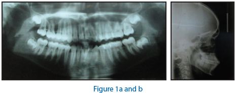
A 13-year-old patient presented with a Class I bimaxillary protrusion malocclusion with a high angle between the maxillary and mandibular planes. The patient requested orthodontic treatment to improve his dental appearance. He had no relevant medical history. Extra oral examination revealed convex profile, high mandibular plane angle and protruded lips (figure 1a and b). Intraoral examination revealed Angles Class I molar and canine relation, curve of spee of 1.5 mm, coincident midlines, good oral hygiene, healthy soft tissue, average maxillary and mandibular arches, normal overjet and overbite, unerupted 3rd molars (figure 2)
Treatment Objectives
- Retraction of Upper and Lower anteriors
- Improvement in facial profile.
- Maintainence Class I molar and canine relation
- Establish normal overjet and overbite

Treatment plan
- Extraction of all first Premolars
- Fixed appliance therapy using simplified Standard edgewise mechanics and high pull head gear.
Treatment progress
Treatment involved the extraction of first premolars in the upper and lower arch to correct the proclination and maintain angle’s Class-I molar relation. Treatment started with the placement of 0.022Ë standard edgewise brackets and molar tubes (Victory series, 3M, UNITEK, Monrovia, CA, USA). In the upper arch banding was done on first molar and molar tube with head gear slot was placed on upper first molar to facilitate head gear use. Patient was asked to wear a high pull head gear with a force of 400 gms for 12 hours daily throughout the treatment period. In the lower arch both first and second molars were banded with an open twin bracket on first molar and molar tube on second molar.
Treatment began with the leveling and alignment with well-coordinated 0.016Ë and 0.018Ë stainless steel (S.S.) arch wires (A.J. Wilcock Premium Plus Australian arch wire, Whittlesea, Victoria, Australia) with molar stops, progressive tip back bends and curve of spee wire in the upper and reverse curve of spee wire in the lower arch (figure 3). Individual canine retraction was started and finished with an elastic chain (3M, UNITEK, Monrovia, CA, USA) on 0.018” stainless steel wire with tip backs. Care was taken to put a light force to avoid tipping of canine.
After canine retraction, 0.019 × 0.025Ë S.S. T loop archwires (Truforce, Ortho Technology, Inc., Tampa, USA) with progressive tip back bends were placed in both arches (figure 4). Progressive buccal root torque was placed in the lower posterior segment, progressive lingual root torque in the upper posterior segment and lingual root torque in the upper and lower anterior segment. Patient was recalled every 6 weeks for reactivation of loops and to check the progress. Finishing was performed on 0.016x0.022” S.S. archwires (Truforce, Ortho Technology, Inc., Tampa, USA) with tip backs and artistic positioning bends.

Results
The first molar was successfully maintained into a Class-I relationship. Treatment time was 2 years. End of treatment, a proper overjet and overbite was established with coincident midlines and no opening of mandibular plane angle. Incisor mandibular plane angle reduced from102° to 88° resulting in better incisal inclination after treatment and consequent profile change. Mandibular plane also reduced with the high pull head gear wear from 31° to 29° adding up to the improvement( figure 5a, 5b, 6, 7, 8).


Figure 8
DISCUSSION
The design of edgewise bracket facilitates bodily movement1 (Angle et al, 1928). Degree of tip back bends on terminal molars is kept such that when arch wire is raised & ligated to first molars, mesial cusps of terminal molars are elevated & first molars are depressed2 (Tweed et al, 1966). Thus, presence of tip backs will enhance anchorage. During the alignment phase tip back bends were kept flush to the mesial wing of the brackets of second premolars and molars. Individual canine retraction then followed on the 0.018 stainless steel wire with elastic chain instead of a coil spring. The force was kept in the range of 50-100 gm to avoid canine tipping as suggested by Natrass et al, 19973. Also, the placement of elastic chain made oral hygiene maintenance better than coil spring. Thus placement of tip back bends, individual canine retraction and head gear in the upper arch reinforced anchorage.
After 10 months of beginning of treatment, anterior retraction was started on 0.019x0.025” stainless steel wire with a T loop. This is in contrast to the standard edgewise mechanics which utilizes a vertical/tear drop loop. In the retraction wire, tip backs were placed flush to the distal wing of the previous bracket so as to facilitate retraction. The advantage of using T loop is a better moment to force ratio (Patel et al, 2014). Wires had to be replaced when tip backs would become flush to the mesial wing of the brackets. The overall duration of treatment was 2 years with no adverse effects. Post-treatment, patient had an improved profile. Upper and lower incisors were retracted to achieve normal overjet and overbite with no anchorage loss. Bilateral Class I molar and canine relation was maintained. Advantages of this technique include no use of elastics contrary to standard edgewise technique where the class II and III elastics have to be used which makes the mechanics extrusive and reduced treatment duration. Disadvantages of this technique would involve canine tipping if not monitored properly as retraction is carried on a round wire.

CONCLUSIONS
Though considered an old technique, Standard Edgewise technique still delivers excellent results if followed thoroughly. In this case report a simplified standard edgewise technique has been demonstrated, which helps reduce the treatment time and also provides stable esthetic result. Thus, this technique has a hint of pre-adjusted edgewise mechanics but maintains its individuality and can be used in high anchorage cases.
NOTE: There is no conflict of interest. This patient was treated after his approval to go ahead with the orthodontic treatment. The patient was informed about all the advantages, disadvantages and limitations before the start of the treatment. Patient also consented to taking pre, post and ongoing stage photographs during the course of treatment.
REFERENCES
- Angle EH. The latest and best in orthodontic mechanisms. Dent Cosmos 1928; 70: 1143-58.
- Tweed CH. Clinical orthodontics Volume 1 and 2, St. Louis , Mosby ,1966.
- Nattrass C, Ireland AJ, Sheriff M. An investigation into the placement of force delivery systems and the initial forces applied by clinicians during space closure. BJO1997;24:127-31.
- Patel AS, Ravindranath VK, Karandikar GR, Malik AS. Comparative assessment of four different designs of retraction loops made of beta titanium arch wire: a finite element study. Journal of contemporary dentistry; Jan-april 2014;4(1):6-9.
Disclaimer from Guident
The views and opinions expressed in articles published in Guident are solely those of the authors and do not necessarily reflect the views of the editors, the editorial board or the publisher, Ivory India. Guident does not endorse any product, technique or treatment described. Clinical content is intended for qualified dental and medical professionals and is not a substitute for individual clinical judgement. Guident, its editors and the publisher accept no liability for any consequence arising from the use of information published in the journal.
How to cite this article
Dr. Siddharth Mehta, Dr. Surendra Lodha, Dr. Anjali Mehta. Standard Edgewise Simplified. Guident. 2014 Sep;7(10). Available from: https://guident.in/articles/orthodontics/standard-edgewise-simplified.html