Management of Traumatically Intruded Incisors by Orthodontic Repositioning
Department of Pedodontics & Preventive Dentistry Post Graduate Institute of Dental Sciences, Rohtak.
ABSTRACT
Intrusive luxation of permanent teeth has great psychologic impact on both parents and children and presents clinical challenge for the dentist. Considering the degree of severity of the injury, different clinical modalities for intrusive luxation treatment may be used: passive repositioning, surgical management, and active repositioning by means of orthodontic traction. The main goal of these modalities of treatment is to achieve a guided repositioning of the tooth without the complications like pulp necrosis, inflammatory root resorption and dentoalveolar ankylosis. This paper presents a case report of management of traumatically intruded maxillary incisors with orthodontic repositioning along with endodontic treatment yielding successful treatment outcomes with no postoperative complications.
Key Words: Intrusive Luxation , Orthodontic Repositioning, Endodontic Treatment
INTRODUCTION
An intrusive luxation is an axial displacement of the tooth into the alveolar socket causing damage to the pulp, to the supporting structures, and to the neurovascular bundle and is often related to impacted fracture of the alveolar bone. This type of injury is more common in primary teeth with the rare occurrence of 0.3 -1.9% in the permanent dentition.1 Since intrusion is a complicated and severe luxation injury, the healing process subsequent to trauma is complex. Complications include pulp necrosis, inflammatory root resorption, dentoalveolar ankylosis, loss of marginal bone support, calcification of the pulp tissue, paralysis or disturbance of root development and gingival retraction.2 Depending on the severity of the injury, different clinical approaches for treatment of intrusive luxation may be used: (i) waiting for passive repositioning,(ii) surgical management, (iii)active repositioning by means of orthodontic traction. Despite the variety of treatment modalities, rehabilitation of intruded teeth is always a challenge and a multidisciplinary approach is important to achieve a successful result.
This case report describes the treatment management of a traumatically intruded mature permanent central incisor by orthodontic repositioning.
CASE REPORT
An eleven year old girl reported to Department of Pedodontics & Preventive Dentistry, PGIDS, Rohtak with a history of fall from stairs 1 day back. The initial impression at the local clinic was probable avulsion of both maxillary central incisors. She was in good general health with no history of unconsciousness or bleeding from nose or ear after trauma. There were no extraoral signs of injury, including swelling, changes in the color of the skin and asymmetry of the face and head. The facial bones and mandible were palpated to assess the mouth opening. Inspection of the nostrils revealed no perforation. Areas of ecchymosis, crepitus or pain upon palpation were not observed, which removed the suspicion of underlying fractures.
Intraoral examination revealed absence of the both maxillary permanent central incisors.
(Fig 1). The intraoral periapical radiograph revealed intrusion of the both maxillary permanent central incisors, with concomitant crown fracture with pulp involvement (Fig 2). The roots of the both central incisors were almost completely developed. Radiographic examination did not reveal any root fracture.
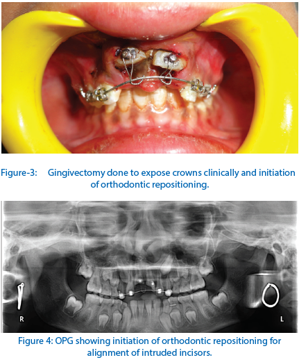
After taking informed consent of patient’s guardian active repositioning by orthodontic traction was planned for the patient. The gingivectomy was performed to expose the labial surface of crowns of both maxillary incisors. The lingual buttons were bonded on labial surface of both crowns ,brackets were bonded on two adjacent teeth on either side of the intruded incisors and orthodontic traction was applied using ligature wire.(Fig 3,Fig 4) The endodontic access was made after 2weeks and calcium hydroxide dressing was given.
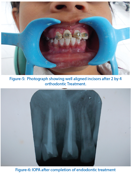
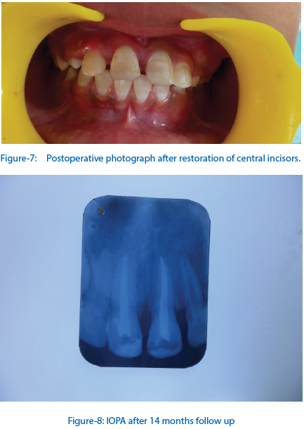
After the crowns of both incisors sufficiently erupted in about 2 months, alignment of incisors to the level of occlusion plane was done by two by four orthodontic treatment with bands on permanent first molars and brackets on all four maxillary incisors using sequential 012,014 and 016 NiTi wires.(Fig 5) Due to high chances of external root resorption in such cases calcium hydroxide dressing was changed every month. It took about 6 months for incisors to align to their normal position. The final obturation was done with gutta-percha (Fig 6) and composite buildup of both crowns was done.(Fig 7) The postoperative course was uneventful with clinical and radiographic success up to 6 months and 14 months of follow up.( Fig 8).
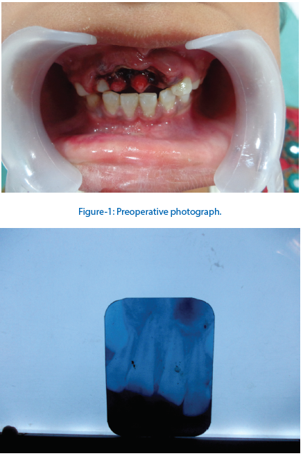
Figure-2: Preoperative IOPA showing intruded maxillary central incisors.
DISCUSSION
Luxative intrusion is a serious kind of injury of maxillary incisors and such an occurrence is found to be most frequent between 6 and 12 years of age and generally affecting 1.9% of traumatic injuries involving permanent teeth.
Factors determining treatment choice are stages of root development, age and intrusion level. According to IADT (International association of Dental Traumatology) 2012, guidelines are:

Spontaneous eruption is the treatment of choice suggested by Ellis3 in 1940 for permanent teeth with incomplete root formation with minor or moderate intrusion to prevent further disturbance to the apical and marginal periodontal tissues. But recently Faria et al4 have shown that even teeth with mature apices re-erupt spontaneously. In teeth with mature root development it is only recommended for teeth with minor intrusion. It usually takes 6 months for completion (range 2-14 months). This treatment seems to lead to fewer healing complications than orthodontic and surgical repositioning. If no movement is observed within one month , initiate orthodontic or surgical repositioning before ankylosis can develop. The disadvantages of this approach are two-fold: One, periodontal surgery- e.g, gingivectomy is needed to gain access to the root canal while waiting for spontaneous re-eruption .Second, root resorption or ankylosis may occur during the observation period.
Orthodontic repositioning enables repair of marginal bone in the socket along with the slow repositioning of the tooth.
Surgical repositioning recommended by Skieller5 in intrusion with major dislocation of the tooth (more than 7 mm) . Surgical repositioning involves a reduced number of visits and allows rapid access to the root canal for any root canal therapy but may increase the risk of loss of marginal bone support.
The choice between orthodontic and surgical repositioning remains an area of debate.
An animal in vitro study6 reported that surgical repositioning of severely intruded permanent teeth with complete root development resulted in more normal orientation of the periodontal fibres and consequently less replacement resorption as the fibres are under less tension with respect to the cementum and bone walls.
According to Andreasen and Andreasen7, in the majority of the cases, the treatment of choice for traumatically intruded permanent teeth with complete root formation should be the orthodontic repositioning rather than the surgical repositioning to prevent the additional trauma that may be caused to the periodontal structures during the surgical repositioning procedures and could increase the possibility of postoperative complications, such as external root resorption and loss of marginal bone support. Hence in our case orthodontic repositioning was chosen as a treatment alternative which might allow for remodelling of bone and the periodontal apparatus.
Andreasen8 advocates that the time interval between the occurrence of an intrusive injury and the surgical repositioning of the intruded tooth is a decisive factor accounting for the development of external root resorption. The author reported that teeth repositioned within 90 min after trauma showed less root resorption than those replanted lately. Kirinos and Sutcliffe9 observed that the retention rate of traumatically intruded teeth that were surgically repositioned more than 24 h after suffering an intrusive injury was significantly lower than that of teeth whose repositioning was not delayed.
With regards to endodontic treatment, Kristerson and Andreasen10,11 reported that surface root resorption could be stopped only if the tooth received endodontic treatment before pulpal necrosis. This treatment should be considered in all cases with completed root formation where the chance of pulp revascularization is unlikely. Endodontic therapy should preferably be initiated within 3-4 weeks post-trauma. A temporary filling with calcium hydroxide is recommended. Teeth with incomplete root development should be monitored closely with root canal treatment being indicated only following diagnosis of pulp necrosis. Where root canal treatment is required, an apical barrier should be achieved prior to obturation. Hence owing to the high incidence of pulp necrosis i.e., 100% in mature teeth we decided to go for prophylactic root canal therapy to prevent other complications arising from the pulp necrosis.
Intrusive luxation in permanent teeth has been associated with severe complications, especially pulp necrosis, external root resorption and marginal bone loss. Parents and patients can be informed of the range of clinical outcomes associated with intrusive luxation.
CONCLUSION
Intrusion of immature permanent anterior teeth presents a great dilemma due to variety of treatment options. Orthodontic repositioning and root canal treatment using calcium hydroxide were successful in repairing the potential damage caused by the impact of an intrusive injury and successful prevention of any post-operative complication commonly associated with these injuries.
REFERENCES
- Andreasen JO, Bakland LK, Matras R, Andreasen FM. Traumatic intrusion of permanent teeth. Part 1. An epidemiological study of 216 intruded teeth. Dental Traumatology 2006; 22: 83 – 89.
- Andreasen FM, Pederson VB. Prognosis of luxated permanent teeth – the development of pulp necrosis. Endod Dent Traumat 1985;1:207–20.
- Chan AW, Cheung GS, Ho MW. Different treatment outcomes of two intruded permanent incisors: A case report. Dent Traumatol 2001;17:275-80
- Faria G, Silva RA, Fiori JM, Nelson FP. Re-eruption of traumatically intruded mature permanent incisor: Case report. Dent Traumatol 2004;20:229-32
- Skieller V.The prognosis for young teeth loosened after mechanical injuries. Acta Odontol Scan 18: 171-81, 1960.
- Cunha RF, Pavarini A, Percinoto C, Lima JE. Influence of surgical repositioning of mature permanent dog teeth following experimental intrusion: a histological assessment. Dental Traumatology 2002; 18: 304 – 308.
- Andreasen JO, Andreasen FM. Essentials of traumatic injuries to the teeth: a step-by-step treatment guide.Copenhagem: Munksgaard; 2000.
- Andreasen JO. Traumatic injuries of the teeth. Copenhagen:Munksgaard; 1972.
- Kirinos MJ, Sutcliffe J. Traumatically intruded permanent incisors: a study and outcome. Brit Dent J 1991;170:144–6.
- Andreasen JO, Borum MK, Jacobsen HL, Andreasen FM. Replantation of 400 avulsed permanent incisors. 4. Factors related to periodontal ligament healing. Endod Dent Traumatol 11: 76-89, 1995.
- Kristerson L.Autotransplantation of human premolars - a clinical and radiographic study of 100 teeth. Int J Oral Surg 14:200-213, 1985.
Disclaimer from Guident
The views and opinions expressed in articles published in Guident are solely those of the authors and do not necessarily reflect the views of the editors, the editorial board or the publisher, Ivory India. Guident does not endorse any product, technique or treatment described. Clinical content is intended for qualified dental and medical professionals and is not a substitute for individual clinical judgement. Guident, its editors and the publisher accept no liability for any consequence arising from the use of information published in the journal.
How to cite this article
Dr. Mitali Jain, Dr. Ritu Namdev, Dr. Samir Dutta. Management of Traumatically Intruded Incisors by Orthodontic Repositioning. Guident. 2014 Dec;8(1). Available from: https://guident.in/articles/endodontics/management-of-traumatically-intruded-incisors-by-orthodontic-repositioning.html