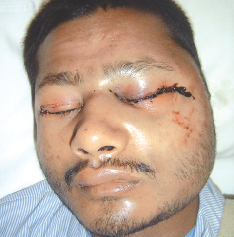
Bilateral Sagittal Split Osteotomy
Department of Oral & Maxillofacial Surgery, Department Of Orthodontia, Department of Oral & Maxillofacial Surgerym, A.B. Shetty Memorial Institute of Dental Sciences, Mangalore (Kar.)
ABSTRACT
Background: A bilateral sagittal split osteotomy for mandibular prognathism is a very popular and most versatile procedure performed on the mandibular body and ramus. The osteotomy splits the ramus and the posterior body of the mandible sagitally which allows either setback or abvancement.
Method: This case report describes management of a skeletal Class III malocclusion with increased reverse overjet. Bilateral sagittal split osteotomy and mandibular setback was performed.
Conclusion: The clinical outcome in the present case indicates that the management of skeletal class III malocclusion with a bilateral sagittal split osteotomy with mandibular setback gives promising results
KEYWORDS : Bilateral sagittal split osteotomy, Skeletal class III, mandibular setback
INTRODUCTION
Currently , bilateral sagittal split osteotomy (BSSO) is the most commonly performed orthognathic surgical procedure which has gained popularity because of its versatility and high cosmetically corrective abilities. It is performed intraorally resulting in no extraoral scar and broad bony contact of the osteotomised segments ensuring good healing and subsequent stability.
In 1957, Trauner and Obwegeser first described the sagittal split osteotomy of the ramus region, only by placing buccal and lingual cortical cuts. The procedure underwent numerous modifications over the years after that. In 1961, Dal Pont changed the lower horizontal cut to a vertical cut on the buccal cortex between the first and second molars. This gave a broader contact surface requiring minimal muscular displacement with improved access. In 1968 Hunsuck further modified the technique, advocating a shorter , horizontal medial cut, just posterior to the lingual to minimize soft tissue dissection. In 1977 Epker suggested various modifications which included minimal stripping of the masseter muscle and limited medial dissection. These modifications were found to minimize post operative oedema, hemorrhage & swelling.
CASE REPORT
This is a case of a 24-year-old student who had been referred to A.B.Shetty Memorial Institute of Dental Sciences for combined orthodontic and surgical treatment. The patient’s chief concern was that her teeth didn’t fit well together properly and had a forwardly placed lower jaw since childhood. On clinical examination she was found to have an increased facial height with a protruded mandible. Intraoral examination revealed anterior crossbite with a reverse overjet of about 2mm. She had missing lower first molars bilaterally as she had undergone extractions due to previous infections in those regions. Acute nasolabial angle was also found in this paitient. Radiographs were taken that confirmed presence of a prognathic mandible. The treatment plan was decided after a cephalometric evaluation.
CEPHALOMETRIC FINDINGS
- SNA= 82
- SNB= 86
- Wits = 13mm Bo ahead
- Mandibular plane angle=37
- Y axis =67%
- Upper incisor to NA = 25
- Lower incisor to NB = 18
- Lower incisor to MP = 70
- Nasolabial angle= 109
- Upperincisor to s line =1
- Lower incisor to s line = -2
- Upper incisor to E line -= 5
- Lower incisor to E line= 0

The incision was placed at the anterior aspect of the ramus of the mandible at a midpoint between upper and lower molars running down the lateral crest of the external oblique ridge and the ends of facial vestibule to the first molar region.

Sharp dissection of the mucosa and buccinator muscle over the ramus was made. Subperiosteal reflection was done and it continued posteriorly over the anterior aspect of the ramus. Dissection is then done subperioateally above the mandibular foramen to prevent damage to the neurovascular bundle.
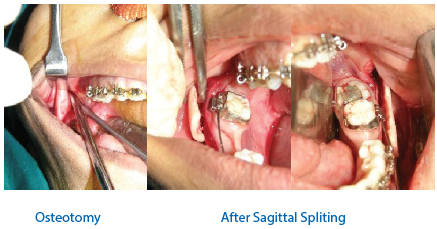
Initial osteotomy cuts were placed uniformly on both the sides of the mandibular angle area. It is started from the medial side of the ramus extending downwards into the anterior border of the ramus onto the superior aspect of the body of the mandible and curved inferiorly through the lateral cortical plate including the inferior border of the ramus.
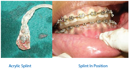
After the osteotomy and completion of sagittal splitting, a prefabricated interocclusal acrylic wafer splint is placed and the patient is put on maxillomandibular fixation to avoid any shift of occlusion after plates and screws are placed. Fixation was done using miniplastes and screws.


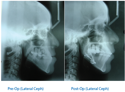
POST SURGICAL CEPHALOMETRICS
- N-B Pre-op = +5.5mm, Post-op = 5mm
- N-Pg Pre-op = +4mm, Post-op = 4mm
- Go-Pog Pre-op = 83, Post-op = 74
- Reduction in lower incisor proclination-Pre-op = 88°, Post-op = 85°
POST SURGICAL MANAGEMENT
- Class III elastics for maintaining the corrected occlusion
- Settling elastics were given
- Mouth opening was evaluated for a period of 4-6 weeks
- At the end of 4-6 weeks patient had a mouth opening of 40mm.
DISCUSSION
Mandibular prognathism is a condition wherein the mandibular arch is farther protruded with respect to the maxillary arch. The term mandibular excess has its historical roots in the 10th century , when members of the Austrian Royal Habsburg family showed mandibular prognathism prevalent enough to become known as Habsburg jaw.
In 75-80% of patients the condition of mandibular excess represents a combination of maxillary deficiency and mandibular protrusion. Only 20-25% of patients are found to have isolated mandibular prognathism Mandibular setback with BSSO is quite a preferred procedure performed with an objective of correcting the mandibular prognathism for shortening the mandibular body.
Indications for a BSSO is both mandibular advancement and mandibular setback.
Advantages and characteristics of BSSO
- Great degree of 3 dimensional flexibility in repositioning the distal fragments
- Broad bony overlap of the osteotomised segements
- Minimal alteration of the natural position of the muscles of mastication and Temperomandibular joint
A prefabricated joint positioning splint can intraoperatively optimize the condyle position in an orthognathic surgery. Use of such splints have shown significantly reduced postoperative dysfunction, disc dislocations, changes to the condylar translation, and incidence of skeletal relapse at 24-month follow-up.
COMPLICATIONS
Any type of surgical treatment conveys some risk of complication. More or less severe type of complications are reported following BSSO. These may occur either at the time of surgery or later. Most complications like hemorrhage and unanticipated bone splits can be corrected sufficiently during the surgical procedure itself. The most common post operative problem is neurosensory disturbance of the inferior alveolar nerve followed by TMJ problems and relapse.
Although inferior alveolar nerve (IAN) injury has been reported as a result of several mandibular operations, its association with the bilateral sagittal split osteotomy (BSSO) is well documented. It is well accepted that the lingual nerve is occasionally injured during BSSO. Unfortunately, the incidence of lingual nerve injury is poorly documented.
Relapse may be defined as a postoperative movement either toward the preoperative position or farther away from it. Relapse is usually three dimensional, with vertical, horizontal, and sagittal components that may occur concurrently. Relapse of mandibular ramus osteotomies may be caused by mandibular condyle positioning intraoperatively, condylar
remodeling or resorption, surface remodeling, or osteotomy slippage. Relapse rates have been reported from 6 months up to several years. 12 months seems to be a minimal period over which to evaluate this complication, although additional relapse may occur for many years thereafter.
Some instances of rare complications reported with bilateral sagittal split osteotomy are, Intraoperative hemorrhage that required special intervention. Aseptic necrosis and facial nerve palsy are also found to occur rarely as complications following BSSO.
Foltan et al assessed the impact of orthognathic surgery for class III malocclusion on ventilation during sleep using a comparison of pre- and post-surgical respiratory parameters. Generally, the mandibular setback procedure worsened breathing function during sleep, as reflected in significant increase of index of flow limitations and decrease in oxygen saturation. The posterior airways space decreased to 75% of its original volume. A young person would probably be able to balance such a decline in respiratory function using different adaptive mechanisms, but the potential impact of orthognathic surgery on the upper airways should be incorporated in a treatment plan.
REFERENCES
- Takahashi H. et al . Assessment of three bilateral sagittal split osteotomy techniques with respect to mandibular biomechanical stability by experimental study and finite element analysis simulation. Med. Bull. Fukuoka Univ. 36(3) , 181-192, 2009.
- Robert A. Bays. Et al. Complications of orthognathic surgery. Oral Maxillofacial Surg Clin N Am 15 (2003) 229–242
- Timothy A Turvey. Et al. Oral and Maxillofacial Surgery. Vol- III , 2nd Edition
- Constantin A. Landes et al. Proximal Segment Positioning in Bilateral Sagittal Split Osteotomy: Intraoperative Controlled Positioning by a Positioning Splint. J Oral Maxillofac Surg 61:1423-1431, 2003.
- Foltán R. et al. The impact of Le Fort I advancement and bilateral sagittal split osteotomy setback on ventilation during sleep. Int. Journal of Oral and Maxillofacial Surg 38 : 1036-1040, 2009
- Landes et al. Proximal segment positioning in bilateral sagittal split osteotomy: intraoperative dynamic positioning and monitoring by sonography. J Oral Maxillofac Surg 62:22-28, 2004
- Nishioka et al. Neurosensory disturbance with rigid fixation of the bilateral sagittal split osteotomy. J Oral Maxillofac Surg 45:20-26, 1987
Disclaimer from Guident
The views and opinions expressed in articles published in Guident are solely those of the authors and do not necessarily reflect the views of the editors, the editorial board or the publisher, Ivory India. Guident does not endorse any product, technique or treatment described. Clinical content is intended for qualified dental and medical professionals and is not a substitute for individual clinical judgement. Guident, its editors and the publisher accept no liability for any consequence arising from the use of information published in the journal.
How to cite this article
Dr. Muralee Mohan C., Dr. Prajwal Shetty, Dr. Shrey Pandya. Bilateral Sagittal Split Osteotomy. Guident. 2014 Dec;8(1). Available from: https://guident.in/articles/oral-surgery/bilateral-sagittal-split-osteotomy.html