
Hemicoronal Approach - a Versatile Approach for Complex Zygomatic Fractures
Delhi
INTRODUCTION
Forty-five percent of trauma to the midface constitutes fractures to the zygomatic complex (ZMC).1 Various approaches and surgical incisions have been described to treat fractures of the zygomatic complex. Traditional closed techniques (intra oral and gillies temporal approach) and total exposure of all fracture lines by conventional multiple incisions2 have been routinely used. All these approaches or a single routine hemicoronal incision are always in a constant state of contention. The ideal and the best surgical approach to treat ZMC fractures should provide maximum necessary exposure of the fracture segments, minimize potential for injury to facial structures, and enable good cosmetic results.3
In 1969, the coronal scalp approach was first described by Tessier, who used then-novel technology to surgically widen the orbit.4 Hemicoronal approach is a variation of the coronal approach where the incision line stops mid way on the scalp and providing access to the supra orbital rim, lateral orbital rim, body of zygoma and the zygomatic arch.
INDICATION FOR REDUCTION OF ZYGOMATIC FRACTURES
Malunited ZMC fractures cause inadequate mouth opening because of the encumbrance of the zygomatic arch to the coronoid process of the mandible, which brings about functional impairment, thereby justifying surgical management. Facial esthetics also features as one of the absolute indications for surgical management of these fractures. When they are involved with the concomitant existence of the zygomatic arch, the situation becomes more challenging and adequate planning needs to be exercised to achieve superior results.
The absolute indications for hemicoronal incisions for treating zygomatic complex fractures are5:
a. Multiple fractures of zygomatic complex or other midfacial bones;
b. Comminuted fractures of zygomatic complex;
c. Old fractures of midfacial bones with mal- or non- union.
ADVANTAGES OF HEMICORONAL APPROACH
1. Complete and uninterrupted visualization of the whole zygomatic complex including the frontozygomatic and zygomaticotemporal sutures.
2. Plating and grafting can be accomplished without any limitations relating to exposure.
3. The Hemicoronal approach can also provide an opportunity to harvest cranial bone through the same incision when immediate bone grafting is indicated, and eliminates the need for a second donor site.
4. This technique provides for reconstruction of all fractures or missing bony fragments under direct vision, which is a great advantage over the conventional techniques of reduction in zygomatic fractures. The complications were divided into two categories6, early and late.
EARLY COMPLICATIONS
a. Haemorrhage and haematoma,
b. Infection,
c. Swelling,
d. Temporary nerve injury
LATE COMPLICATIONS
a. Alopecia,
b. Obvious scarring in bald patients,
c. Permanent paralysis of the facial nerve,
d. Depressions of the temporal fossa.
e. Parietal scalp pain
f. Numbness and paresthesia
g. Temporal fossa depression
h. Nasal orbital hypertrophy.7,8
CASE REPORT
A 25 years old patient presented in the emergency with a history ofroad traffic accident hit by a truck while riding a bike, on examination the patient had a history of LOC for 20 min with bleeding from the mouth and nose, swelling on the left side of the face, circumorbital edema and ecchymosis with bleeding from the lateral canthus of the left eye and edema on the right eye was also noted (figure 1), mouth opening was restricted though occlusion was normal. Eye movements could not be assessed during the initial assessment dueto severe edema
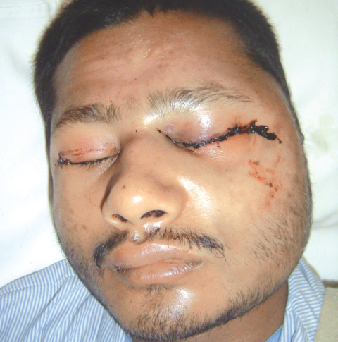
Fig. 1- Pre Operative
On palpation bony crepitus and tenderness was felt left infraorbital rim and the fronto zygomatic suture. A non contrast 3 dimensional CT scan of the face and mandible suggested fracture of left zygomatic complex involving the arch, infra orbital rim, fronto zygomatic suture and the maxilla including the lateral wall of maxillary sinus (figure 2,3,4).
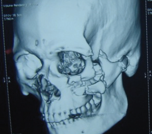
Fig. 2- 3 D CT Scan
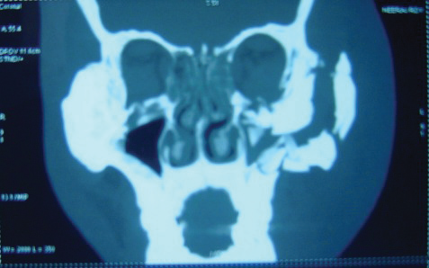
Fig. 3- Coronal View CT Scan

Fig. 4- Axial View CT Scan
Following neurological clearance the patient was posted for surgery after 5 days for open reduction and internal fixation of the zygomatic complex fracture
SURGICAL PROCEDURES
A 2cm strip was shaved in the immediate area of the incision for surgical convenience. Temporary tarsorrhaphy done in the left eye. The hemicoronal scalp incision was initiated at the tragus, extended preauricularly in front of the lobe and transversely upto the midline of the vault of the skull. The incision was parallel to the direction of the hair follicle. The line of incision is infiltrated with 10–15 ml of 2% lidocaine containing 1:2,000,000 adrenalin and the flap containing skin, subcutaneous tissue and galea was raised leaving the periosteum intact. Skin incision below the superficial temporal line was to the depth of glistening superficial layer of temporal fascia (figure 5), to insure a correct plane blunt scissor dissection is done from above towards the zygomtic arch upto the preauricular lobule (figure 6). The lateral portion of the flap is dissected inferiorly atop the outer surface of the temporalis fascia above the zygomatic arch. At that point, an incision running anterosuperiorly at 45 degree was made through the superfacial layer at the root of the zygomatic arch just in front of the ear. Once a plane of dissection was established deep to the superficial layer of the temporal fascia, the dissection was continued inferiorly until the periosteum of the zygomatic arch was reached. The periosteum was then incised and reflected laterally over the arch, the body of zygoma, and the lateral orbital rim. Fracture segments were visualized (figure 7) reduced and fixed using Titanium osteosynthesis (figure 8). Other incisions, such as subciliary and intraoral vestibular incisions were added for reduction and fixation of the infraorbital rim and maxilla respectively. Vacuum drain was applied with pressure dressing for 72hours.
Post operative PNS Waters view X ray (figure 9) showing rigid osteosynthesis. Three month follow up (figure 10, 11) with well hidden scar.
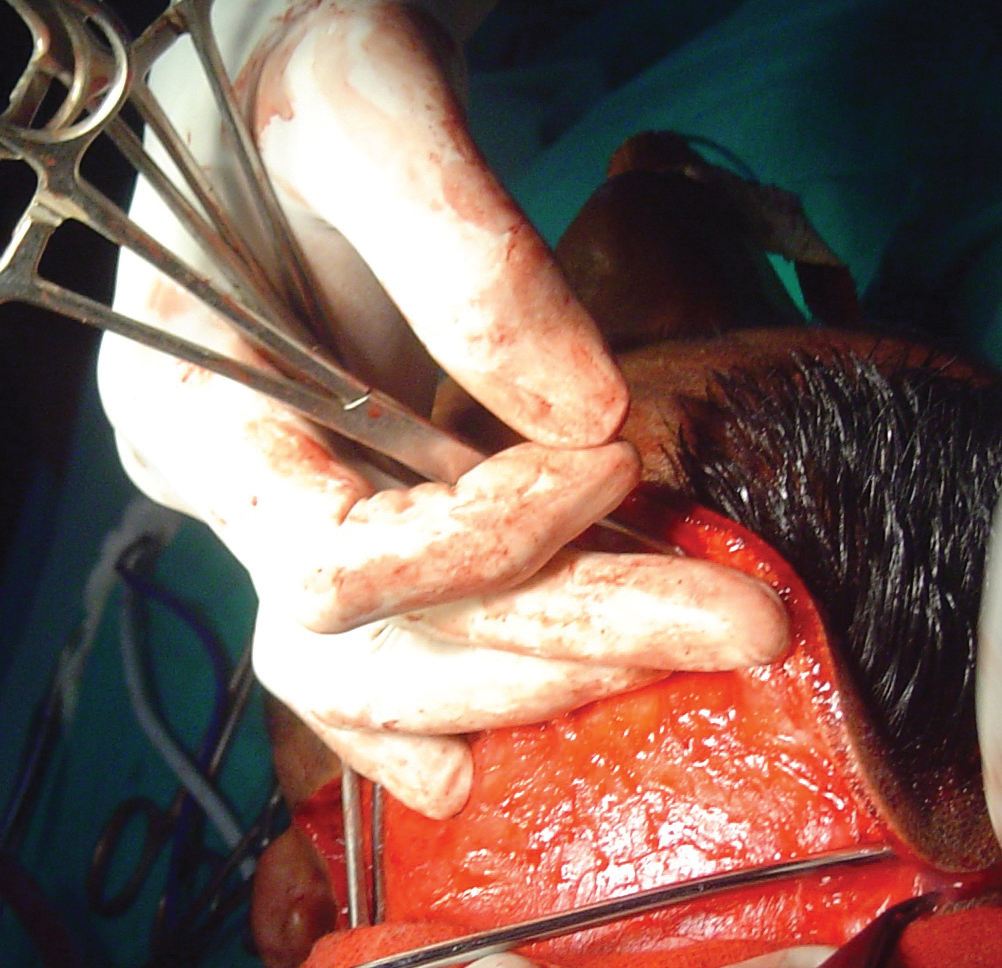
Fig. 5- Superficial layer of temporal fascia

Fig. 6- Scissor dissection flap above the temporal fascia
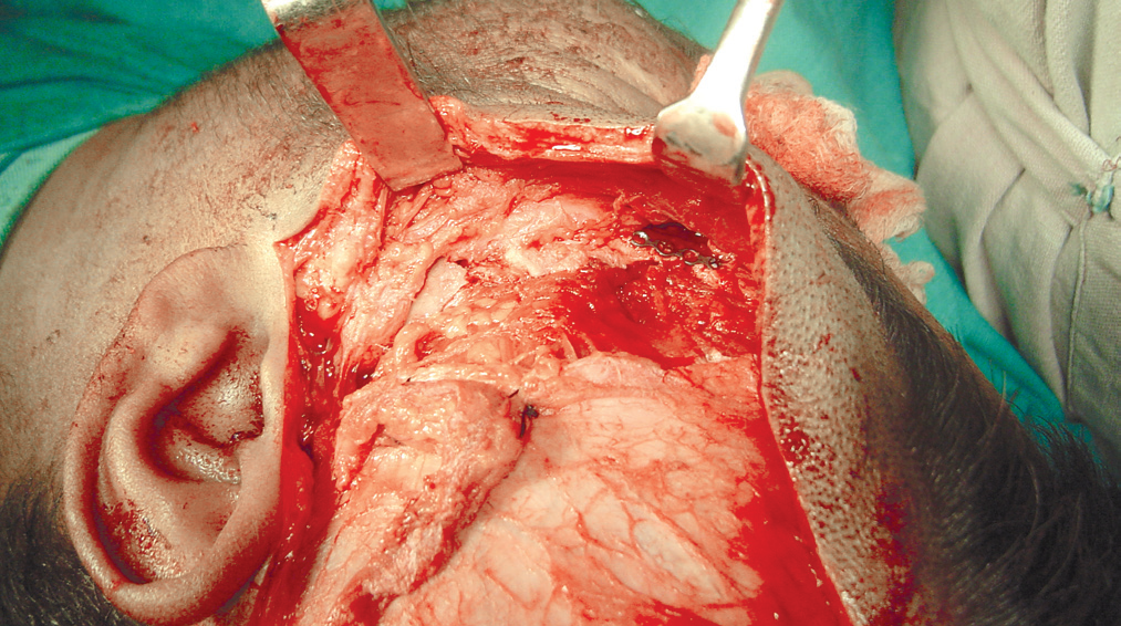
Fig. 7- Fractured Arch
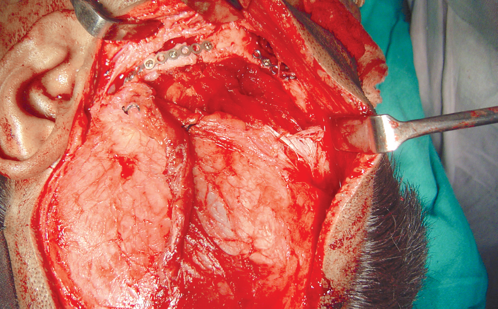
Fig. 8- Plate Osteosynthesis
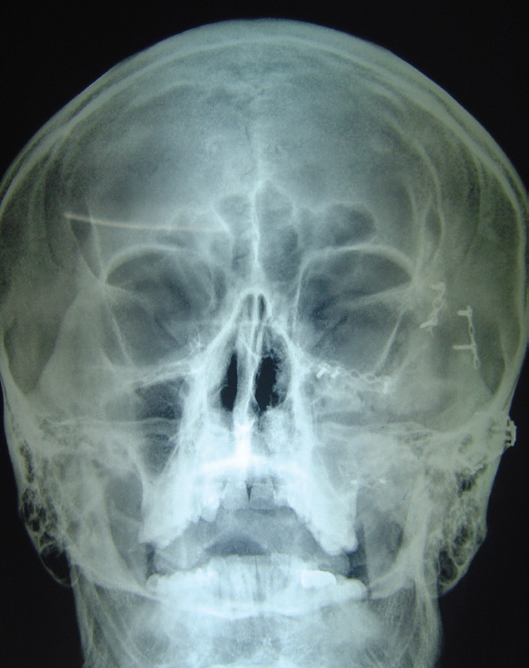
Fig. 9- Post Op PNS view

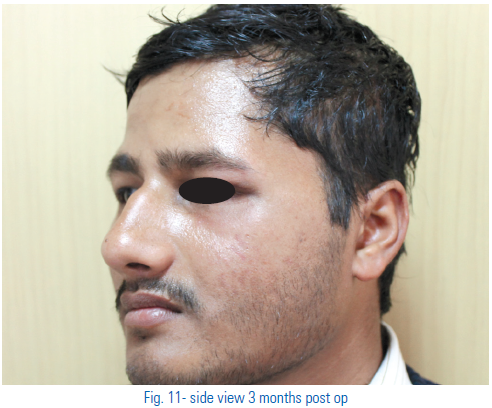
DISCUSSION
There are still unsolved questions regarding the approaches for reducing zygomatic complex fractures. The surgical approach could influence the ease of reduction and fixation of the fragments, the length of operating time, and the length of hospital stay. The approaches for zygomatic complex fractures are generally divided into two categories: local, and coronal scalp incisions.
Hemicoronal approach initially may appear as a radical approach to the treatment of zygomatic complex fractures, it provides excellent access to the orbits, zygomatic bodies and zygomatic arches with virtually no complications if done the right way.8
It is an extremely useful incision where there is combination of the supraorbital and lateral orbital rims, and zygomatic body and arch. The scar produced is hidden within the hairline and is therefore invisible. This approach can be combined with other approaches like subsciliary to access the infraorbital rim and the maxillary vestibule for the maxilla.
REFERENCE
- Kovacs AF, Ghahremani M. Minimisation of zygomatic complex fracture treatment. Int J Oral Maxillofac Surg 2001;30:380-3. 2. Manson PN, Crawley WA, Yaremchuk GM, Rockamnn J,
- Hoopes E, French JH. Midface fractures: advantages of imme- diate extended open reduction and bone plating. Plast Reconstr Surg 1985;76:1-10.
- Manson PN, Crawley WA, Yaremchuk GM, Rockamnn J, Hoopes E, French JH: Midface fractures: advantages of immediate extended open reduction and bone plating. Plast Reconstr Surg 76: 1–10, 1985
- essier P. Surgical widening of the orbit. Ann Chir Plast 1969; 14:207- 14.
- Qing-Bin ZHANG ;Coronal incision for treating zygomatic complex fractures; Journal of Cranio-Maxillofacial Surgery (2006) 34, 182–185.
- Abubaker AD, Sotereanos G, Patterson GT: Use of the coronal surgical incision for reconstruction of severe craniofacial injuries. J OralMaxillofac Surg 48: 579–582, 1990.
- Baek RM, Heo CY, Lee SW. Temporal dissection technique thatprevents temporal hollowing in coronal approach. J Craniofac Surg2009;20:748-51.
- Matic DB, Kim S. Temporal hollowing following coronal incision:a prospective, randomized, controlled trial. Plast Reconstr Surg 2008;121:379e-85e.
- Shepherd DE, Moos KF: Morbidity of bcoronal flaps in maxillofacial surgery. Br J Oral Maxilofac Surg 23:1 1953
Disclaimer from Guident
The views and opinions expressed in articles published in Guident are solely those of the authors and do not necessarily reflect the views of the editors, the editorial board or the publisher, Ivory India. Guident does not endorse any product, technique or treatment described. Clinical content is intended for qualified dental and medical professionals and is not a substitute for individual clinical judgement. Guident, its editors and the publisher accept no liability for any consequence arising from the use of information published in the journal.
How to cite this article
Dr. Gagan Sabharwal. Hemicoronal Approach - a Versatile Approach for Complex Zygomatic Fractures. Guident. 2014 Apr;7(5). Available from: https://guident.in/articles/oral-surgery/hemicoronal-approach-a-versatile-approach-for-complex-zygomatic-fractures.html


