The Anatomy Dilemma: Cracking the Code of Complex Root Canals- Two Cases Report
Department of Conservative Dentistry and Endodontics, Seema Dental College And Hospital, Rishikesh
ABSTRACT
Endodontic therapy aims to treat infected or damaged pulp tissue, preserving the tooth's function. A common reason for treatment failure is missed canals due to unrecognized anatomical variations. This case series presents two unique cases: one involving a mandibular canine with two roots and a lateral incisor with a split canal, and another with an S-shaped curvature in a maxillary premolar. Successful management requires a thorough understanding of root canal morphology and careful radiographic evaluation. Early detection and appropriate treatment strategies, including the use of CBCT and precise instrumentation, are essential for reducing complications and improving treatment outcomes.
INTRODUCTION
Endodontic therapy focuses on the root canal system to treat infected or damaged pulp tissue within the tooth, ultimately preserving the tooth and restoring its function. The root canal space should be cleaned with the use of proper instruments and chemical disinfectants to eradicate infected pulpal tissue and its remnants.1
One of the most common reasons for failure of endodontic treatment is a missed canal due to lack of knowledge on anatomical variations.2
The Present Case series demonstrates two cases; one with two roots in mandibular canine and split canal lateral incisor; one with a S-shaped curvature in maxillary premolar.
CASE REPORT 1
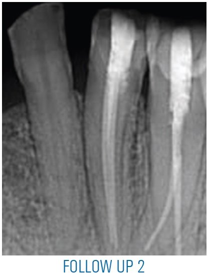
A 34 year old patient reported to the department with a chief complaint of pain in lower left back tooth region since 2-3 months. Pain was dull intermittent in nature and aggravated on chewing food. Past dental history revealed initiation of root canal therapy irt 32,33.Clinical examination revealed temporary restoration irt 32,33 Radiographic examination revealed unusual anatomy irt 33 with two roots and two canals, also split canal was noted irt 32.
A provisional diagnosis of previously initiated endodontic therapy irt 32,33 was made and treatment plan was non surgical root canal treatment.
Straight line access was established followed by determination of working length using apex locator and confirmed radiographically. Cleaning and shaping was done using 5.25% NaOCl and normal saline followed by selection of master cone corresponding to master apical file size 30/.04 and obturation was done using warm vertical compaction technique followed by post endodontic composite restoration.


CASE REPORT 2
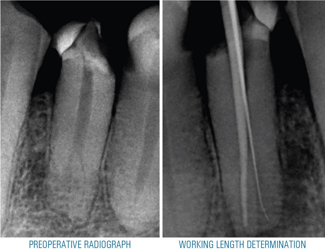
A 40 year old patient reported to the department with a chief complaint of pain in lower right back tooth region since 1 week. Pain was dull intermittent in nature and aggravated on chewing food. .Clinical examination revealed restoration irt 44. Radiographic examination revealed apical split irt 44.
Straight line access was established followed by determination of working length using apex locator and confirmed radiographically. Cleaning and shaping was done using 5.25% NaOCl and normal saline followed by selection of master cone corresponding to master apical file size 30/.04 and obturation was done using warm vertical compaction technique followed by post endodontic composite restoration.

DISCUSSION
The mandibular canine is a strategically important tooth in the dental arch. The root being long and stable, it is useful for prosthetic support and because of its proprioceptive properties, regulate or guide masticatory function, combined with its role in occlusal guidance during the eccentric movements and posterior disocclusion.3
Therefore, considerable effort should be directed to its preservation, inspite of occasional diverse morphologic challenges.
Early detection of a mandibular canine with anatomical aberration favourably influences the success rate of endodontic treatment. Therefore, it is crucial to carefully inspect the diagnostic radiographs taken in different horizontal angulations or if intraoral periapical radiographs won’t suffice, a CBCT may be advised to accurately determine the number of roots, curvatures, and bifurcations in both sagittal and axial planes.4,5
In clinical conditions, two curves can be present in the same root canal trajectory. This type of geometry is denoted as the “S” shape, and it is a challenging condition.6
There is a consensus in the literature that instrumentation in curved canals considering a great degree of curvature predisposes higher risks of accidents.7/p>
The S-curve causes increased strain on nickel-titanium instruments during cleaning and shaping.8
A study conducted by Al-Sudani, et al. (2012), found that instrument fatigue occurs very quickly once the file encounters a double curvature.9
Varela-Patiño, et al. (2005), found that fewer fractures occurred when a wide and smooth-walled glide path was created, and the canal was pre-flared before the introduction of rotary files into the root canal.10
CONCLUSION
A thorough knowledge of the tooth and root canal morphology, clinical exploration and detailed radiographic interpretation may be helpful in detecting root canal aberrations and to achieve success in their management.
REFERENCES
- Manik K, Ikhar A, Patel A, Chandak M, Bhopatkar J, Rathod P, Bhojwani PR. Anatomical Variations in Mandibular Molars: Focus on Midmesial Canals. Cureus. 2024 Jun 5;16(6):e61711. doi: 10.7759/cureus.61711. PMID: 38975529; PMCID: PMC11226225.
- S Tabassum F R Khan Failure of endodontic treatment: the usual suspectsEur J Dent20161011445110.4103/1305-7456.175682
- J Abduo M Tennant J Mcgeachie Lateral occlusion schemes in natural and minimally restored permanent dentition: a systematic reviewJ Oral Rehabil2013401078880210.1111/joor.12095
- S C White M J Pharoah Intraoral radiographic examinations. Oral radiology principles and interpretation7th200812165
- A Soleymani N Namaryan E Moudi A Gholinia Root canal morphology of mandibular canine in an Iranian population: a CBCT assessmentIran Endod J2017121788210.22037/iej.2017.16
- Machado R, Chaniottis A, Vera J, Saucedo C, Vansan LP, Silva EJ. S-Shaped Canals: A Series of Cases Performed by Four Specialists around the World. Case Rep Dent. 2014;2014:359438. doi: 10.1155/2014/359438.
- Setzer FC, Kwon T, Karabucak B. Comparison of apical transportation between two rotary file systems and two hybrid rotary instrumentation sequences. Journal of Endodontics. 2010;36(7):1226–1229.
- Pruett JP, Clement DJ, Carnes DL Jr. Cyclic fatigue testing of nickel-titanium endodontic systems. J Endod. 1997;23(2):77-85.
- Al-Sudani D, Grande NM, Plotino G, et al. Cyclic fatigue of nickel-titanium rotary instruments in a double (S-shaped) simulated curvature. J Endod. 2012;38(7):987-989
- Patiño PV, Biedma BM, Liébana CR, Cantatore G, Bahillo JG. The influence of a manual glide path on the separation rate of Ni-Ti rotary instruments. J Endod. 2005;31(2):114-116.
Disclaimer from Guident
The views and opinions expressed in articles published in Guident are solely those of the authors and do not necessarily reflect the views of the editors, the editorial board or the publisher, Ivory India. Guident does not endorse any product, technique or treatment described. Clinical content is intended for qualified dental and medical professionals and is not a substitute for individual clinical judgement. Guident, its editors and the publisher accept no liability for any consequence arising from the use of information published in the journal.
How to cite this article
Dr. Siddhi Goyal, Dr. Ayushi Rawat, Dr. Anil Dhingra. The Anatomy Dilemma: Cracking the Code of Complex Root Canals- Two Cases Report. Guident. 2024 Nov;17(12). Available from: https://guident.in/articles/endodontics/the-anatomy-dilemma-cracking-the-code-of-complex-root-canals-two-cases-report.html