Immediate Implant Placement Using Super-Line Dentium Implant in Freshly Extracted Canine Region-a Case Report
Department-Conservative Dentistry and Endodontics, Department of Prosthodontics and Crown & Bridge, K.D. Dental College & Hospital, Mathura, UP, India.
ABSTRACT
Aiming to reduce the process of alveolar bone resorption and treatment time, the immediate endo-osseous implant placement into extraction sockets is known to achieve a high success rate between 94-100%, compared to the delayed placement.
INTRODUCTION
The success of implant placement immediately onto the extraction site is completely dependent over the phenomenon of osseointegration with the alveolar bone into the surrounding tissue.1
In present world immediate implant placement becomes an accepted as well as profitable manner, simply all the fundamentals of implant placement are followed, differently it may lead to damage. Literature suggested that primary dental implant was fitted in the period of pre colunbian.2,3
In the year of 1952, Branemark introduced threaded endosteal implant with cylindrical design and get acceptance to the system by ADA in the year 1986.4
The literature stated that placement of implant immediately in the extraction site shows positive and significant results.5
Indications
- Fracture of the root
- Resorbed root
- Peri-apical pathology
- Endodontically failed tooth
- Root perforation
- Psychological advantage to the patient
- Reduced treatment time interval
Contraindications
- Alveolar wall resorption
- Periodontal disease
- Acute peri-apical infection
- If anatomy of the alveolar bone not favorable
- Any risk of infection
- Bisphosphonates therapy.6,7
Literature revealed that the immediate implantation is successful in limited or otherwise ideal extraction cases in terms of aesthetics or in otherwise compromised cases the aesthetic results of immediate implant placement is unpredictable.8,9
Aim of this case report is to place the immediate implant after the extraction of the maxillary left canine with cement-retained prosthesis.
CASE REPORT
A 55-year-old male patient visited to the Department of the Prosthodontic Crown and Bridge with the chief complaint of replacement of decayed tooth in maxillary left anterior region of mouth due to unpleasant aesthetics. Clinical and radiographic evaluation revealed adequate alveolar bone, absence of periapical pathology. Preoperative OPG was advised (fig 1) which revealed significant bone height and width in canine region. So, it was decided to extract and place endo-osseous implant immediately. A super line dentium implant system was used. An implant size of 4.0x10.0mm was selected before the surgical procedure the patient was informed about the procedure and written consent was taken.
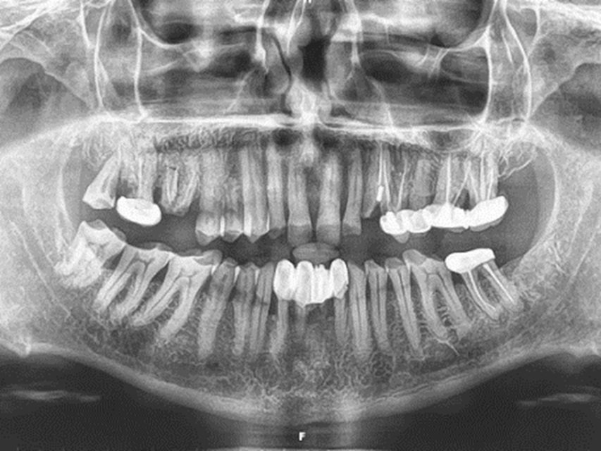
Figure 1-OPG
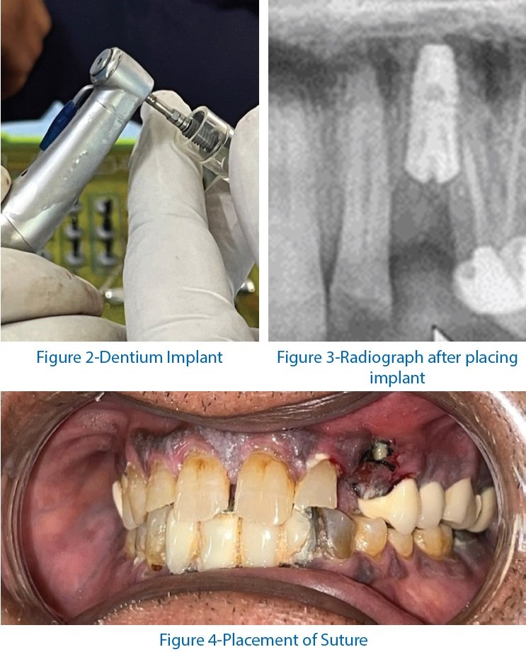
After administering appropriate antibiotic and analgesic, induction of local anesthesia was carried out using lignocaine with adrenaline. As preservation of alveolar bone is the key to success of immediate implants, extraction of tooth must be atraumatic, so using periotomes and small periosteal elevators, the fragment was luxated without excessive enlargement of the socket.
Primary stability was achieved by anchoring into the bone beyond the apex of the socket, BIO-OSS bone graft was packed between the implant and bone and radiograph was taken (Fig.2,3) The cover screw was placed and interrupted sutures were placed. (Fig-4)
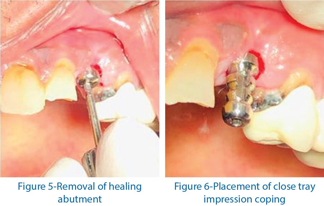
Post operative instructions were given to the patient. After 15 days sutures were removed. The patient was recalled after 4 months for the prosthetic procedures. Implant site was open using round bur after induction of local infiltration. The cover screw was extracted along with the soft tissue covering it. Later on healing abutment was placed at the site. After 15 min the tray was selected for close tray impression. Healing abutment was removed (Fig-5) and close tray impression coping was placed and tightened with a hex driver. (Fig-6) Impression was taken by using light body and PVS putty impression material and loosen the coping.
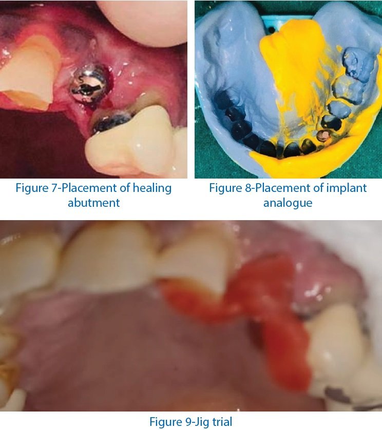
After removal of the impression again healing abutment was placed over the implant in the patient's mouth. (Fig-7) The implant analogue was attached to the coping onto the impression. (Fig-8) Gingival mask was made and the impression was poured. After the removal of the cast, place the coping and make a jig over the cast and the trial was done in the patient's mouth. (Fig-9) The patient was recalled after 15 days.
Prosthetic procedure
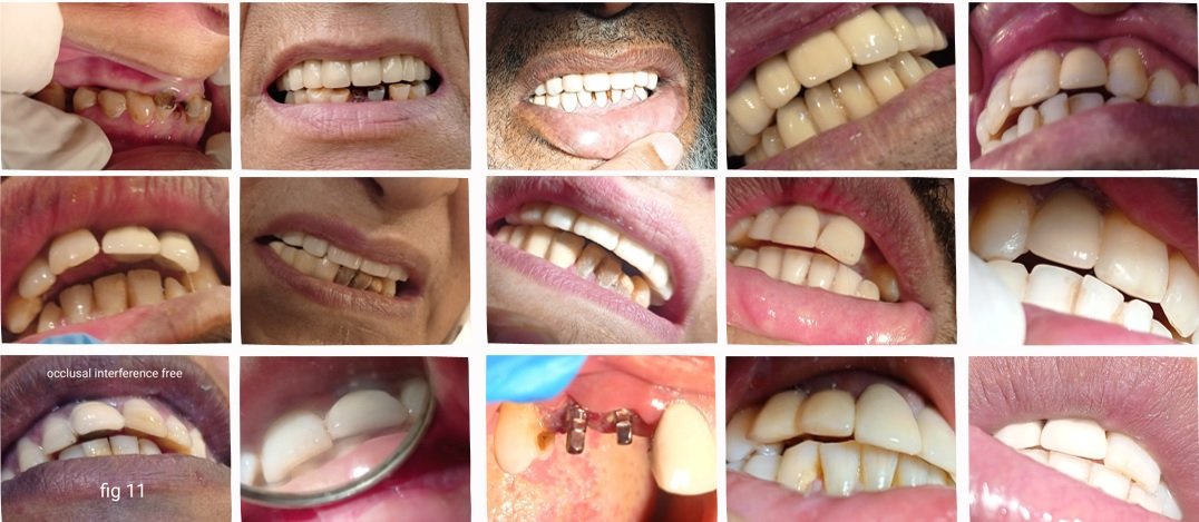
After 15 days a cement retained porcelain fused to metal crown was fabricated according to the selected shade of the adjacent teeth. The final restoration was checked. (Fig-10) The patient was satisfied with the results. The clinical and radiographic appearances at 6 months show good esthetic, osseointegration and maintenance of bone around the implant. (Fig.11)
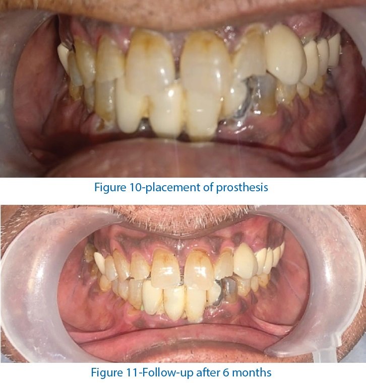
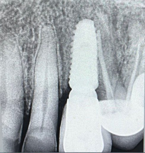
Figure 12
DISCUSSION
Implant therapy must fulfil both functional and esthetic requirements to be considered a primary treatment modality. Aiming to reduce the process of alveolar bone resorption and treatment time, the immediate placement of endo-osseous implants into extraction sockets is known to achieve a high success rate of between 94 and 100 %, compared to the delayed placement. 10
The flapless technique provides a minimally invasive approach to extraction with socket grafting or immediate implant placement. Because the interdental papilla remains intact, there is less disruption of blood supply. The flapless approach involves inserting the implant without raising any flaps. It simplifies the procedure, reducing operative time and patient discomfort and favoring acceptance of the implant protocols.11,12
From a biologic point of view, the main advantage of a flapless procedure is preservation of the periosteum and supraperiostal and as a result the blood supply to the alveolar bone is maintained.13,14
The main objective of immediate implant placement is to provide an osseointegrated fixture suitable for an aesthetic and functional restoration. Bone fills gap between the implant and the peripheral bone is important. The buccal aspect of an implant is of great concern, especially in the aesthetic zone, because the buccal bony plate is usually thin15,16 and its resorption can result in soft tissue recession.17
Socket preservation is a relatively new term to implant dentistry. It implies that placement of varying implantable materials within the sockets alone or with barrier membranes maintains socket anatomy.18
Literature revealed that the immediate implantation is successful in limited or otherwise ideal extraction cases in terms of aesthetics or in otherwise compromised cases the aesthetic results of immediate implant placement is unpredictable.19,20
CONCLUSION
Immediate implant placement is a reliable technique with implant success rates comparable to those obtained by conventional protocols. It allows a significant comfort to the patient, a reduction of the healing duration and a preservation of the gingival architecture; which optimises the aesthetic outcomes.
REFERENCES
- Budhiraja D, Sheoran L, Sharma S, Loli AC, Udhey C, Sehrawat M. A literature review on immediate implant placement IP Annals of Prosthodontics and Restorative Dentistry 2021;7(2):68–71
- Scotta A, Guelcher HJ. An introduction to biomaterials. vol. 396. CRC Press;(2007).
- Cranin AN. The anchor oral endosteal implant. J Biomed Mater Res. (2008);73:235–49.
- Branemark PI, Zarb GA, Albrecktsson T. Tissue integrated prosthesis; Osseo integration in Clinical dentistry. (1985). Chicago, III: Quintessence Publishing company. p. 11.
- Grunder U, Pollizzi GA. A 3-year prospective multicentric follow up report on the immediate and delayed immediate placement implants. Int J Oral Maxillofac Implants. (1999);14:210–226.
- Yong TL. Case Series of Single Stage Immediate Implant Placements into Fresh Extraction Sockets of Mandibular Molars. Proceedings of Singapore Healthcare. 2012;21(1):65–72.
- Cartsos VM, Zhu S, Zavras AI. Bisphosphonate Use and the Risk of Adverse Jaw Outcomes. J Am Dent Assoc. (2008);139(1):23–30.
- Kissa J, Kholti WE, Laalou Y, Farouki ME. Augmentation of keratinized gingiva around dental implants. J Stomatol Oral Maxillofac Surg. 2017;118(3):156–60.
- Juodzbalys G, Wang HL. Soft and hard tissue assessment of immediate implant placement: a case series. Clin Oral Implant Res. 2007;18(2):237–43.
- Sekar C, M. Praveen, Saxena A,Gautam. Immediate Implant Placement: A Case Report (2012) ;Ind J Prosth0Soc (2):120–122
- Lin GH, Chan HL, Bashutski JD, Oh TJ, Wang HL. The effect of flapless surgery on implant survival and marginal bone level: a systematic review and meta-analysis. J Periodontol. 2014:8:91–103.
- Arisan V, Karabuda CZ, Ozdemir T. Implant surgery using bone- and mucosa-supported stereolithographic guides in totally edentulous jaws: surgical and post-operative outcomes of computer-aided vs standard techniques. Clin Oral Implants Res. 2010:21: 980–8.
- Costich ER, Ramfjord SP. Healing after partial denudation of the alveolar process. J Periodontol. 1968;39:127–34
- Staffileno H. Significant differences and advantages between the full thickness and split thickness flaps. J Periodontol. 1974;45:421–5.
- Januário AL, Duarte WR, Barriviera M, Mesti JC, Araújo MG, Lindhe J. Dimension of the facial bone wall in the anterior maxilla: A conebeam computed tomography study. Clin Oral Implants Res. 2011;22(10):1168–71.
- Katranji A, Misch K, Wang HL. Cortical bone thickness in dentate and edentulous human cadavers. J Periodontol. 2007;78(5):874–8.
- Chen ST, Darby IB, Reynolds EC. A prospective clinical study of non-submerged immediate implants: Clinical outcomes and esthetic results. Clin Oral Implants Res. 2007;18(5):552–62.
- Iasella J M et al. Ridge preservation with freeze-dried bone allograft and a collagen membrane compared to extraction alone for implant site development: a clinical and histologic study in humans. J Periodontol 2003; 74: 990-999.
- Kissa J, Kholti WE, Laalou Y, Farouki ME. Augmentation of keratinized gingiva around dental implants. J Stomatol Oral Maxillofac Surg. 2017;118(3):156–60.
- 20. Juodzbalys G, Wang HL. Soft and hard tissue assessment of immediate implant placement: a case series. Clin Oral Implant Res. 2007;18(2):237–43
Disclaimer from Guident
The views and opinions expressed in articles published in Guident are solely those of the authors and do not necessarily reflect the views of the editors, the editorial board or the publisher, Ivory India. Guident does not endorse any product, technique or treatment described. Clinical content is intended for qualified dental and medical professionals and is not a substitute for individual clinical judgement. Guident, its editors and the publisher accept no liability for any consequence arising from the use of information published in the journal.
How to cite this article
Dr. Juhi Dubey, Dr. Abhishek Sharma, Dr. Siddharth Sisodiya. Immediate Implant Placement Using Super-Line Dentium Implant in Freshly Extracted Canine Region-a Case Report. Guident. 2024 Nov;17(12). Available from: https://guident.in/articles/implantology/immediate-implant-placement-using-super-line-dentium-implant-in-freshly-extracted-canine-region-a-case-report.html