
Treating a Collapsed Bite with Full Mouth Reconstruction-a Case Report
Desai Dental Clinic, Vadodara, Guj., India
A STEP-BY-STEP SYSTEMATIC APPROACH FOR GRADE 3 WEAR
Patient Information:
Age/Gender: 52 years old, Male
Chief Complaint (C/O): Patient presented with difficulty eating due to extensive tooth wear, flattening, and generalized cold sensitivity.
DIAGNOSTIC EVALUATION
No clicking in the temporomandibular joint (TMJ) or history of associated pain or discomfort.
Negative load test on both jaws; normal range of mandibular movement.
Significant loss of occlusal anatomy impeding proper mastication.
Teeth #37 and #47 had prior endodontic treatment, radiographically satisfactory.
Widespread wear facets and nearly complete loss of occlusal enamel with substantial dentin exposure.
Significant material loss on mandibular anteriors and premolars; upper anteriors relatively less affected.
Lower anterior compensatory eruption resulted in a deep bite with minimal lower incisor visibility in maximum intercuspation (MIP).
Reduced mandibular tooth display due to incisal plane deficiency and irregularity.
No notable periodontal concerns; good bone support with no bleeding on probing for mandibular anteriors.
Upper anteriors were well-aligned for phonetic comfort.
Collapsed bite evident intraorally, yet only mildly apparent extraorally at rest.
Orthopantomogram (OPG) revealed no additional abnormalities.
(Refer to Diagnostic Images: Pics 1-5)

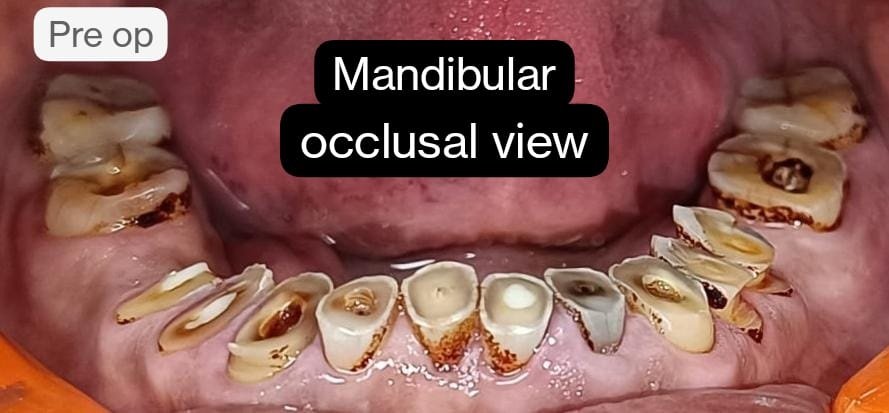
Figure 5
TREATMENT PLANNING:
Primary Objectives:
Restore masticatory function with optimal posterior cuspal anatomy and anterior guidance.
Resolve lower anterior sensitivity through endodontic treatment.
Enhance anterior aesthetics.
Diagnostic Steps:
Impressions taken with A-silicone for pre-treatment records.
A 4 mm vertical bite opening was planned for an additive wax-up. (Images 6-8)

Facebow Record obtained to capture the spatial orientation of the maxilla. (Images 9, 10)
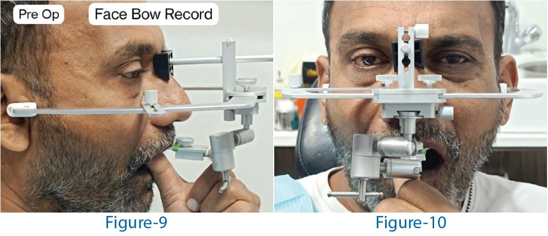
Deprogramming with customized Unwind MD device using self-curing acrylic resin. (Image A)

Image A
Mountographic Evaluation:
Diagnostic mounting revealed a centric relation (CR) contact point on the right first molar.
Articulator sent to the lab for 3D mock-up design and printing. (Images 11-14)


Verification Process:
After getting 3D mock up putty indices were made from C silicone. Figure 15

Figure 15
3D mock-up verified via test drive using chairside temporization material. (Images B, C)

In subsequent appointments, endodontic treatment was performed on lower anteriors and premolars, with post and core build-up and crown lengthening using electrocautery as needed (fiber posts and Wonder Edge DC from Wizdent utilized). (Images 16)
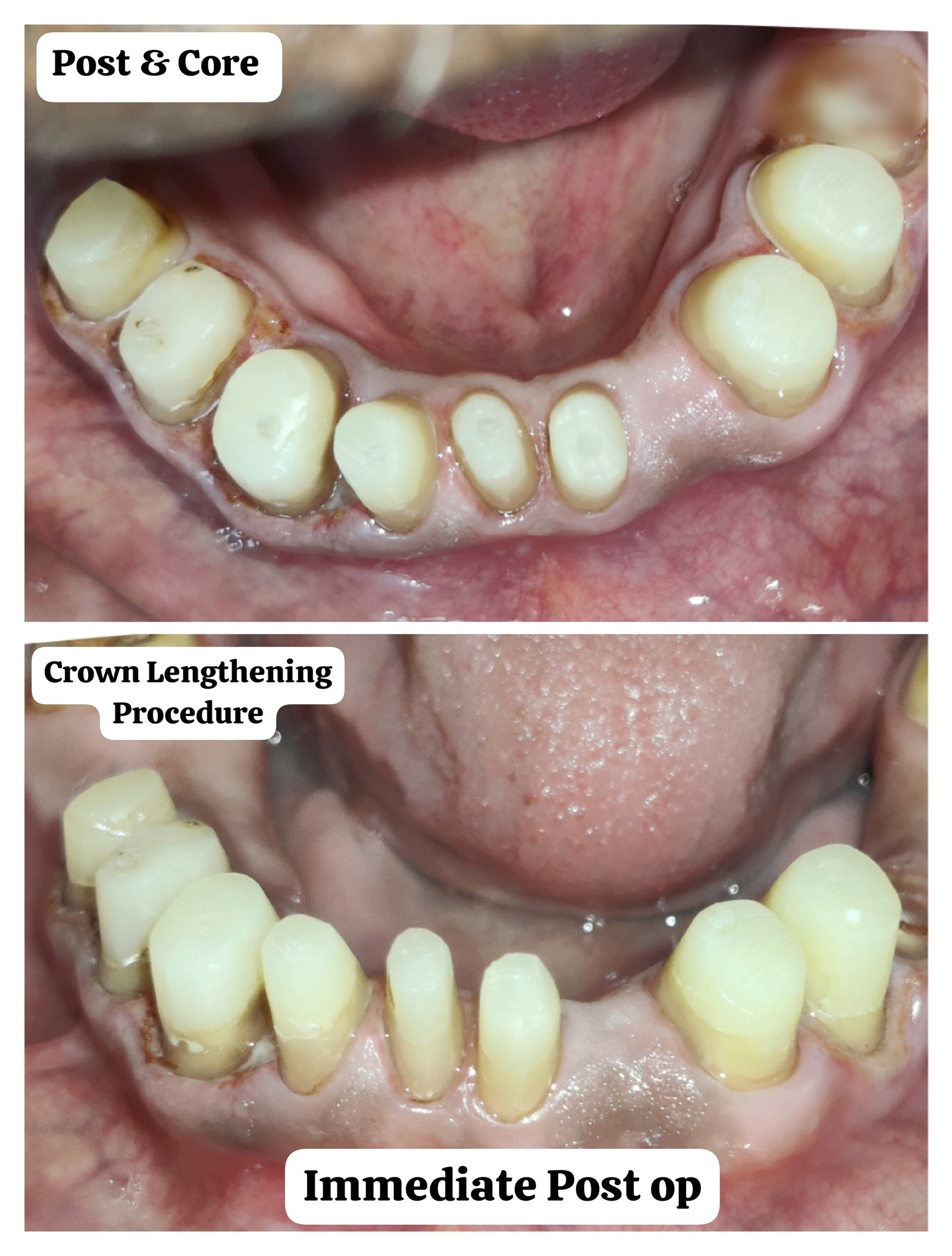
Figure 16
Preparations and Impressions:
Volumetric reduction carried out on maxillary teeth and mandibular molars; full-coverage crown prep on lower anteriors. (Images D, E, K)
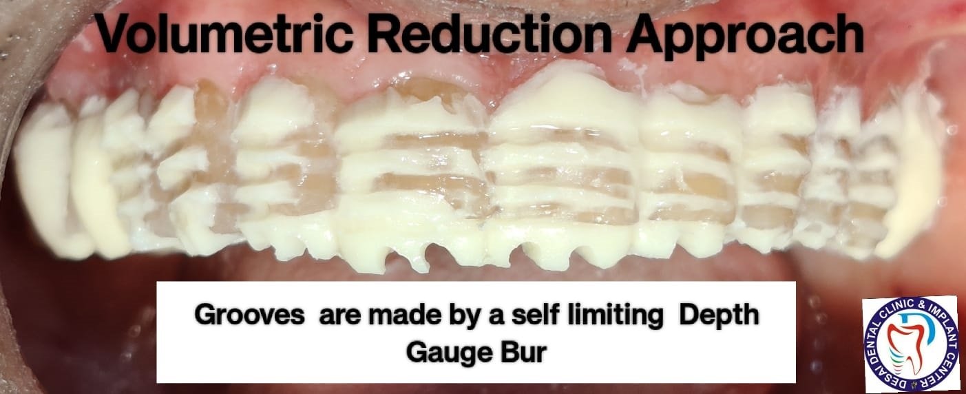
Image D
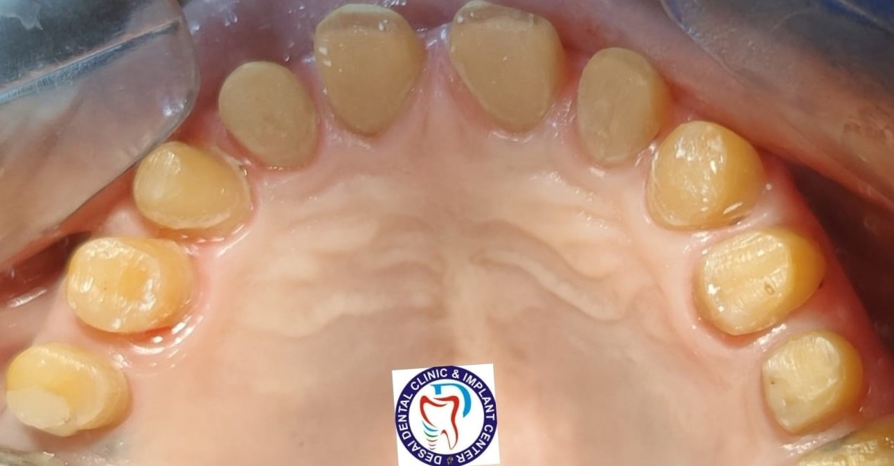
Image E

Image K
Both arches impressed with single-step A-silicone putty-light body technique. (Images 17, 18)

Figure 17 & Figure 18
Bite and Occlusion Records:
A Facebow record taken for prepared teeth. (Image 19)
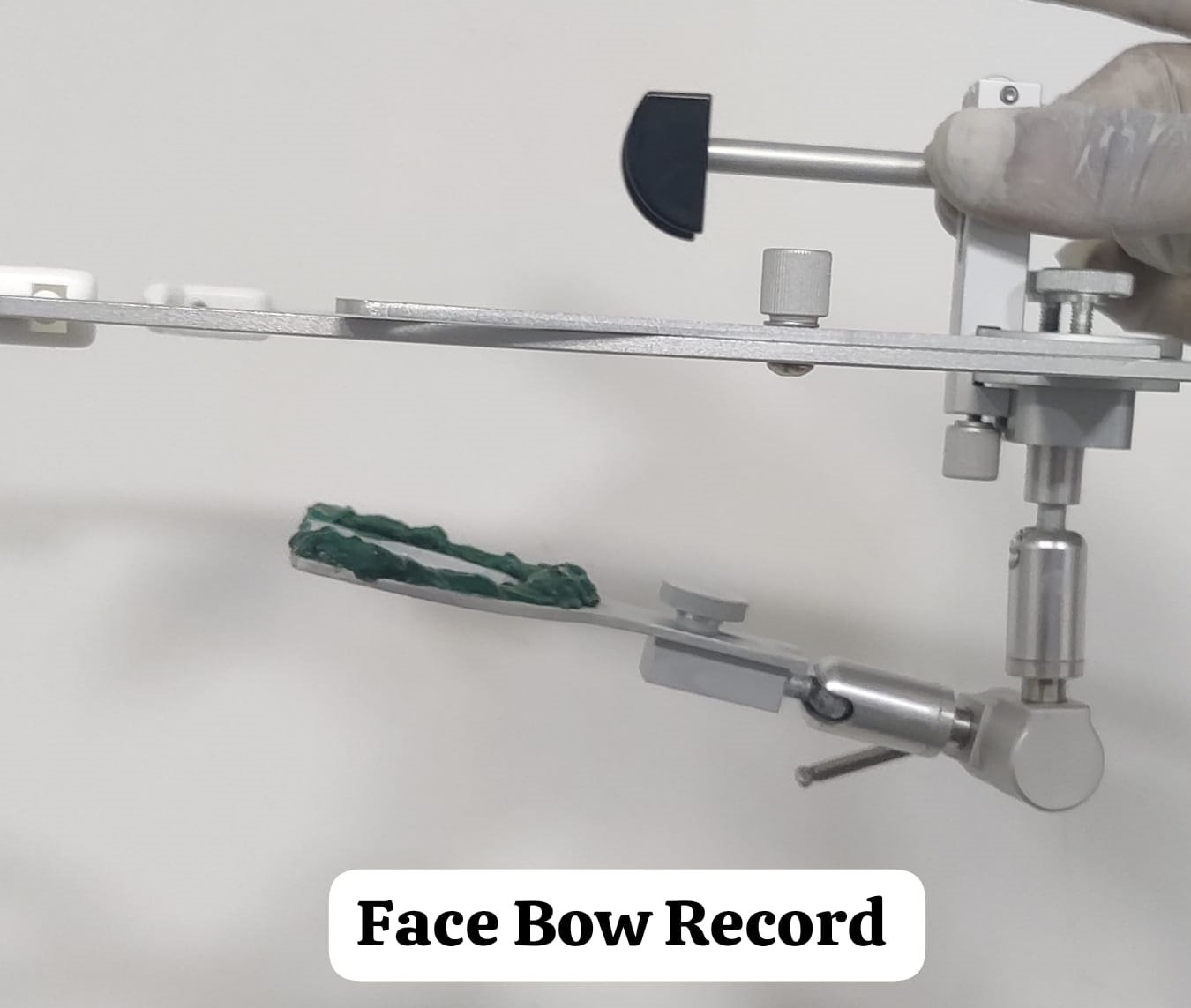
Figure 19
Centric relation (CR) bite recorded using Aluwax and bimanual manipulation at an arbitrary vertical dimension.
Temporary Restorations:
PMMA single-piece provisionals fabricated in the lab and luted with temporary cement for a 4-week trial period. (Images 20-24)


Figure 24
Canine and anterior guidance checked. (Images 25-27)
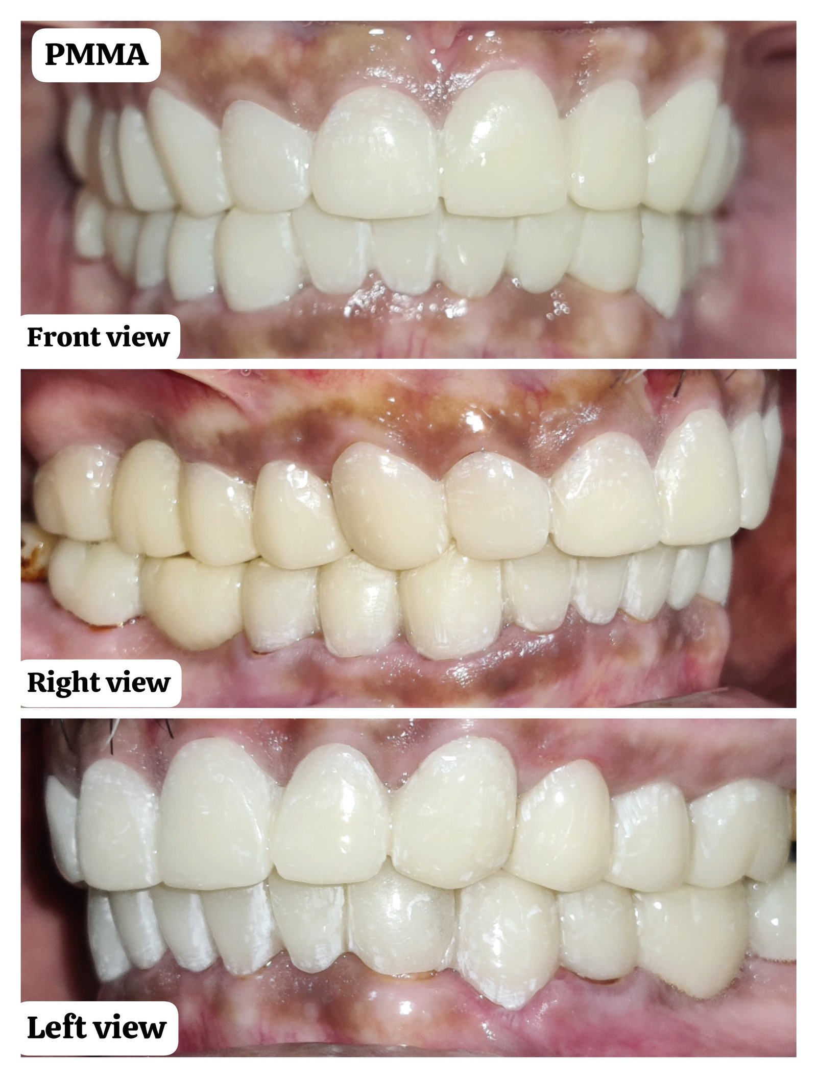
Figure 25

Figure 26

Figure 27
Final Restorations and Adjustments:
After 4 weeks, DMLS (direct metal laser sintered) restorations were tried, with adjustments at metal and bisque stages. (Images 28, 29, F, G, H, I)

Figure 28
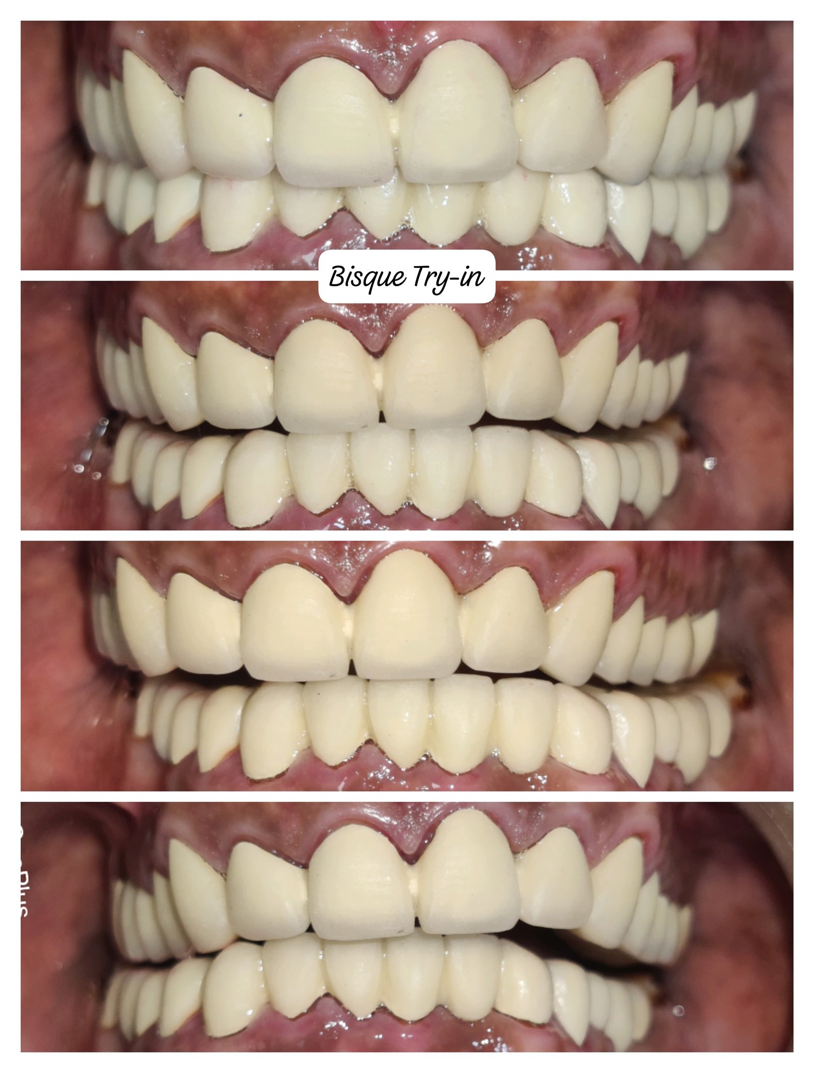
Figure 29
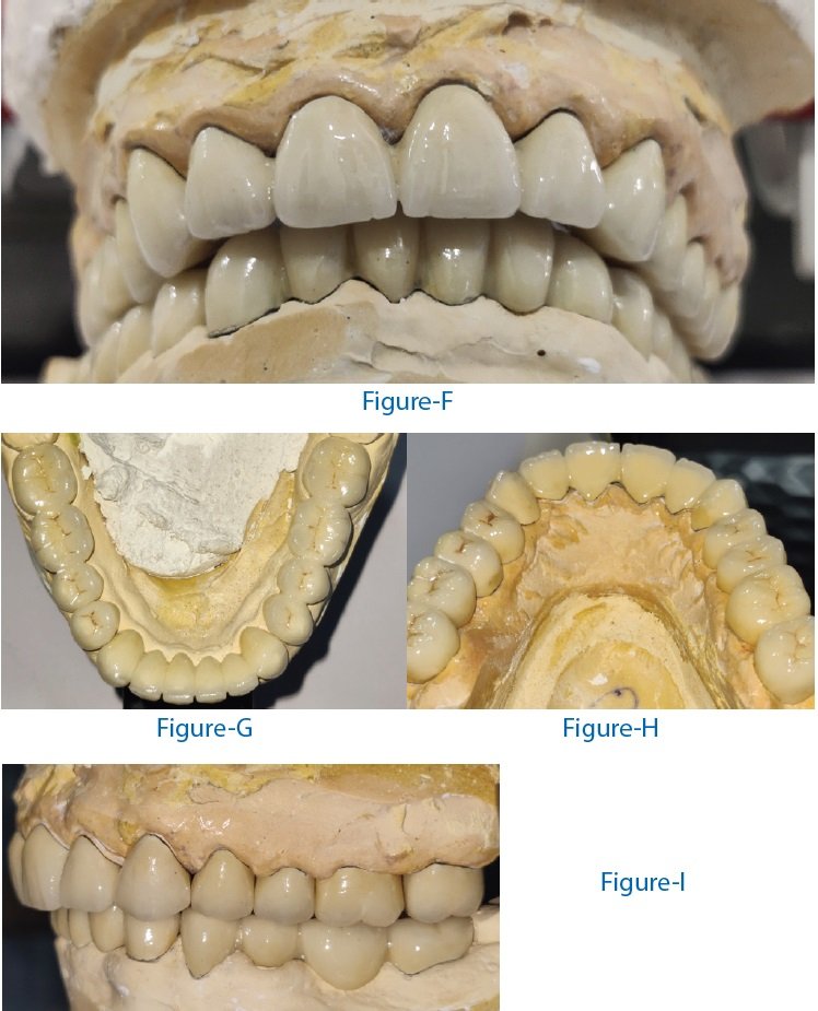
Definitive full-coverage ceramic crowns luted with RMGI (3M), following a sequence of lower anteriors, upper anteriors, lower posteriors, and upper posteriors.
Initial occlusal adjustments completed in this appointment, with final adjustments at one-week follow-up. (Images 30-32)

Figure 30
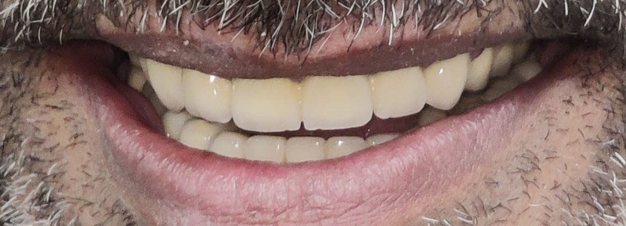
Figure 31

Figure 32
POST-TREATMENT MAINTENANCE
Prescribed hard acrylic splint for long-term stability. (Image 33)
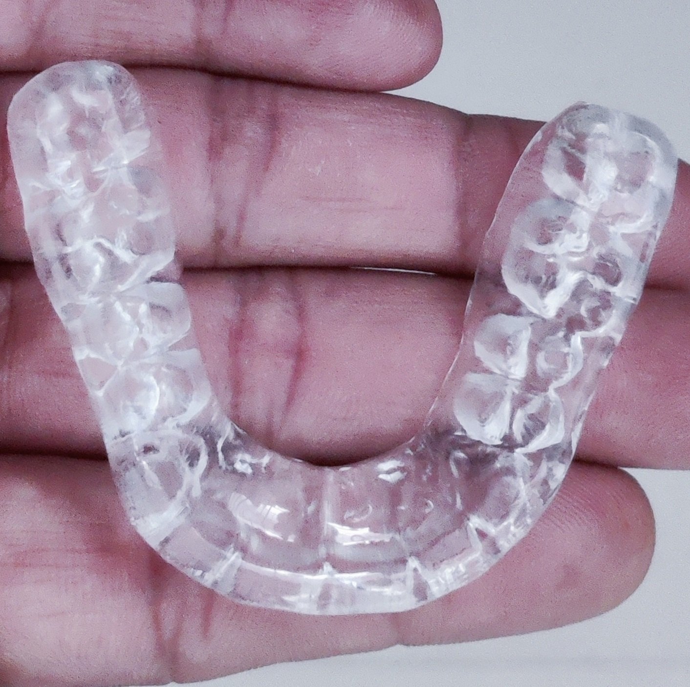
Figure 33
CONCLUSION
This methodical, step-by-step reconstruction significantly improved the patient’s function, comfort, and aesthetics, restoring a harmonious occlusal relationship and stable bite alignment.
Disclaimer from Guident
The views and opinions expressed in articles published in Guident are solely those of the authors and do not necessarily reflect the views of the editors, the editorial board or the publisher, Ivory India. Guident does not endorse any product, technique or treatment described. Clinical content is intended for qualified dental and medical professionals and is not a substitute for individual clinical judgement. Guident, its editors and the publisher accept no liability for any consequence arising from the use of information published in the journal.
How to cite this article
Dr. Kinnar Desai. Treating a Collapsed Bite with Full Mouth Reconstruction-a Case Report. Guident. 2024 Nov;17(12). Available from: https://guident.in/articles/prosthodontics/treating-a-collapsed-bite-with-full-mouth-reconstruction-a-case-report.html


