
Autologus Tooth Grafts: An Innovative Approach in Ridge Preservation and Socket Management
Department of Prosthodontics and Crown & Bridge, K.D. Dental College & Hospital, Mathura, UP, India.
ABSTRACT
The utilization of extracted natural teeth as an autologous bone graft material is gaining attention in the field of oral and maxillofacial surgery and prosthodontics. This innovative approach is supported by growing evidence suggesting that demineralized dentin matrices (DDM) derived from extracted teeth can provide excellent osteoconductive and osteoinductive properties. This article aims to explore the rationale, methods, clinical outcomes, and potential challenges of using extracted natural teeth as bone graft materials.
INTRODUCTION
The concept of utilizing natural teeth as a bone graft material was first introduced in the 1960s. However, only in recent years has this method gained clinical relevance due to advancements in demineralization and processing techniques. Teeth are composed of hydroxyapatite, collagen, and non-collagenous proteins, many of which have a structure similar to alveolar bone. Given the high percentage of hydroxyapatite in dentin (approximately 70%) and its favorable biological properties, teeth serve as a promising source of autologous graft material.
DENTIN MATRIX COMPONENTS
The mineral content of dentin is primarily hydroxyapatite, similar to bone, with a collagen-rich matrix that can facilitate new bone formation. Dentin also contains growth factors such as bone morphogenetic proteins (BMPs) and transforming growth factor-beta (TGF-β), which contribute to osteoinductive properties.
OSTEOCONDUCTIVE AND OSTEOINDUCTIVE POTENTIAL
Dentin, particularly when demineralized, exhibits osteoinductive characteristics due to the release of these growth factors during the demineralization process. Studies have shown that the dentin matrix can promote bone regeneration by acting as a scaffold (osteoconduction) and stimulating the differentiation of progenitor cells into osteoblasts (osteoinduction) of Tooth-Derived Graft Material
SELECTION OF TEETH
Types of Teeth
The process typically begins with the extraction of teeth from the patient, as this autologous approach eliminates the risk of immune rejection. Any type of extracted tooth can potentially be used, including molars, premolars, canines, and incisors, but molars are preferred due to their larger size and higher dentin content.

Contraindications
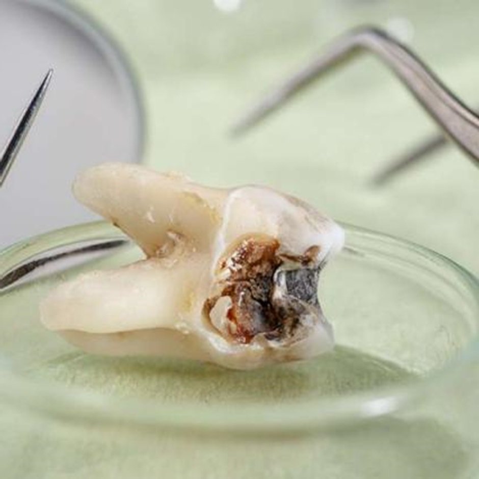
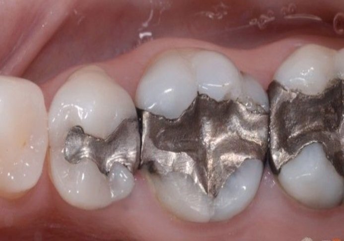
Teeth that are grossly decayed, contain large restorations, or are affected by infections such as cysts or granulomas are not suitable for use in graft preparation. These conditions compromise the structural integrity and biological activity of the dentin matrix. Teeth with root canal treatments are also avoided due to concerns about sterility and the potential alteration of their chemical composition.
TOOTH CLEANING AND DECONTAMINATION
Removal of Soft Tissue
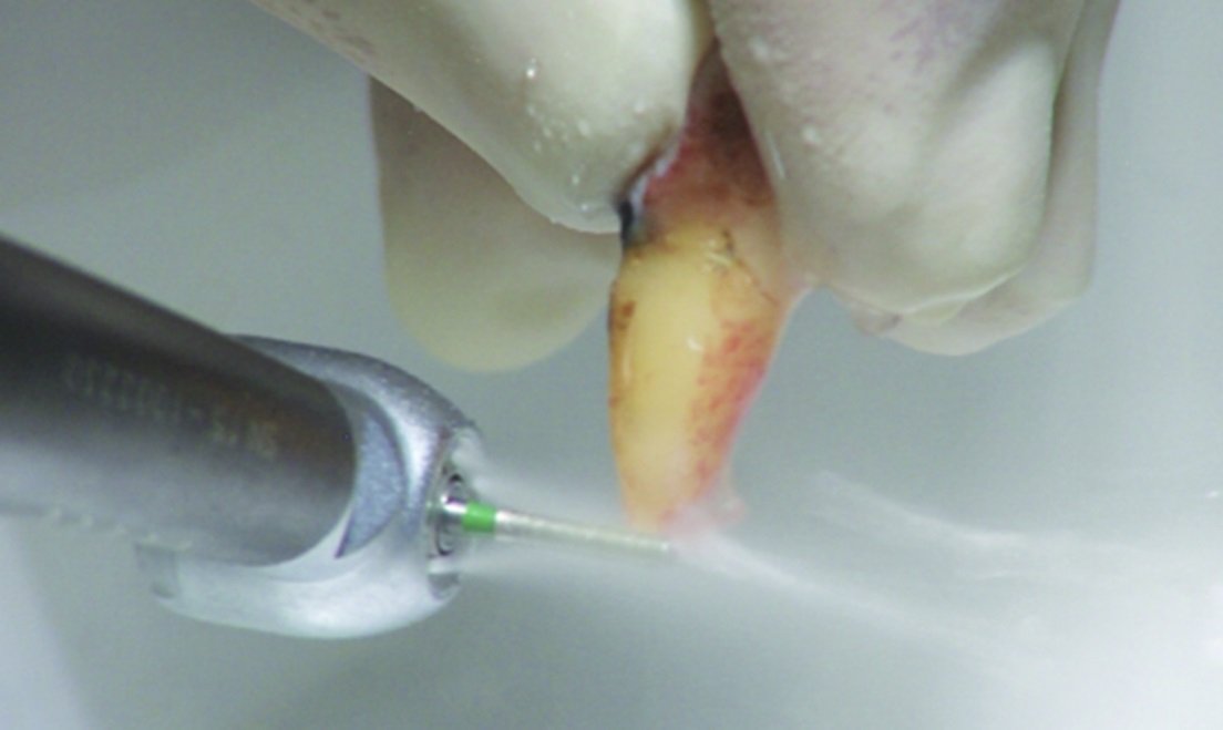
Once the tooth is extracted, all soft tissues (e.g., periodontal ligaments, pulp, gingival attachments) are carefully removed using hand instruments or ultrasonic scalers. Any residual organic material on the tooth surface, including plaque or calculus, is also thoroughly cleaned to prevent contamination.

Disinfection
Following the mechanical cleaning, the tooth is disinfected by placing it in a solution of hydrogen peroxide or another disinfecting agent for 5–10 minutes. This step ensures that all remaining bacteria and debris are eliminated.

SECTIONING AND GRINDING OF THE TOOTH
Sectioning the Tooth
The tooth is then divided into smaller sections. Typically, the crown is removed because it contains enamel, which has a low organic content and is highly mineralized, making it less suitable for grafting purposes. The dentin, which makes up the bulk of the tooth’s structure, is what will be processed into the graft material.
Grinding the Tooth
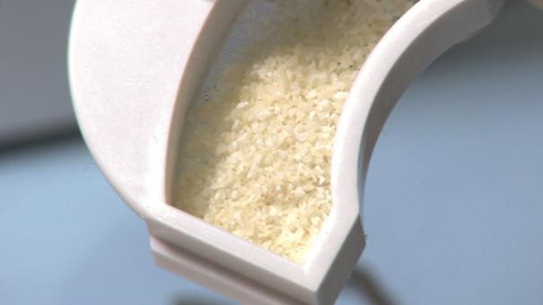
The dentin is ground into small particles using a tooth-grinding device. Several commercially available devices can be used, such as the Smart Dentin Grinder. The particles are typically ground to a size of 300–1,200 microns, as this particle size provides an ideal balance between surface area and handling properties during the grafting procedure. Larger particles provide better scaffold function, while smaller particles may degrade more quickly, releasing growth factors to aid in bone regeneration.


DEMINERALIZATION OF DENTIN
Acid Treatment
Once the dentin is ground into particles, it undergoes a demineralization process. This is done using a weak acid, typically a 0.6N solution of hydrochloric acid (HCl), which removes the inorganic components (hydroxyapatite) from the dentin. The demineralization step is crucial as it exposes the collagen matrix and enhances the release of bioactive growth factors, such as bone morphogenetic proteins (BMPs) and transforming growth factor-beta (TGF-β), which contribute to the material's osteoinductive properties.
Timing and Monitoring
The demineralization process typically takes between 20 and 30 minutes, but the exact time may vary depending on the thickness of the dentin particles and the concentration of the acid solution. Over-demineralization should be avoided as it can compromise the structural integrity of the graft material.
Neutralization
After demineralization, the acid is neutralized using phosphate-buffered saline (PBS) or another neutralizing solution to ensure the pH of the graft material is returned to a physiologically acceptable range. This step is essential for ensuring the graft material is biocompatible and will not cause adverse reactions when implanted into the body.
Sterilization of Graft Material
Chemical Sterilization
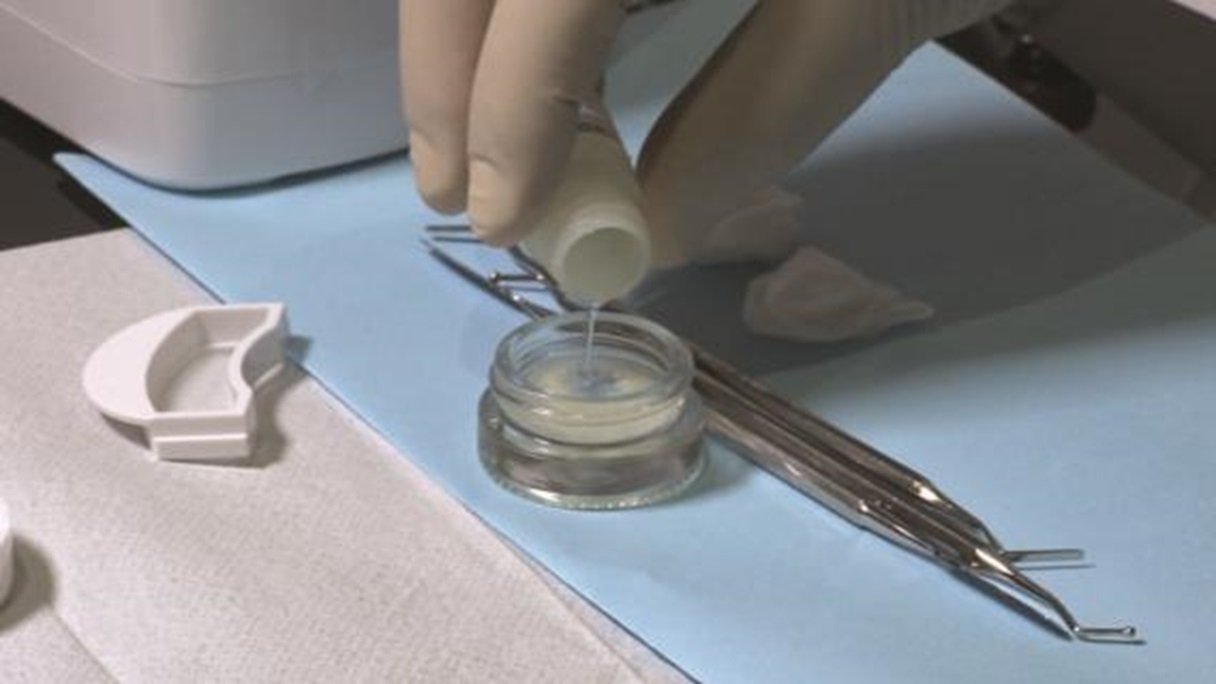
Following demineralization and neutralization, the graft material must be sterilized to ensure it is free from any microorganisms. This can be achieved using chemical sterilization methods such as immersion in 70% ethanol or ethylene oxide gas. Sterilization is critical to minimize the risk of infection and other complications post-surgery.
UV or Autoclave Sterilization
In some cases, ultraviolet (UV) light or autoclaving may be used for additional sterilization. However, autoclaving must be done cautiously, as high temperatures can denature the collagen matrix and reduce the osteoinductive potential of the graft material.
STORAGE AND PRESERVATION
Storage Conditions
Once the tooth graft material has been prepared, it can either be used immediately or stored for future use. If storage is required, the material should be kept in sterile conditions, ideally in a refrigerated environment (4°C to 8°C). For longer-term storage, the material can be freeze-dried and kept at low temperatures (-20°C) without compromising its biological properties.
Commercialization
Some commercial products are based on this technology, such as Tooth Transformer or Smart Dentin Grinder, which offer systems to create dentin graft material from patients’ extracted teeth in a dental clinic. These systems provide controlled environments for tooth preparation and standardize the processing protocol, ensuring consistent outcomes.
CLINICAL APPLICATION
Ridge Preservation and Alveolar Augmentation
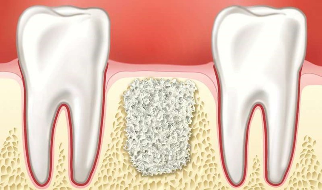
One of the most common applications of tooth-derived bone grafts is in alveolar ridge preservation following tooth extraction. The placement of demineralized dentin graft material into extraction sockets can prevent alveolar bone resorption, maintaining the contour of the ridge for future prosthodontic or implant placement .
Sinus Lift Procedures
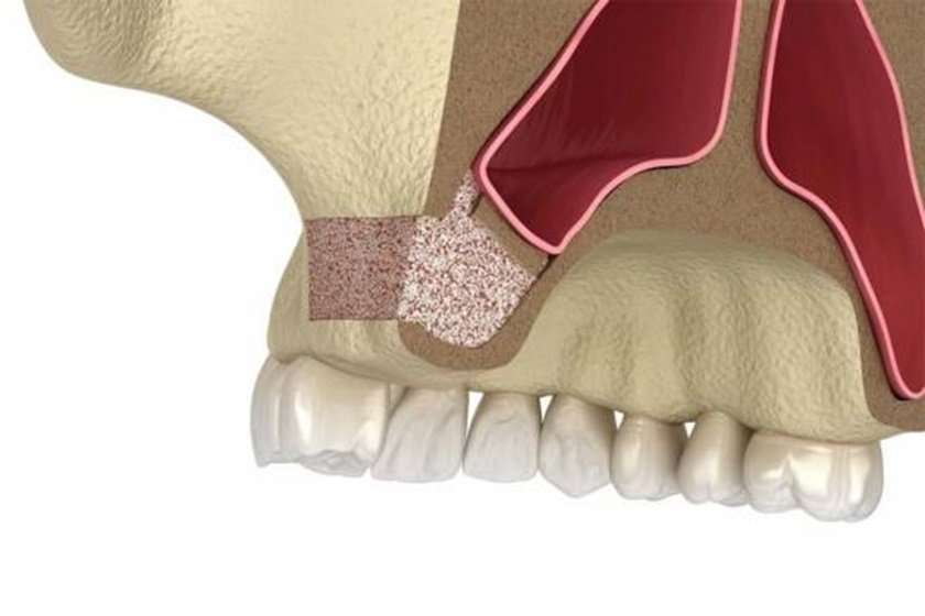
Tooth bone grafts have also been used successfully in sinus lift procedures, where the graft material is placed in the maxillary sinus to promote bone growth in preparation for dental implant placement. Studies have reported positive outcomes, showing bone regeneration and implant stability comparable to traditional bone graft materials such as autologous bone or allografts .
Treatment of Bone Defects
In cases otal bone defects or traumatic bone loss, tooth-derived graft materials have been applied with promising results. The ability of dentin matrices to promote bone regeneration makes them a viable option for reconstructive procedures in both small and large defects .

ADVANTAGES OF TOOTH-DERIVED GRAFT MATERIAL
Autologous Nature
One of the key advantages of using teeth as graft material is that they are autologous, meaning they come from the patient’s own body. This eliminates the risk of immune rejection or disease transmission, a concern with allogeneic or xenogeneic grafts .
Cost-Effectiveness and Sustainability
Using extracted one graft material can be cost-effective as it repurposes a by-product that would otherwise be discarded. Furthermore, this method reduces the need for harvesting autogenous bone from other parts of the body, which can reduce patient morbidity and surgical time .
Challenges and Limitations
Availability of Extracted Teethation of this approach is the availability of sufficient extracted teeth, especially in patients who have already experienced extensive tooth loss or have dental conditions that may compromise the integrity of their teeth as graft material .
Standardization of Processing
The lack of standardized protocols for proth into graft material poses a challenge. Variations in particle size, demineralization techniques, and sterilization methods can affect the clinical performance of the graft material . However, increasing efforts are being made to develop commercially available tooth graft streamline and standardize the process.
CONCLUSION
The use of extracted natural teeth as bone graft material is a promising and innovative approach in dental and maxillofacial reconstructive surgery. With their osteoconductive and osteoinductive properties, teeth provide a unique and autologous alternative to traditional graft materials. While challenges remain regarding standardization and availability, ongoing research continues to validate the efficacy and long-term outcomes of this approach, potentially revolutionizing how clinicians manage bone defects and preserve alveolar ridges.
REFERENCES
- Kim, Y. K., et al. (2010). "Tooth-derived bone graft material." Journal of Clinical Periodontology, 37(11), 985-992.
- Minamizato, T., et al. (2018). "Osteoinductive potential of demineralized dentin matrices and its applications in dental implantology." International Journal of Oral and Maxillofacial Surgery, 47(9), 1218-1225.
- Binderman, I., et al. (2014). "The use of demineralized dentin matrix for bone regeneration." Clinical Implant Dentistry and Related Research, 16(4), 569-578.
- Schwarz, F., et al. (2016). "Autogenous tooth-derived bone graft material in alveolar ridge augmentation procedures." Journal of Periodontology, 87(5), 544-552.
- Nampo, T., et al. (2010). "Autogenous demineralized dentin matrix for ridge preservation: A preliminary report." Journal of Clinical Periodontology, 37(6), 571-577.
- Sohn, D. S., et al. (2014). "Autogenous demineralized tooth graft in sinus augmentation." Implant Dentistry, 23(3), 299-308.
- Yamada, Y., et al. (2011). "Tooth-derived bone graft materials in periodontal surgery." Journal of Clinical Periodontology, 38(10), 929-934.
- Lee, J. H., et al. (2014). "Use of autogenous tooth bone graft material for alveolar ridge augmentation: Case report." Journal of the Korean Association of Oral and Maxillofacial Surgeons, 40(2), 90-95.
- Gomes, M. F., et al. (2020). "Autogenous dentin grafts: A sustainable approach for alveolar bone regeneration." Oral and Maxillofacial Surgery Clinics of North America, 32(1), 97-109.
- Kim, Y. K., et al. (2014). "Bone regeneration using tooth-derived graft material." Journal of the Korean Academy of Prosthodontics, 52(2), 140-148.
- Pang, K. M., et al. (2019). "Clinical applications of tooth-derived bone graft materials." Journal of Craniofacial Surgery, 30(4), 1237-1243.
Disclaimer from Guident
The views and opinions expressed in articles published in Guident are solely those of the authors and do not necessarily reflect the views of the editors, the editorial board or the publisher, Ivory India. Guident does not endorse any product, technique or treatment described. Clinical content is intended for qualified dental and medical professionals and is not a substitute for individual clinical judgement. Guident, its editors and the publisher accept no liability for any consequence arising from the use of information published in the journal.
How to cite this article
Dr. Shikha Shahi, Dr. Yamini Bhushan, Dr. Manesh Lahori. Autologus Tooth Grafts: An Innovative Approach in Ridge Preservation and Socket Management. Guident. 2024 Nov;17(12). Available from: https://guident.in/articles/prosthodontics/autologus-tooth-grafts-an-innovative-approach-in-ridge-preservation-and-socket-management.html


