Surgical Management of a Periapical Lesion in Lower Anteriors Using MTA
Department of Conservative Dentistry and Endodontics, Seema Dental College And Hospital, Rishikesh
INTRODUCTION
Apicoectomy is the excision of the apical portion of the tooth and the attached soft tissues during periapical surgery.1 The surgery goal is periapical lesion removal and the apical third sealing, allowing soft and hard tissue regeneration. According to the meta-analysis of Tsesis et al, paraendodontic surgery success rate is 91.6%, while failure rate is 4.7%.2 In periradicular surgery with retrograde root-canal obturation, several surgical steps are essential to achieve healing: surgical exposure of the root-end, debridement of pathological tissue, root-endresection, root-end cavity preparation, root-end filling,and wound closure.3
Mineral trioxide aggregate (MTA) was introduced to create an effective seal between the root canal system and the periradicular tissues. Comparative studies with other root canal filling materials have shown less leakage as well as excellent periradicular healing when used as a rootend filling material or apical plug.4
Indications for apical surgery have been updated in the past by the ESE (European Society of Endodontology, 2006) and it includes: Radiological findings of apical periodontitis, Extruded material with clinical or radiological findings of apical periodontitis, Persisting or emerging disease following root-canal treatment when root canal re-treatment is inappropriate, Perforation of the root or the floor of the pulp chamber and where it is impossible to treat from within the pulp cavity.5
The success rate of apicectomised teeth ranges from 46.7010(Hirschet aI., 1979) to 90.0010(Harty,Parkins and Wengraf,1970). Clinical evaluation in conjunction with peri-apical radiographs has been used to assess healing. Microscopic study of biopsies taken from healing or healed peri-apical tissues has been suggested as a better means of assessment (Hirschet).6
This article describes a clinical case in which root canal treatment was performed followed by root resection surgery with MTA as retrograde filling material.
CASE REPORT
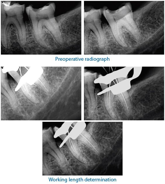
A 28 year old female patient reported to the Department of Conservative Dentistry and Endodontics with a chief complaint of pain in lower front tooth region since 1 month. Pain was dull, throbbing in nature. She also complained of trauma in lower front tooth region 2 years back. She reported that an attempt to root canal treatment was also performed in lower left lateral incisor 3 months back. Medical history was non contributory. Radiographic examination revealed diffuse periapical radiolucency involving teeth i.r.t,31,41. Cold test was negative i.r.t 31 and lingering response i.r.t 31,41,42. Teeth presented a delayed response to electric pulp testing i.r.t 31,41,42 and no response i.r.t 32. The treatment planned was root end resection w.r.t 31 41 with retrograde filling of MTA. It was explained to the patient and consent was obtained. After the access opening of 31, 32, 41, 42 was done the working length was determined using k file and thorough shaping and cleaning was done. The teeth were then obturated using warm vertical condensation.

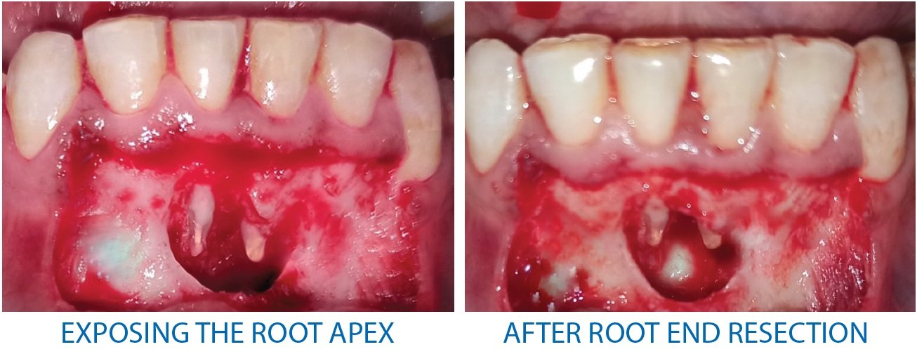
Raising Ochsenbein – Luebke (O-L) Flap, a periapical surgery was performed and periapical pathosis was enucleated. 3mm root end resection and retrograde cavity was prepared using ultrasonic tip. MTA was placed as retrograde restorative material of 3mm thickness. The placement of the retrograde filling material was confirmed using a radiograph and the sutures were placed. The patient was recalled after 10 days for suture removal.
The patient was kept under observation for the purpose of postoperative evaluation of healing of the periapical intervention in intervals of 1, 3 months.




DISCUSSION
Pulpal response to dental trauma is variable. In some cases, the pulp remains normal, whereas in some cases it becomes necrotic. As a consequence to dental trauma, the pulp loses its ability to protect itself from bacterial invasion and the bacteria penetrates through the dentinal tubules, colonizes in the necrotic pulp, and leads to the development of periapical lesion.7
Apical surgery is the standard endodontic surgical procedure to maintain a tooth with significant periapical lesion5 better results were achieved when root filling and apicectomy were carried out simultaneously (Nordenram and Sverdstrom, 1970). In this case, apicoectomy would be the treatment of choice to preserve the teeth. Hence,in the present caseroot end resection was performed and prepared.6
Owing to the improvements in surgical techniques and suture materials, oral surgery has become more conservative and delicate, and the Luebke–Ochsenbein flap with submarginal incisions is now more widely used. In the present case it was justified Ochsenbein – Luebke (O-L) Flap have advantage over the Trapezoidal flap as it is less aggressive with the gingival tissue than an intrasulcular incision flap, and it is easy to make the incision slightly triangular or angled in order to secure precise repositioning, it is easy to reflect, has good visibility, less bleeding duration and is easy to handle during surgery.11,12
In the past, many materials such as amalgam, zinc phosphate cement were used as retrograde filling material. But because of their limitations like marginal leakage, lack of corrosion resistance and irritation to periapical tissues, many newer materials have been introduced for retrograde filling, the important among them is MTA.13
Mineral trioxide aggregate (MTA) was introduced to create an effective seal between the root canal system and the periradicular tissues.2 MTA appears to be equal or superior to other root-end filling materials with respect to biocompatibility, dye and bacterial leakage, marginal adaptation, solubility, and compressive strength. Interestingly, this material also appears to induce cementogenesis with new cementum deposition on the surface of the retrofilling materia.8
Disadvantage of MTA is its delayed setting time of 170 mins which will lead to an increased risk of partial material loss and alteration of the interface during finishing. This came as a limitation to this case.10
CONCLUSION
With a high percentage of successful treatment outcomes of conventional endodontics together with high success of surgical endodontics almost all teeth with endodontic lesions can be successfully treated9. The surgical technique applied in this case, apicoectomy, was appropriate. This was proven by both clinical and radiographic results.
REFERENCES
- Ajayi JO, Abiodun-Solanke IMF, Olusile OA, Oginni AO, Esan TA. COMPARATIVE STUDY OF TREATMENT OUTCOME IN APICECTOMIES WITH OR WITHOUT ROOT-END FILLING. Annals of Ibadan Postgraduate Medicine. 2018 Dec;16(2):109-114.
- Pedroche, L.O., Barbieri, N., Tomazinho, F.et al. (2013). Apicoectomy after conventional endodontic treatment failure: case report.
- von Arx T, Gerber C, Hardt N. Periradicular surgery of molars: a prospective clinical study with a one-year follow-up. Int Endod J. 2001 Oct;34(7):520-5. doi: 10.1046/j.1365-2591.2001.00427.x. PMID: 11601769.
- Asgary, Saeed & Ehsani, Sara. (2013). Periradicular Surgery of Human Permanent Teeth with Calcium-Enriched Mixture Cement. Iranian endodontic journal. 8. 140- 4.
- Avinash, Shashant & Agrawal, Eiti & Mushtaq, Iqra & Bhandari, Anuva & Khan, Farheen. (2019). Apicoectomy: An elucidation to a hitch. Journal of Dental Specialities. 7. 28-32. 10.18231/j.jds.2019.006.
- Oginni, Adeleke & Olusile, Adeyemi. (2002). Follow-up study of apicectomised anterior teeth. SADJ : journal of the South African Dental Association = tydskrif van die Suid-Afrikaanse Tandheelkundige Vereniging. 57. 136-40.
- Garlapati, Roopadevi & Venigalla, Bhuvan Shome & Patil, Jayaprakashet al. (2014). Surgical Management of Mandibular Central Incisors with Dumbbell Shaped Periapical Lesion: A Case Report. Case reports in dentistry. 2014. 769381. 10.1155/2014/769381.
- von Arx, Thomas. (2005). Failed Root Canals: The Case for Apicoectomy (Periradicular Surgery). Journal of oral and maxillofacial surgery : official journal of the American Association of Oral and Maxillofacial Surgeons. 63. 832-7. 10.1016/j.joms.2005.02.019.
- Bhattacharjee, Arka & Prathigudupu, Raja & Singh, Shalini & Sharma, Yesh & Sehrawat, Kritika & Tiwari, Rahul. (2018). Endodontic Surgery -A Review. 10.13140/RG.2.2.32965.29929.
- Insha Showkat, Ashish Amit Sinha et al Surgical intervention: Saviour of a failed root canal treatment International Journal of Applied Dental Sciences 2019; 5(4): 91-94
- Diago, M.P., Ballestera, J.C., & Oltraa, D.P. (2017). Flap design: New perspectives in periapical surgery.
- Bruce J. Kramper, Edward J. Kaminski et al A comparative study of the wound healing of three types of flap design used in periapical surgery,Journal of Endodontics, Volume 10, Issue 1,1984,Pages 17-25
- Torabinejad M, Pitt Ford TR. Root end filling materials: a review. Endod Dent Traumatol. 1996 Aug;12(4):161-78. doi: 10.1111/j.1600-9657.1996.tb00510.x. PMID: 9028180.
Disclaimer from Guident
The views and opinions expressed in articles published in Guident are solely those of the authors and do not necessarily reflect the views of the editors, the editorial board or the publisher, Ivory India. Guident does not endorse any product, technique or treatment described. Clinical content is intended for qualified dental and medical professionals and is not a substitute for individual clinical judgement. Guident, its editors and the publisher accept no liability for any consequence arising from the use of information published in the journal.
How to cite this article
Dr. Deepika Rawat, Dr. Sugandha Sati, Dr. Annil Dhingra. Surgical Management of a Periapical Lesion in Lower Anteriors Using MTA. Guident. 2021 Apr;14(5). Available from: https://guident.in/articles/endodontics/surgical-management-of-a-periapical-lesion-in-lower-anteriors-using-mta.html