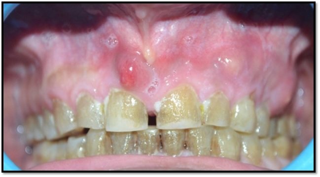
Twin Mesiodens-a Case Report
Burhani Hospital, Surat, Gujarat (INDIA)
ABSTRACT
The most common supernumerary tooth is a Mesiodens, which is a malformed, peg-like tooth that occurs between the maxillary central incisors.
Mesiodens is a supernumerary tooth present in the midline between the two central incisors.
It usually results in oral problems like food lodgement, malocclusion/poor aesthetics, midline diastema, displacement or rotations and can lead to cyst formations too. The treatment of supernumerary teeth depends on their type, position, and possible complications, detected clinically and radiographically.
Key words: Supernumerary teeth, Mesiodens, impacted teeth, hyperdontia, permanent teeth, deciduous teeth.
INTRODUCTION
Supernumerary tooth can be defined as one that is additional to normal series and can be found in almost any region of the dental arch. Though the etiology of this condition is not well understood, it has been suggested that the interference with the stage of the initiation, results in either missing tooth or extra tooth. Supernumerary tooth is found more often in permanent dentition than in the primary.
Supernumerary teeth classification:
They can be classified on the basis of position-
- Mesiodens-present in the incisor region.
- Paramolar-present beside molars.
- Distomolar-present distal to last molar.
- Parapremolar-present beside premolar.
Depending upon their size and shape there are 2 types of Mesiodens-
- DYSMORPHIC TOOTH- Tooth differs in shapes and sizes and are further classified as-
- Tuberculate, conical, odontomas and supplemental.
- EUMORPHIC TOOTH- Tooth is having regular morphology and are similar to the central incisors.
Occurrence may be single or multiple, unilateral or bilateral, erupted or unerupted/impacted and in one or both the jaws.
Sometimes conditions associated with an increased prevalence of supernumerary teeth include cleft lip and palate, Cleidocranial dysplasia, Gardner’s syndrome.
Mesiodens can be diagnosed through clinical and radiographic examinations using the maxillary anterior periapical and occlusal radiography. In addition to these, orthopantomograph (OPG) and lateral cephalograph can also be taken for diagnosis.
CASE REPORT
A 23 years old male patient came with a chief complaint of difficulty in chewing along with irregularly placed teeth in upper front teeth region of the mouth. Patient concern was esthetics with proper functionality.
Medical, dental and familial history was found non-contributory towards the complaint.
The clinical examination revealed permanent dentition with malalignment of the upper anterior region along with double conical supernumerary teeth erupted palatally [Figure1] and [Figure 2].
Intraoral tissues were normal and healthy.
Mandibular arch showed missing left 1st premolar with rotated canine on the same side.
Radiographically with OPG it got confirmed as-Twin Mesiodens.
TREATMENT
Intra-alveolar extraction (forceps technique) of erupted supernumerary teeth was done with uneventful healing.
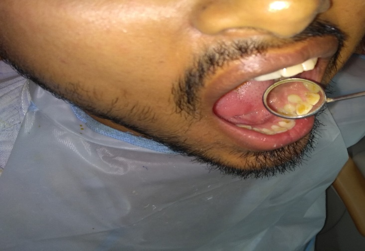
Figure 1- Both erupted palatally Mesiodens (Twin Mesiodens)

Figure 2- Extracted Mesiodens

OPG showing 2 Mesiodens
DISCUSSION
The etiology of supernumerary teeth is unknown. Many of us has the view that they develop from a third tooth bud arising from the dental lamina near the permanent tooth bud or probably from splitting of the permanent bud itself. This view is supported by the fact that a supernumerary tooth usually closely resembles the teeth of the group to which it belongs. The most common supernumerary tooth is the 'Mesiodens' a tooth situated between the maxillary central incisors.
Mesiodens may usually be diagnosed because of delayed eruption of central incisors. Unilateral persistence of deciduous incisor, wide diastema or rotation of erupted permanent incisors are other common presentations. Rarely, it manifests as dentigerous cyst. Mesiodens is seen as an additional tooth lying cranial to the central incisors. It may occur singly or in pairs, may be erupted or impacted. The direction of the crown of mesiodens may be normal, inverted or horizontal. Mesiodens is known to have a cone shaped crown and a short root as seen in our patient. It is a rare entity with a reported incidence of 0.15 to 1.9% and has a slight male predominance.
The presence of erupted mesiodens is best diagnosed by clinical examination and the unerupted mesiodens can be diagnosed by both clinical and radiographic evaluation. Panoramic, maxillary occlusal and periapical radiographs are recommended to assist the diagnosis of mesiodens and the bucco-lingual position of the unerupted mesiodens can be determined using parallax technique. Early diagnosis of the entity helps to reduce the problems that might occur resulting in impaired esthetics and malocclusion. Clinically, presence of unerupted mesiodens can be suspected in case if dental asymmetry or if delayed eruption of adjacent teeth exists.
The classification of supernumerary teeth is usually based on their morphology. According to the shape and size, the mesiodens teeth are classified as eumorphic that resembles to a normal sized central incisor and dysmorphic type that presents different shapes and sizes. Various complications might occur as a result of the presence of mesiodens, including delayed eruption, alteration in the path of eruption of permanent incisors, impaction of permanent incisors, crowding, spacing, median diastema, rotation and root resorption of the adjacent teeth or even eruption of incisors in the nasal cavity, cystic lesions and other intraoral pathological problems.Management of supernumerary teeth depends on the type, position of the tooth and the stage of dentition. Munn’s recommended earlier removal of the mesiodens for achieving better prognosis. Extraction of mesiodens is usually not advocated in primary dentition since they often erupt into the oral cavity and thus risk of damaging the permanent incisor during surgical removal of mesiodens can be avoided. However, at early mixed dentition stage, the permanent central incisors erupt spontaneously after the extraction of mesiodens. This also promotes better alignment of the teeth and minimizes the need for orthodontic treatment. Close monitoring of the dentition is required after the extraction of a mesiodens.
Clinical and radiographic reassessment is recommended after 6 months of mesiodens extraction and if the permanent incisor does not erupt averagely after 12 months of extraction of mesiodens, closed eruption with orthodontic mechanotherapy is recommended. Delay in extraction of mesiodens might result in failure of spontaneous eruption of permanent incisor due diminished eruptive forces, arch perimeter loss, midline shifting and mesial drifting of lateral incisors into central incisor space, which might require comprehensive orthodontic treatment with surgical exposure of the unerupted teeth.
In order to avoid complications, the orthodontists recommend early removal of the supernumerary teeth.
CONCLUSION
Mesiodens is the most prevalent form of supernumerary teeth in permanent dentition that occurs as a result of genetic and environmental factors and hyperactivity of dental lamina. Males are affected two folds than the females. Early diagnosis of a mesiodens reduces the treatment required and prevents development of associated problems. Diagnosis of mesiodens can be done by clinical and radiographic examination and extraction of mesiodens in the early mixed dentition helps spontaneous alignment of the adjacent teeth. If the permanent incisors fail to erupt spontaneously, further surgical and orthodontic treatment may be required.
REFERENCES
- Shafer’s Textbook of Oral Pathology 6th edition
- Sharma A. Familial occurance of mesiodens – A case report. J Indian Soc Pedod Prev Dent 2003; 21: 84-85
- Munns D. A case of partial anodontia and supernumerary tooth present in the same jaw. Dent Pract Dent Rec 1967;18:34-7
- Double mesiodens in mixed dentition period: Report of 3 cases
Sukhvinder Singh Rana, Neera Ohri
Department of Dentistry, Dr RPGMC, Kangra, Himachal Pradesh, India.
Disclaimer from Guident
The views and opinions expressed in articles published in Guident are solely those of the authors and do not necessarily reflect the views of the editors, the editorial board or the publisher, Ivory India. Guident does not endorse any product, technique or treatment described. Clinical content is intended for qualified dental and medical professionals and is not a substitute for individual clinical judgement. Guident, its editors and the publisher accept no liability for any consequence arising from the use of information published in the journal.
How to cite this article
Dr. Kalpana Sinsinbar. Twin Mesiodens-a Case Report. Guident. 2021 Apr;14(5). Available from: https://guident.in/articles/general/twin-mesiodens-a-case-report.html