Prosthodontic Management of Anterior Disc Derangement with Reduction-a Case Report
307, Shivanagar, Kewra Badi, Raigarh (C.G.), Department of Prosthodontics, Maitri College Of Dentistry & Research Centre, Anjora, Durg (C.G.)
ABSTRACT
TMD is often viewed as a repetitive motion disorder of the structures of mastication, with many similarities to musculoskeletal disorders of other parts of the body, and is therefore treated with similar therapeutic approaches. The combination of splinting, physical therapy, medication therapy, and behaviour modification as first-line conservative strategies has been reported as effective in up to 86% of these patients with disc displacement who become pain-free and regain acceptable function with this strategy. Following is a case report of a 55-year-old female presenting with progressive disc derangement tendencies managed using anterior positioning splint. This case report potentiates the practice of employing occlusal splints for initial non-surgical management of disc derangement.
INTRODUCTION
Temporomandibular Joint (TMJ) is considered to be the most complex synovial articulation in the human body. Articular surfaces within this joint are in contact with the articular disc, a nonvascular structure and nourished by synovial fluid, a secretion of the internal layer of the articular capsule, the synovial membrane. In sagittal view, the articular disc is a flexible and biconcave structure, constituted by dense connective tissue, usually positioned between the posterior slope of the articular tubercle and the anterior medial surface of the mandibular condyle. It transversally splits the TMJ in two distinct compartments, which are completely separate under normal conditions. It is possible to identify three different regions in the articular disc: the posterior band (the thickest portion), the intermediate (central) band (the thinnest part) and the anterior band (with intermediate thickness).
Temporomandibular dysfunction (TMD) is a musculoskeletal disorder with many sub diagnoses, including myofascial pain, temporomandibular joint (TMJ) inflammatory conditions (traumatic, arthritides, or ankylosis), and disc derangement. TMD is often viewed as a repetitive motion disorder of the structures of mastication, with many similarities to musculoskeletal disorders of other parts of the body, and is therefore treated with similar therapeutic approaches.1 The various clinical conditions are characterized by pain in the preauricular area, TM joint, or muscles of mastication; limitation or deviation in mandibular range of motion; and TM joint sounds (clicking, popping, and crepitus) during mandibular function.2 Common patient complaints include headache, neck ache, face ache, and earache.3 Other unexplained associated complaints include tinnitus, ear fullness, and perceived hearing loss. Controversy exists because of the limited knowledge regarding the etiology and natural history or course of TMD.
Pathology that may require a more intensive approach is anterior disc derangement with or without reduction, secondary to posterior ligament insufficiency. These ligamentous injuries can be either acute or chronic, and most commonly result in anterior/anteromedial disc displacement due to elongation of the posterior discal ligaments. Chronic ligamentous injuries generally represent chronic instability from failure of acute ligament healing, often involving fiber stretching or tearing and joint laxity, and may have an inflammatory component.4 Restoring the mechanics of the affected region, reducing inflammation when possible, and restoring stability are important first-line conservative measures. The combination of splinting, physical therapy, medication therapy, and behaviour modification as first-line conservative strategies has been reported as effective in up to 86% of these patients with disc displacement who become pain-free and regain acceptable function with this strategy.5 Once the environment has been optimized, symptoms are more likely to resolve; however, in stubborn cases, there may be an argument for the use of platelet-rich plasma (PRP) to synergistically assist the inflammatory cascade and regenerative processes in healing injured tissue.
CASE PRESENTATION
A 55-year-old female patient reported to the department with a chief complaint of pain in front of the ears with severe discomfort while mouth opening in the last 6 months (Fig. 1). Since past 4-5 years she had been experiencing clicking noise and occasional pain in both her temporomandibular joint (TMJ). Symptoms originally presented as an asymptomatic click, which became painful and progressively deteriorated, eventually resulting in a sustained pain. She also mentioned locking of jaws few times previously which resolved without an active intervention. Patient gives a history of receiving stabilization splint therapy 6 months ago, which she continued to use for 3 months and was relieved of symptoms for that duration of time. But the symptoms returned within a month of discontinuation of the splint therapy. Patient also gives a history of physical therapy 2 weeks ago, which also provided her short-term benefit, but returned later with more frequent clicking and pain. Patient reported no history of emotional distress or parafunctional behaviors.

Figure 1: Intraoral photographs of maxillary and mandibular arch
Physical examination revealed no obvious extraoral swelling, facial asymmetry or occlusal discrepancies. Bilateral TMJ, masseter and temporalis muscles were tender on palpation. she reported the onset of headaches into the temporal and frontal region. Range of motion was limited to 25 mm of opening (defined as maximal opening of incisor distance), 1 to 2 mm protrusion. No pain was reported on left and right deviation.
DIAGNOSIS
Following the initial assessment of the case this individual seems to be presenting with progressive disc derangement tendencies with likely development of posterior ligament insufficiency.
TREATMENT
Disorder was explained to the patient, patient was taught to function in a manner to reduce the pain and joint sound, i.e. restrict wide mouth opening and position mandible slightly anteriorly while opening. For the treatment maxillary anterior positioning splint was constructed. Centric relation was established by bilateral mandibular manipulation.
TREATMENT GOALS
The anterior repositioning appliance is an occlusal device that encourages the mandible to assume a position anterior than the intercuspal position. Its goal is to provide a better condyle-disc relationship in the fossae so that tissues have a better opportunity to adapt or repair. The goal of the treatment is not to alter the mandibular position permanently but only to change the position temporarily so as to enhance adaptation of the retrodiscal tissues. Once tissue adaptation occurs, the appliance is eliminated, allowing the condyle to assume the musculoskeletally stable position and painlessly function on the adaptive fibrous tissues.
Patient was asked to wear the appliance during night time and occasionally during day to reduce pain.
She was prescribed Ibuprofen 400 mg + Paracetamol 325 mg TDS and was recalled every week for check-ups.
On the first week itself patient reported considerable relief in pain and sound, subsequently in three weeks time patient was normal with no pain and clicking sound and increased mouth opening, the appliance was continued for a week, after which patient was asked to discontinue the appliance. No need was felt for a stabilization appliance and follow up of the patient was done for a couple of months with no signs of pain and discomfort.
FABRICATION TECHNIQUE 3
An alginate impression of both the arch was made and poured immediately with a type III gypsum product. When the stone was adequately set, the cast was withdrawn from the impression.
Face-bow record was established and the casts were mounted on the semi adjustable articulator (Fig. 2).
The anterior guidance pin was raised by 2mm to create an even space for the splint
The outline of the appliance was then marked on the cast.
Undercuts in the maxillary arch were blocked out with dental plaster, and the appliance was developed in wax (Fig. 3).
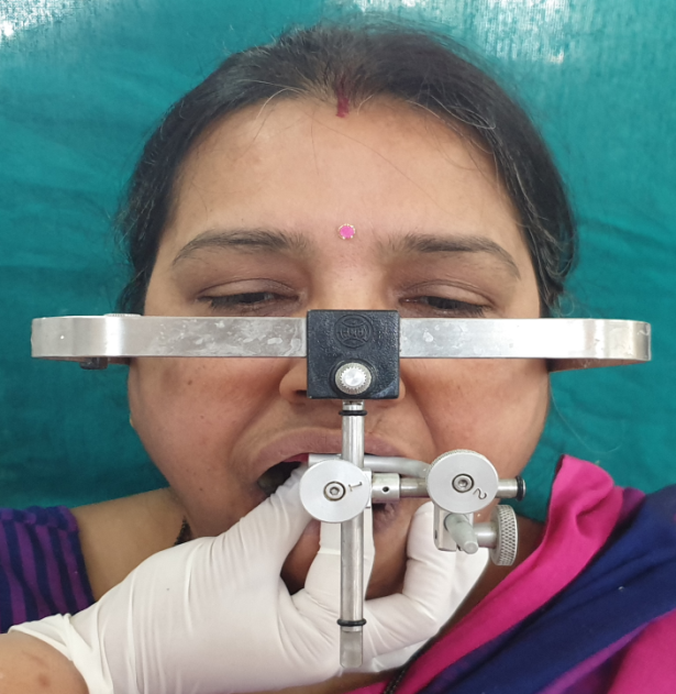
Figure 2: Facebow record made for fabrication of splint
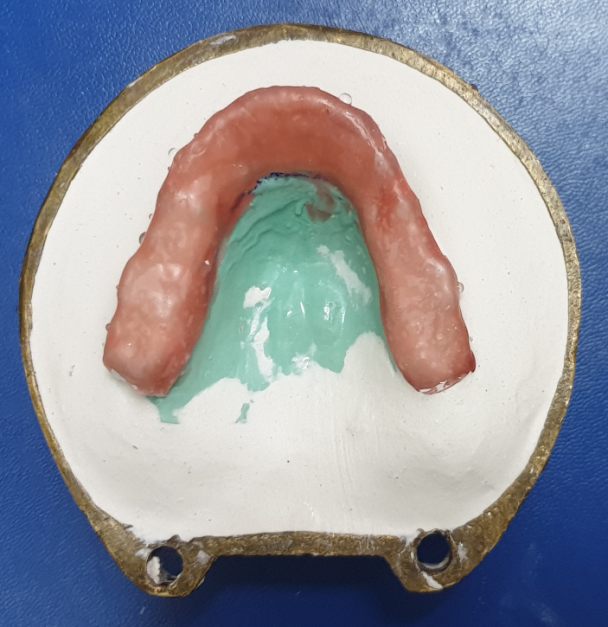
Figure 3: 2mm wax sheet adapted on the maxillary cast for even thickness spnit fabrication
The waxed appliance was invested and processed with self-cure clear acrylic. After the initial fabrication process, the fit of the appliance was checked and the anterior stop was constructed (Fig. 4).
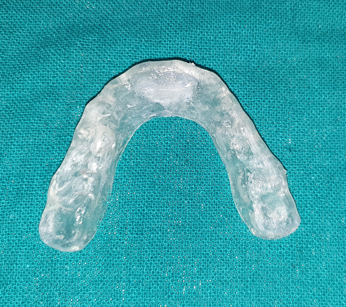
Figure 4: Clear acrylic splint with anterior stop
LOCATING THE CORRECT ANTERIOR POSITION
The anterior stop is used to locate the anterior position. The surface of the stop was adjusted to be flat and perpendicular to the long axes of mandibular incisors.
The patient was made to repeatedly open and close on the stop. When incisors contact with it the posteriors were close but did not contact the posterior portion of appliance.
The joint is evaluated for the symptoms and the anterior position that stopped the clicking was marked with blue marking paper as the patient tapped on the stop (Fig.5).
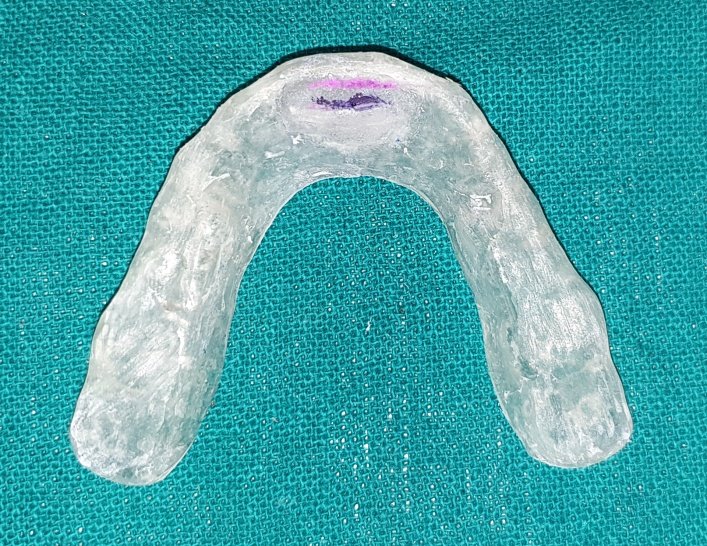
Figure 5: 2mm anterior point marked and grooved
Once this was marked the appliance was removed and the area of contact was grooved approximately 1mm deep with small round bur.
The appliance was then returned to the patient’s mouth, and the position was verified.
Once the proper location was found the joints were revaluated for symptoms. There was no joint sound on opening and closing. Joint pain on clenching was also reduced.
When the joints symptoms had been eliminated and verified by the anterior stop, the appliance was taken out of the patient’s mouth and self curing acrylic was added to the remaining occlusal surface to establish occlusal contacts and seated again in the mouth with the petroleum jelly applied to the mandibular teeth. And the patient was instructed to close on the groove (Fig.6).
Excess acrylic was placed lingual to the groove and the device was returned to the mouth (Fig.7).
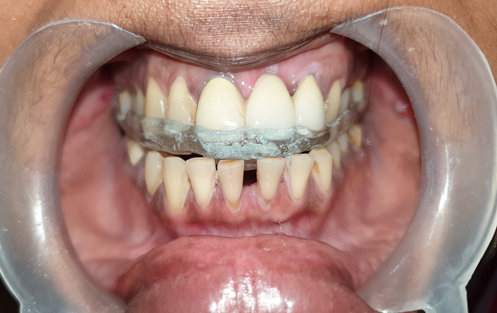
Figure 6: Splint in place with mouth closed at antetior groove
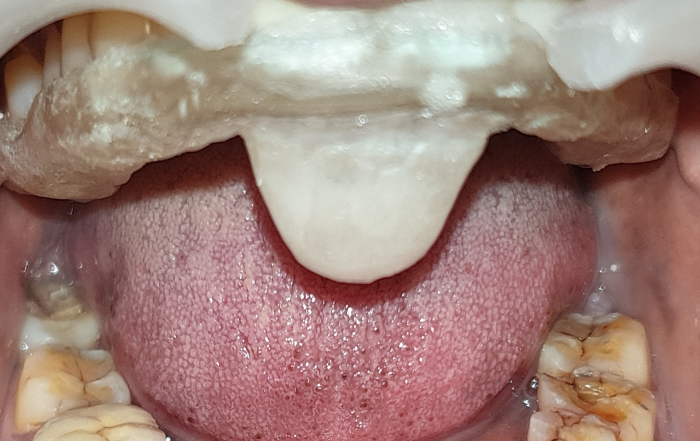
Figure 7: Anterior guiding ramp created with acrylic distal to the groove marked
The patient was asked to slowly close into the grooved area by encouraging her to close more anteriorly thereby not disturbing the acrylic that will form guiding ramp.
When the resin became firm and before the heat was produced, the appliance was removed and allowed to bench cure.
DISCUSSION
The anterior positioning appliance is sometimes called an orthopedic repositioning appliance because its goal is to change the position of the mandible in relationship to the cranium.6
It is an interocclusal device that encourages the mandible to assume a position more anterior than the intercuspal position. Its goal is to provide a better condyle-disc relationship in the fossa so that the tissues have a better opportunity to adapt or repair.7
The goal of treatment is not to alter the mandibular position permanently but only to change the position temporarily to enhance adaptation of the retrodiscal tissues.7
Once tissue adaptation has occurred, the appliance is eliminated, allowing the condyle to assume the MS position and painlessly function on the adaptive fibrous tissues.
INDICATIONS
- Disc derangement disorders.
- Joint sounds (e.g. single or reciprocal click).
- Intermittent or chronic interlocking of the TMJ.
- Some inflammatory disorders such as retrodiscitis.
The anterior positioning appliance is a full-arch acrylic device that can be used in either arch. However, the maxillary arch is preferred because a guiding ramp [Figure 4] can be more easily fabricated to direct the mandible into the desired forward position; with a mandibular appliance, the guiding ramp does not achieve this forward position easily.8 Hence, the mandible is not controlled as well.
The patient is instructed to wear the appliance only at night. It should not be worn during the day so that the normal function of the condyle will promote the development of fibrotic connective tissue in the retrodiscal tissue.9 If the patient reports pain during the day, the appliance may be worn for short periods during daytime to reduce the pain.10
REFERENCES
- Wright EF, North SL. Management and treatment of Temporomandibular disorders: A clinical perspective. J Man Manip Ther. 2009;17(4):247–54.
- Issacson G, Linde C, Isberg A (1989) Subjective symptoms in patients with temporomandibular joint disk displacement versus patients with myogenic craniomandibular disorders. J Prosthet Dent 61:70–77
- Okeson JP (ed) (1996) Orofacial pain: guidelines for assessment, diagnosis, and management. Quintessence, Chicago
- Paoloni J, DeVos RJ, Hamilton B, Murrell GA, Orchard J. Platelet-rich plasma treatment for ligament and tendon injuries. Clin J Sport Med. 2011;21(1):37–45.
- Navi F, Motamedi MHK, Talesh KT, Lasemi E, Nematollabi Z. Diagnosis and Management of Temporomandibular Disorders. In: Motamedi MHK, editor. A Textbook of Advanced Oral and Maxillofacial Surgery. Croatia: InTech; 2013. ISBN: 978-953-51-1146-7.
- Vaughan EN. Bite relief. Br Dent J 2007; 203:668-9.
- Moufti MA, Lilico JT, Wassell RW. How to make a well-fi tting stabilization splint. Dent Update 2007; 34:398-400.
- Woodside DG, Metaxas A, Altuna G. Th e infl uence of functional appliance therapy on glenoid fossa remodeling. Am J Orthod Dentofacial Orthop 1987; 92:181-98.
- John MT. Splint therapy over a time period of 3 months does not provide additional benefit for TMD pain relief over dentist prescribed self-care alone. JEvid Based Dent Pract 2007; 7:1601.
- Stohler C. Temporomandibular joint disorders--the view widens while therapies are constrained. J Orofac Pain 2007; 21:261.
Disclaimer from Guident
The views and opinions expressed in articles published in Guident are solely those of the authors and do not necessarily reflect the views of the editors, the editorial board or the publisher, Ivory India. Guident does not endorse any product, technique or treatment described. Clinical content is intended for qualified dental and medical professionals and is not a substitute for individual clinical judgement. Guident, its editors and the publisher accept no liability for any consequence arising from the use of information published in the journal.
How to cite this article
Dr. Preksha Jain, Dr. Gulab Chand Baid. Prosthodontic Management of Anterior Disc Derangement with Reduction-a Case Report. Guident. 2021 Apr;14(5). Available from: https://guident.in/articles/prosthodontics/prosthodontic-management-of-anterior-disc-derangement-with-reduction-a-case-report.html