Dental Complications After COVID-19 Mucormycosis Understanding the Pathophysiology-Case Series Part-2
Wisdom Dental Clinics, Jaipur, India
ABSTRACT
Covid-19 a pandemic disease affected population around the world, came into existence during late 2019.It caused mild to severe symptoms majorly affecting upper respiratory system, in some individuals post complications were also reported. Patients who were severely affected with covid-19 are found to be associated with abnormalities which increase the risk for thromboembolic events in the body. Currently there is very less data available highlighting the thromboembolic events in covid-19 infected individuals. In this we emphasize three cases which were suspected for thromboembolic event having unique presentation with literature review and showing importance of radiological and pathological findings that becomes highly important for health workers to consider while treating and diagnosing their patients. This is first ever pilot study we were making to diagnose thromboembolic event and planned for CT study of face angiography.
Key words: Thromboembolic event, Necrosis, Covid-19,CT study of face angiography
INTRODUCTION
Around December 2019,a virus emerged into the population of Wuhan, a city of china causing a cluster if pneumonia cases which now is named as severe acute respiratory syndrome corona virus -2(SARS-CoV-2).1 This virus took no time to spread affecting the world thus becoming pandemic.2 We continue our series of case report further:
To investigate the incidence of thromboembolic events in covid-19 infected patients dedicated imaging is carried out in patients to look for emboli. For the initial identification of coronavirus related arterial or venous thromboembolic events prompted for the expanded use of vascular imaging studies such as CT angiography and venous duplex ultrasounds.3
D-dimer is a fibrin degradation product which is being generated in the final step in the process of thrombus formation. It is elevated in acute VTE. D-dimer is elevated in various clinical conditions where thrombosis and fibrinolysis are increased like malignancy, trauma, surgery, stroke, acute coronary syndrome, infection, pregnancy and advancing age.4
Generally viral infections cause an imbalance between pro and anticoagulant states causing disruption of vascular endothelium during the course of disease.5 Mobility associated with bed for bed rest in severe cases along with activation of systemic coagulation increase the risk for venous thromboembolism in covid -19 patients.5
The thromboembolic event occurs by invading the arteries leading to thrombosis that result in necrosis of both hard as well as soft tissues. Medical literature does not have much data available for cases of post covid-19 dental complications.
However, specific mechanism and pathological pathway have been noticed linking periodontal diseases with systemic diseases, showing translocation of bacteria to blood, induced auto immune damage an systemic inflammation.6
After presenting with our previous dental complication a case of osteomyelitis related to post covid-19 infection in part 1 here. In this we discuss about three case reports presenting with similar condition with slight variation which were suspected to be cases of thromboembolic event associated with post covid-19 infection. As covid infection involve cardiac and renal problems and also invading the arteries leading to thrombus formation so here we suspected it can even involve maxillary arteries and lead to thromboembolic event.
In case report 2 and 3 all relevant radiological and pathological tests were carried which included OPG, CBCT, KOH mount test, culture and sensitivity to look for any co relating lead affecting bone or teeth then moving on to our 4th case report we decided to go for CT study of face angiography and D-dimer test to rule out thromboembolic event.
This is the first ever pilot study we were planning to know any thromboembolic movement is present there or not. Early and prompt diagnosis can definitely reduce the rate of morbidity and mortality of diseases which are lethal.
CASE REPORT-2
A male patient aged 60 years visited to the clinic with the chief complaint of swelling in upper jaw and difficulty in chewing.
On intraoral examination multiple draining sinus were found in maxillary arch with generalised occlusal attrition in the teeth.(Figure 1).
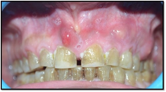
Figure 1. Multiple draining sinus in maxillary arch
Palatal mucosa was found to be affected showing oedema and teeth were non carious.(Figure 2)
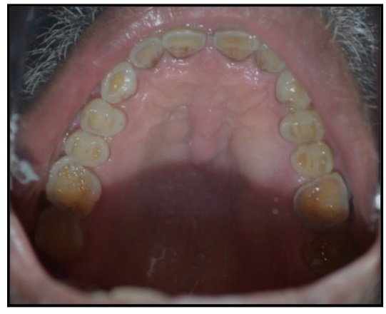
Figure 2. oedematous palatal mucosa with occlusal attrition
Diffuse gingival oedema was seen involving attached gingiva all around.(Figure.3)
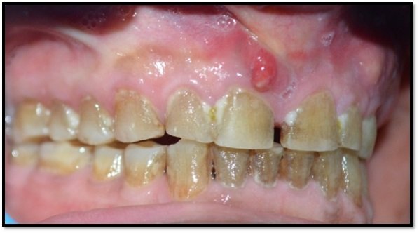
Figure 3. Diffuse Gingival oedema
Now, further OPG, CBCT as well as other tests were preferred to rule out any other pathology related to bone or teeth.
OPG report revealed that no pathology is associated with bone or teeth.(Figure.4)

Figure 4. OPG of maxillary arch
Further for pathologic evaluation in maxilla and maxillary sinus CBCT scan was performed which showed:
Osteomyelitis of the maxillary jaw predominantly involving the left side and right side till 15 mesial aspect. (Avascular osteo-necrosis of the maxilla) (Figure 5.a,b,c,d)

Figure 5a,b. CBCT of maxilla axial view

Figure 5c,d. CBCT maxillary sinus showing avascular osteo-necrosis
Looking at the condition of the case, patient was referred for haematological, microbiological and radiological tests. Here report of patient showed increase in platelet count.(Figure.6)

Figure 6. Increase in platelet count
KOH mount revealed absence of fungus.(Figure.7)
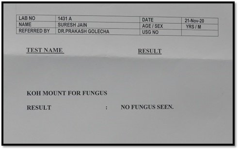
Figure 7. KOH mount test- no fungus seen
Culture and sensitivity test report showed presence of coagulase +ve for staphylococci. Sensitivity test report resulted as follows:
SENSITIVE:
Ceftriazone, Cefexime, Piperacillin, Cefperazone, Meropenem, Clindamycin, Netilimycin, Levofloxacin, Ofloxacin, Telcoplanin, Vancomycin, Azithromyci, Clarithromycin, Doxycycline and Linezoid (Figure.8 a,b)
RESISTANT:
Ampicillin, Amoxacillin,Ciprofloxacin, Co-tromoxazole
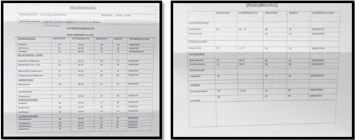
Figure 8a,b. Culture sensitivity findings showing coagulase positive staphylococci growth with antibiotic sensitivity
CASE REPORT-3
A 52 year old male patient reported to our clinic with his chief complaint of swelling along with difficulty in chewing from both the sides in upper jaw. Patient gave history of previous infection of TB 6years back.
After examining intraorally multiple pus discharging sinus were found on both the side of maxillary arch with bilateral inflammation.(Figure 1.)
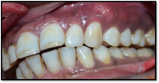
Figure 1. Multiple pus discharging sinus in maxillary arch.
Palatal arch presented with diffuse oedema.(Figure 2)
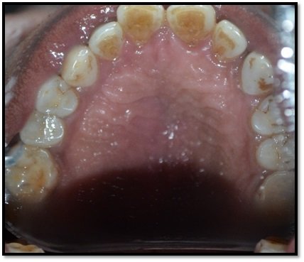
Figure 2. Palatal arch with oedema
To manage the present symptoms patient was prescribed with PPI, antibiotics, NSAIDS and antifungal drugs.
To rule out any pathology involving soft and hard tissue patient was advised for CBCT,CECT study of PNS,CECT of face and neck.
The CBCT of maxilla and maxillary sinus revealed:
- Acute Osteomyelitic changes of the maxillary jaw predominantly involving the left side region (Avascular osteo-necrosis of the maxilla?)
- Right and left maxillary sinus chronic sinusitis? (Figure.3 a,b)

Figure 3a,b. CBCT of maxilla and maxillary sinus
While CECT of face and neck showed:
- extensive permeative osteolysis of hard palate likely infective; fungal or granuloma? Neoplastic? (Figure. 4 a,b,c)

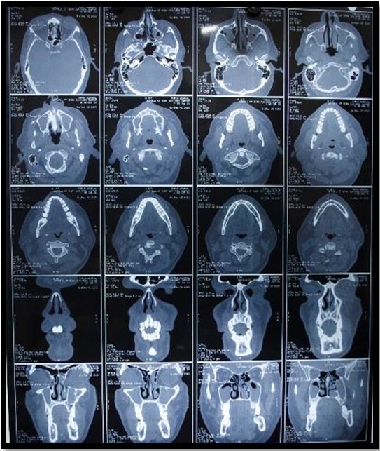
Figure 4 a,b and c. CECT report showing permeative osteolytic lesion likely to be infective.
The CECT study of PNS gave impression of:
Extensive permeative osteolysis of hard palate with soft tissue component consistent with infective /granulomatous disease.(Figure .5)

Figure 5. CECT study of PNS showed permeative osteolysis of hard and soft palate with infective disease
Further MRI of neck was planned to know if the lesion is infective or neoplastic.The report showed :
Multiple enlarged oval shaped lymphnodes with maintained fatty hilum are seen in bilateral level Ib,II largest measuring 20*13*14mm in left level Ib (benign/reactive).(Figure. 6)

Figure 6. MRI report showing enlarged lymphnodes
To rule out any infection culture and sensitivity tests were carried out which showed growth of enterococcus faecalis after 24hrs of incubation.(Figure 7)
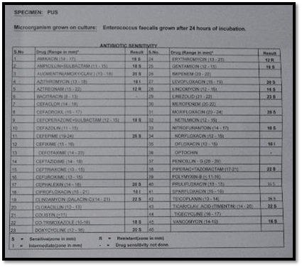
Figure 7. Culture and sensitivity report showing growth of Entrococcus faecalis with antibiotic sensitivity
To find out malignancy punch Biopsy was adviced which revealed acute or chronic inflammation with no evidence of malignancy.(Figure .8)
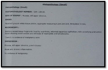
Figure 8. Punch biopsy report showing acute/chronic inflammation with no evidence of malignancy
CASE REPORT-4
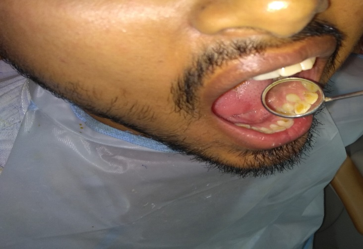
A 38 year old male patient reported to the clinic presenting with chief complaint of swelling in upper teeth region with pus discharge and difficulty in chewing. After examining intraorally tenderness was present upon palpatation, multiple pus discharging sinus were found in maxillary arch.(Figure.1)

Figure 1. Multiple pus discharging sinus in maxillary arch
Palatal mucosa was found to be intact with oedema. (Figure.2)

Figure 2. Intact palatal mucosa
Upon taking history of any previous infections ,patient was found to be infected with covid -19.
Patient also gave medical history of hypertention and diabetes which were controlled by medications since 2yrs. He additionally complained of difficulty in breathing and blocked nose.
For management of present signs and symptoms he was prescribed with NSAIDS, PPI inhibitor, antibiotic(Amoxacillin+clavulanic acid), mucolite, montelucast+levocitrezine and nasal spray with steam inhalation for his breathing issues. Patient was also prescribed with steroids to reduce inflammation.
As patient gave history of hypertension so he was advised for ECG and X-ray chest to look for any underlying chest infection and changes.
Further based on the patient’s clinical presentation other laboratory findings,CT and CBCT of face was adviced which revealed:CBCT report revealed:
Acute Osteomyelitic changes of the maxillary jaw predominantly involving the left side posterior region (Avascular osteo-necrosis of the maxilla)
- Right and left maxillary sinus chronic sinusitis? (Figure.3a,b)

Figure 3a,b. CBCT of maxilla and maxillary sinus
CT study of PNS were suggestive of (Figure 4a,b):
- Bilateral maxilloethamoid sinusitis (sporadic type) with bilateral maxillary retension cyst.
- keros type I ethmoid roof/olfactory groove
- left nasal turbinate hypertrophy
- blocked OMC
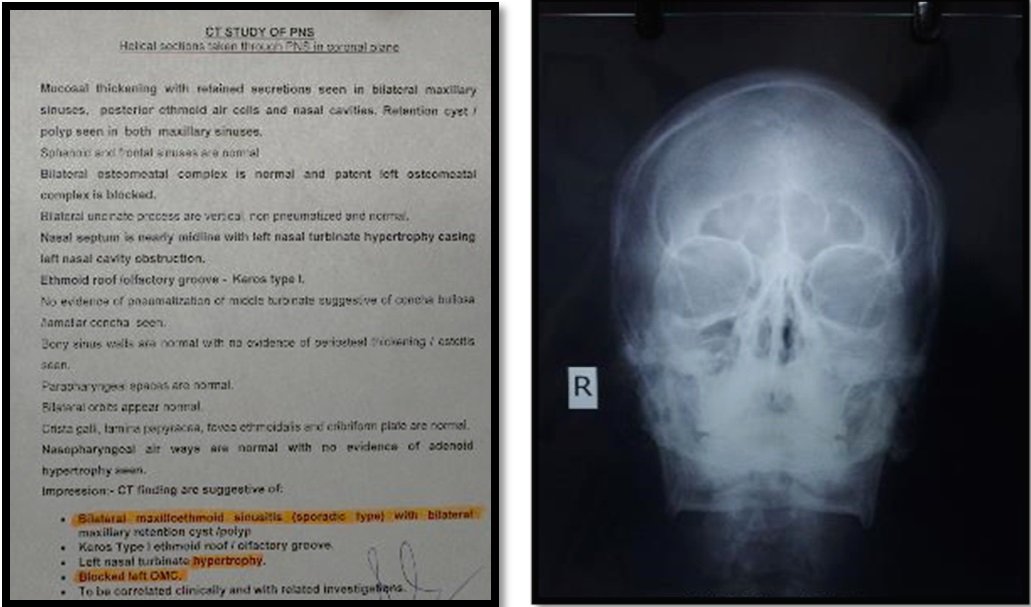
Figure 4a,b. Report and radiograph of CT study of PNS
Culture and sensitivity test showed presence of methicillin sensitive coagulase negative staphylococci.(Figure 5a,b)
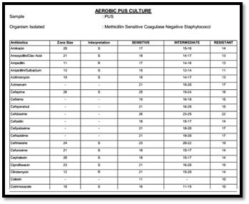
Figure 5a. Methicillin sensitive coagulase negative staphylococci

Figure 5b. antibiotic sensitivity report
Looking at the clinical sign and symptoms the case was suspected to be a thromboembolic event so to rule out D-Dimer test and CT study of face angiography was done.
Value of D-Dimer test came to be 0.3ug/mL. (Figure 6)
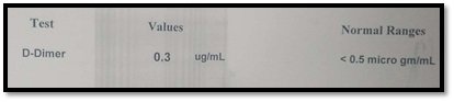
Figure 6. D-Dimer value 0.3ug/mL
Finally CT study of face angiography was done which showed:
Normal study of neck vessels and vessels supplying hard palate as well as gingiva.(Figure 7.a,b,c,d) (Figure.8)

Figure 7.a,b,c,d. CT of face angiography showing normal neck vessels and vessels supplying hard palate and gingiva

Figure 8. CT study of face angiography report
DISCUSSION
As many post covid complications are reported with involvement of arteries leading to thromboembolic events so here we planned for a pilot study based on two similar cases, to detect presence of thrombus formation in maxillary artery. We decided to go with CT study of face angiography and D-dimer test values to look for presence of any thromboembolic movement.
We were reported with three cases presenting almost similar clinical condition first case was of 60 yr old male patient and other was of 38yr old male patient both presented with multiple discharging sinus in maxilla along with difficulty in chewing and another case of 52yr old male patient with similar clinical condition but bilaterally .All three cases were suspected to be of thromboembolic event so radiological and pathological evaluation was done but in first and second case CBCT of maxilla and maxillary sinus showed osteomyelitis of maxillary arch,possibly an avascular necrosis. While in the other case general lab findings could not clear the suspected diagnosis so further d-dimer test and CT study of face angiography was done to see the co relation with thromboembolic event associated with covid -19 infection,as patient clinically recovered from covid-19 but later developed multiple pus discharging sinus in maxillary arch.
This is unfortunate that no such cases are reported in medical literature till yet. Several studies reported for thromboembolic events especially in ICU patients who were previously affected by covid-19 severely.7,8
Few of the studies reported,showed cases of mild covid-19 affected individuals to complicate with thromboembolic events during the active phase of the disease.9-10
The normal immunologic response of patients to infections get altered in comorbid conditions such as uncontrolled diabetes mellitus and hypertension. Such kind of patients have alterations in polymorphonuclear leukocyte response along with decrease in granulocyte phagocytic ability. Few Reports have suggested that low immunity patient’s serum has reduced to inhibit Rhizopusinvitro, which as a result makes them suitable hosts for opportunistic infections.11 In this our third case report, patient was having medical history of diabetes and hypertension which were controlled by medication since two years which made our case more susceptible for post covid-19 complication.
New studies showing evidence of thromboembolism as a complication of covid-19 is very limited, still cases have started emerging.12 Thrombus formation mechanism may be variable, but it is been postulated that Covid -19 can lead to increase in inflammatory reaction, hypoxia as well as disseminated intravascular coagulation (DIC),which in turn can lead to patient’s natural tendency to thromboembolic disease.12
Thromboembolic events not even present with fever,swelling or pain, it is also connected with other findings which may or may not be present such as proptosis,periorbital swelling or palsies. Spread of infection from paranasal, ethmoidal, maxillary and sphenoidal sinus may lead to secondary infection, however dental origin is rare.13
In our part 2 third case, individual presented with breathing difficulty and blocked nose along with atypical facial pain. CT study of PNS showed involvement of maxillary sinus with blocked left osteomeatal complex.
According to one of the studies stated male predilection is more than female, which made one of the possible explanation for male sex to have an increased risk factor for severe covid-19 based upon immune response to SARS-CoV-2 infection.14 The reason for male predilection is in the theory that women produces less no. of inflammatory cytokines after any kind of infection, which id linked with higher survival rate in women.15 It is also been noted that men are much more prone to thrombosis as compared to women affected with covid-19.16 Our all three cases are also dealing with male gender.
Different coagulation pathways causing increase and decrease in coagulation results in increasing of vonwillibrand factor which develop cross linked fibrin clots. During the process of breakdown D-dimer level increase with fibrin degradation levels which is been found connected with poor prognosis of covid-19 affected individuals.17,18 Third case could be found related with such event as patient gave history of covid infection, so D-Dimer test was done which came to be within normal range value.
Thrombosis is found frequently associated with critically ill patients which can be fatal if not diagnosed and managed within time. The health care providers should take into consideration prophylactic versus therapeutic anticoagulation based upon patient’s criteria which include laboratory results,imaging,clinical suspicion. Also making a balance between thrombotic and bleeding risks.19
In 60yr old male patient and 52 yr old male patient case before it was thought to be a case of thrombolytic event but looking upon laboratotay and radiological findings with no such co relating pathologic results were found,later it was considered to be a cases of avascular necrosis of maxilla. But on the other hand our other case of 38yr old male patient who was previously infected with covid-19 and presented with atypical facial pain along with multiple discharging sinuses in maxillary arch so to clear up the dilemma we went further to go for d-dimer values and angiography of face which gave clear picture that it is not the case of thromboembolic event because there were no such laboratory or radiological findings proving it to be a thromboembolic event after covid-19 infection.
This is the first pilot study till now to go for CT study of face angiography to confirm any thromboembolic event involving maxillary artery and its branches. As limited data is present on thromboembolic events associated with dental conditions,still clinicians and dental surgeons should consider thromboembolic events and be aware of its signs and symptoms while treating patients.
Clinical as well as other lab findings were not showing a final case of thromboembolic event still affected soft and hard tissues made them as suspected cases for thrombosis.
CONCLUSION
Thrombosis is one of the complication of covid -19 which may occur late after recovery from covid-19. Such events cause avascular necrosis due to impaired blood supply in certain parts of maxilla. With many of known risk factors associated with thrombotic events like immobility, advanced age, surgery, obesity are increasing in present society, it has become a growing public health issue,yet still till now this condition has received very little attention. In this pilot study looking upon all three cases our theory for thromboembolic event went inaccurate as radiological and pathological findings i.e CT study of face angiography and D-Dimer vale confirmed that cases were not of thromboembolic event. Untill more information is available,routine active surveillance is recommended which is being guided by clinical,pathological and radiological assisment to make prompt diagnosis of thrombotic events which may be beneficial in guidance and management of such cases.
REFERENCES
- World Health Organization. Novel Coronavirus (2019-nCoV): situation report - 1: 21 January 2020. [cited 2020 Aug 12]. Available from: https://www.who.int/docs/default-source/coronaviruse/situation-reports/20200121-sitrep-1-2019-ncov.pdf?sfvrsn=20a99c10_4 [ Links ]
- World Health Organization. Coronavirus disease 2019 (COVID-19): situation report - 51. [cited 2020 Aug 12]. Available from: https://www.who.int/docs/default-source/coronaviruse/situation-reports/20200311-sitrep-51-covid-19.pdf?sfvrsn=1ba62e57_10 [ LiLinks nks ]
- Edison Lee, Adam Krajewski, Cynthia Clarke, David O’Sullivan, Timothy Herbst& Steven Lee.Arterial and venous thromboembolic complications of COVID-19 detected by CT angiogram and venous duplex ultrasound. JEmergRadiol (2021). https://doi.org/10.1007/s10140-020-01884-0
- Hargett CW, Tapson VF: Clinical probability and D-dimer testing: how should we use them in clinical practice? Semin. Respir. Crit. Care Med. 29, 15–24 (2008).
- Atallah B, Mallah SI, AlMahmeed W. Anticoagulation in COVID-19. European heart journal cardiovascular pharmacotherapy, pvaa036. 2020. Advance online publication. https://doi.org/10.1093/ehjcvp/pvaa036
- Schenkein, H. A, Papapanou, P. N, Genco, R, & Sanz, M (2020). Mechanisms underlying the association between periodontitis and atherosclerotic disease. Periodontology 2000, 83(1), 90– 106. https://doi.org/10.1111/prd.12304. Wiley Online Library PubMed Web of Science®Google Scholar
- Poissy J, Goutay J, Caplan M, Parmentier E, Duburcq T, Lassalle F, et al. Pulmonary embolism in patients with COVID-19: awareness of an increased prevalence. Circulation. 2020;142:184-6. [ Links ]
- Nahum J, Morichau-Beauchant T, Daviaud F, Echegut P, Fichet J, Maillet JM, et al. Venous thrombosis among critically ill patients with coronavirus disease 2019 (COVID-19). JAMA Netw Open. 2020;3:e2010478. [ Links ]
- Jasinowodolinski D, Filisbino MM, Baldi BG. COVID-19 pneumonia: a risk factor for pulmonary thromboembolism? J Bras Pneumol. 2020;46:e20200168. [ Links ]
- Nauka PC, Oran E, Chekuri S. Deep venous thrombosis in a non-critically ill patient with novel COVID-19 infection. Thromb Res. 2020;192:27-8. [ Links ]
- Brown OE, Finn R. Mucormycosis of the mandible. J Oral MaxillofacSurg 1986;44:132-6.
- Klok FA, Kruip M, van der Meer N, Arbous MS, Gommers D, Kant KM, Kaptein F, van Paassen J, Stals M, Huisman MV, Endeman H. Incidence of thrombotic complications in critically ill ICU patients with COVID-19. Thrombosis Res. 2020;S0049–3848(20):30120–1. Advance online publication. https://doi.org/10.1016/j.thromres.2020.04.013.
- Gi-Sung Yeo, Hyun Young Kim, Eun-Jung Kwak, Young-Soo Jung, Hyung-Sik Park, and Hwi-Dong Jung.Cavernous sinus thrombosis caused by a dental infection: a case report.Journal of Korean association of oral and maxillofacial surgeons 2014 aug (40)(4):195-198
- Kragholm K, Andersen MP, Gerds TA, Butt JH, Østergaard L, Polcwiartek C, et al. Association between male sex and outcomes of Coronavirus Disease 2019 (Covid-19) – a Danish nationwide, register-based study. Clin Infect Dis. 2020 In Press. [ Links ]
- Nauka PC, Oran E, Chekuri S. Deep venous thrombosis in a non-critically ill patient with novel COVID-19 infection. Thromb Res. 2020;192:27-8. [ Links ]
- Bilaloglu S, Aphinyanaphongs Y, Jones S, Iturrate E, Hochman J, Berger JS. Thrombosis in hospitalized patients with COVID-19 in a New York City health system.JAMA. 2020 In Press. [ Links ]
- Giannis D, Ziogas IA, Gianni P. Coagulation disorders in coronavirus infected patients: COVID-19, SARS-CoV-1, MERS-CoV, and lessons from the past. J ClinVirol. 2020;127:104362. Advance online publication. https://doi.org/10.1016/j.jcv.2020.104362.
- Middeldorp S, Coppens M, van Haaps TF, Foppen M, Vlaar AP, Müller MC, Bouman CC, Beenen LF, Kootte RS, Heijmans J, Smits LP, Bonta PI, van Es N. Incidence of venous thromboembolism in hospitalized patients with COVID-19. J ThrombHaemost. 2020. Accepted author manuscript; https://doi.org/10.1111/jth.14888.
- SamhatiMondal, Ashley L. Quintili,KunalKaramchandani, and somnathbose.Thromboembolic disease in COVID-19 patients: A brief narrative review.J Intensive Care. 2020; 8: 70.Published online 2020 Sep 14. doi: 10.1186/s40560-020-00483-y
Disclaimer from Guident
The views and opinions expressed in articles published in Guident are solely those of the authors and do not necessarily reflect the views of the editors, the editorial board or the publisher, Ivory India. Guident does not endorse any product, technique or treatment described. Clinical content is intended for qualified dental and medical professionals and is not a substitute for individual clinical judgement. Guident, its editors and the publisher accept no liability for any consequence arising from the use of information published in the journal.
How to cite this article
Dr. Gaurav Gupta, Dr. D.K. Gupta, Dr. Neelja Gupta. Dental Complications After COVID-19 Mucormycosis Understanding the Pathophysiology-Case Series Part-2. Guident. 2021 Apr;14(5). Available from: https://guident.in/articles/general/dental-complications-after-covid-19-mucormycosis-understanding-the-pathophysiology-case-series-part-2.html