Endodontic Pecularities-a Case Series
Department of Conservative Dentistry and Endodontics, Seema Dental College And Hospital, Rishikesh
ABSTRACT
Introduction: There are cases in the endodontic treatment which are a real challenge. There are teeth with diferent anatomies of the root canals of diferent teeth and their treatment. Patients usually lose their teeth because these anatomical features are less familiar.
Purpose: The aim of this study is to investigate several cases with different root canal anatomies like extra root canals in maxillary first molar or mandibular premolar with vertucci’s class v anatomy of the root canal system also a Case of management of internal resorption of the root canal of mandibular canine along with a case of radix entomolaris.
Material and methods: one case of extra root canals in maxillary first molar, one case of mandibular pre molar with vertucci’s class v ,a case of management of internal resorption of mandibular canine, one case of S shaped canal and its management in mandibular first premolar along with a case of radix entomolaris.Careful exploration of the grooves between all orifices with magnification, use of ultrasonic irrigation; and a modified filling technique are of particular use.1
Results: Results are observed after several years. In performing a root canal treatment on such teeth, one should appreciate the complexity of the root canal system, canal obliteration and configuration, and the potential for additional root canal systems.
Conclusions: Knowledge of the phenomenon of the complexity of the root canal system, canal obliteration and configuration, and the potential for additional root canal systems and radix entomolaris will improve the medical practice of the general dental practitioner.
CASE-1
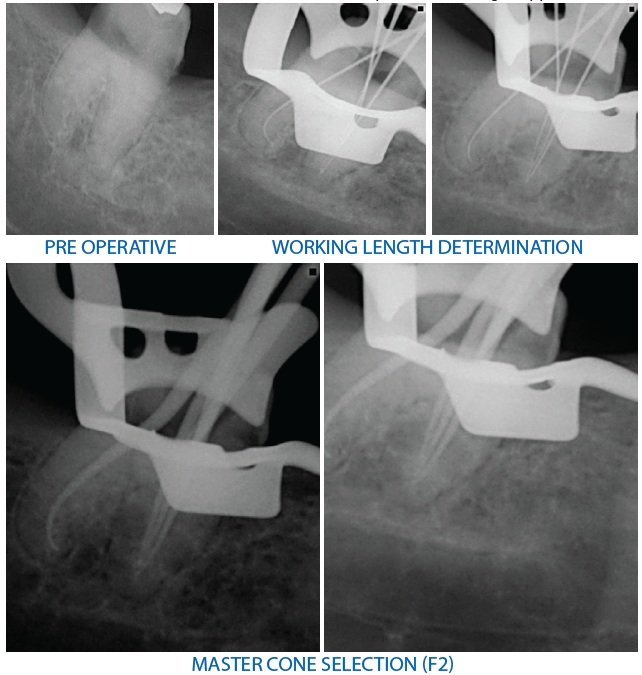
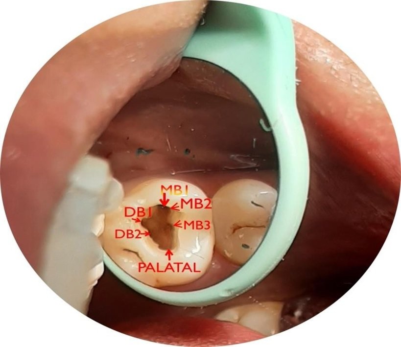
A 37-year-old male with a noncontributory medical history was referred with a complaint of severe discomfort with his right maxillary teeth. The clinical and radiographic examinations revealed a maxillary right first molar with deep occlusal caries with tenderness on percussion. The clinical findings, radiographic findings, and pulp sensibility test led to a diagnosis of irreversible pulpitis with acute apical periodontitis with maxillary right first molar. Radiographi evaluation of the involved tooth did not reveal any unusual anatomy at the first but while negotiating the canals MB1,MB2 along with MB3 on mesial side was found, with DB1 and DB2 on the distal side of the chamber and one palatal canal. Total 6 orifices were found which were confirmed radiographically and prepared with protaper gold rotary files after preparation of the canal the MB1 & MB2 were merged together which were then finally obturated.2
PRE OPERATIVE RADIOGRAPH
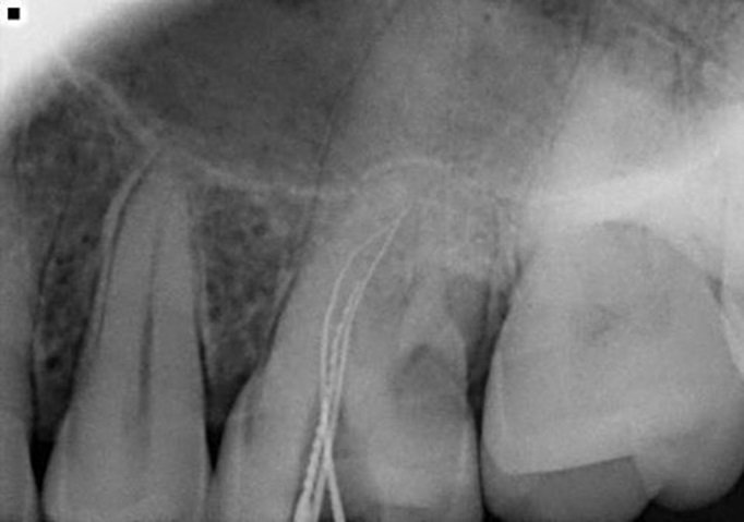
WORKING LENGTH OF MB1 ,MB2,MB3
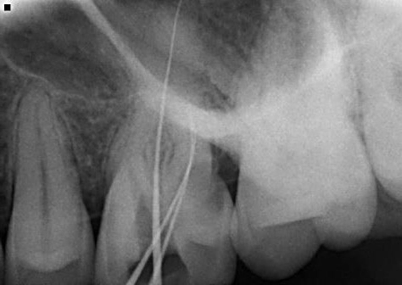
WORKING LENGTH OF DB1&DB2
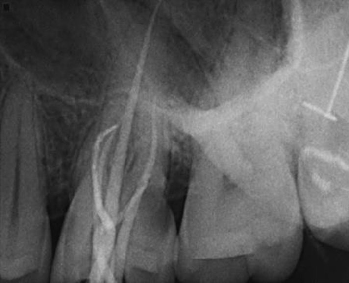
MASTER CONE SELECTED
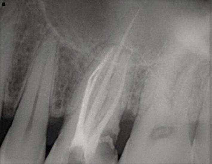
FINAL OBTURATION

POST OPERATIVE RADIOGRAPH
CASE-2
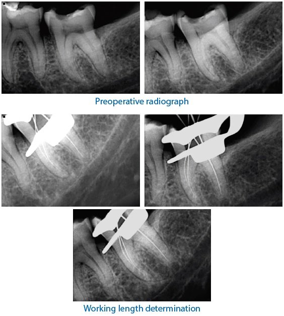
A 33-year-old male patient was referred to the Department of Conservative Dentistry and Endodontics of Seema Dental College and Hospital, Rishikesh, with the chief complaint of sensitivity and occasional pain in the left region of lower back teeth. On clinical examination, the tooth was grossly decayed and The radiograph revealed distoproximal dental caries involving enamel, dentin, and pulp of tooth 34. An electric pulp test suggested symptomatic irreversible pulpitis.3
a second root canal (palatal canal) was seen branching out from the main root canal (buccal canal) at the middle third of the root. The palatal root canal does not joins the buccal root canal at the apical third, just before the exit suggesting Vertucci type V canal configuration .The palatal canal was negotiated with #10 hand K-files. A working length radiograph was taken to confirm the presence of the palatal canal After orifice enlargement with a #1 Gates Glidden drill instrumentation was done till #20 hand K-file followed by preparation of the remaining canal using HYFLEX EDM and the irrigation for chemical debridement with 3.5% sodium hypochlorite during canal preparation.4
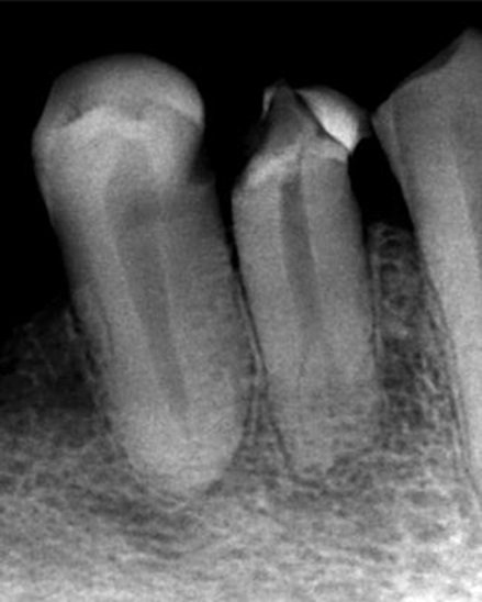
PRE OPERATIVE RADIOGRAPH
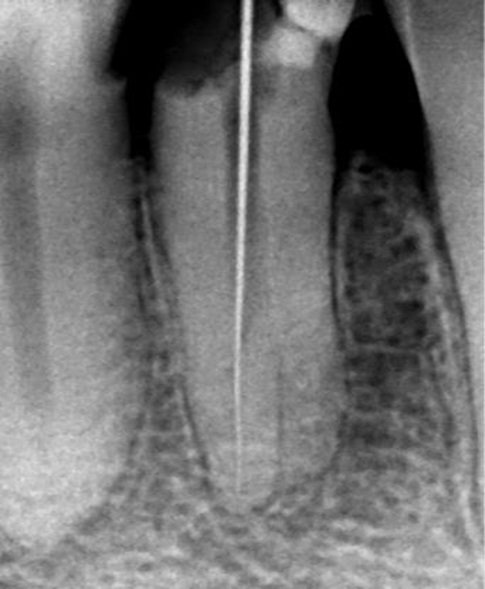
WORKING LENGTH BUCCAL CANAL
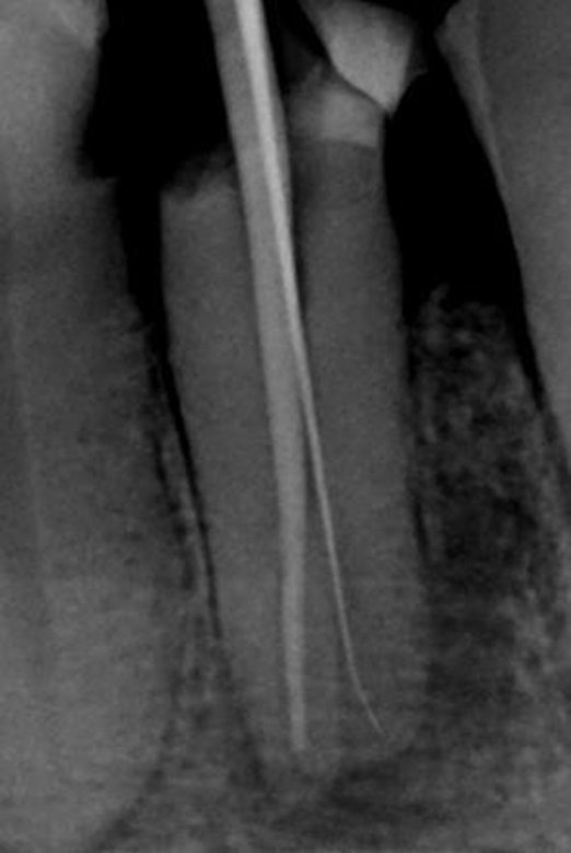
WORKING LENGTH PALATAL CANAL
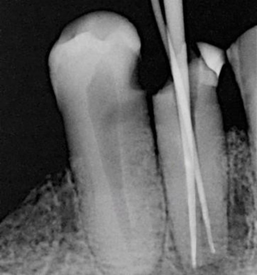
MASTER CONE MESIAL VIEW
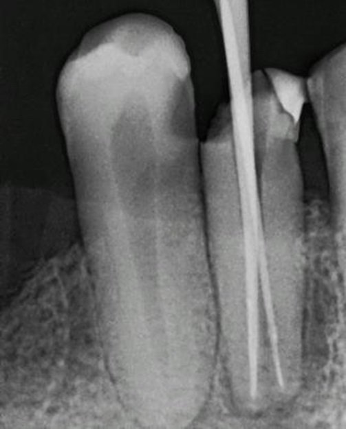
MASTER CONE DISTAL VIEW
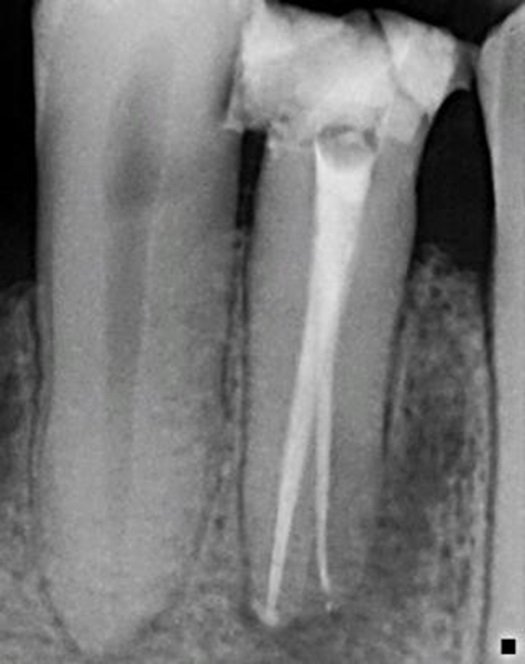
FINAL OBTURATION
CASE-3
A 45-year-old female patient came to the department with a chief complaint of dull pain in lower left front tooth region since one month.The clinical examination showed distal caries wrt33.
Radiographic examination showed radiolucency in middle third of root surface of the canine indicating a case of internal resorption and an associated radiolucency in the periapical area of same tooth Also there was widening of periodontal ligament wrt 33. The diagnosis of asymptomtic apical periodontitis wrt 33 was made.Access opening was initiated Working length radiograph was taken wrt33. Biomechanical preparation of the tooth was done in the first visit. In the next visit, 2 days later the tooth was prepared till 50 no. K file.5
Master cone was then selected corresponding to the required biomechanical preparation. Thick consistency of the sealer (Endomethasone) was mixed and properly applied with lentulospiral into the canal and the resorptive defect followed by obturation of the canal using warm vertical condensation method with thermoplasticised gutta percha. The patient was recalled on a regular basis for upto one year.5
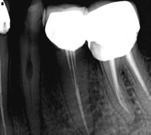
PRE OPERATIVE RADIOGRAPH
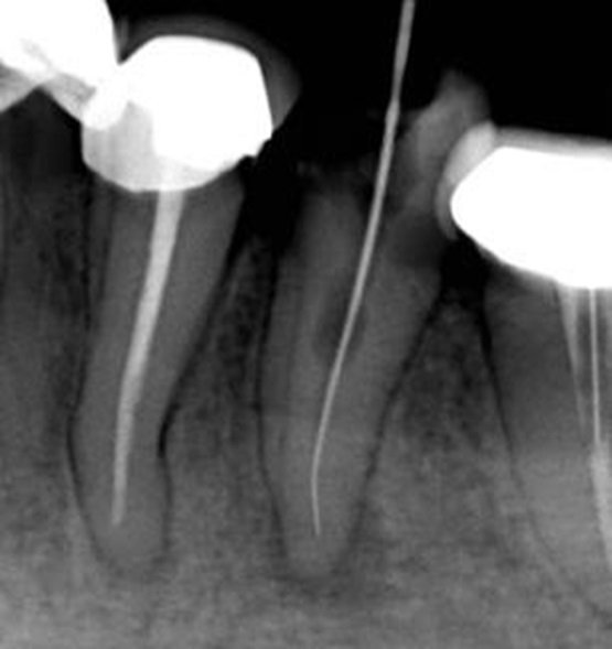
WORKING LENGTH DETERMINED
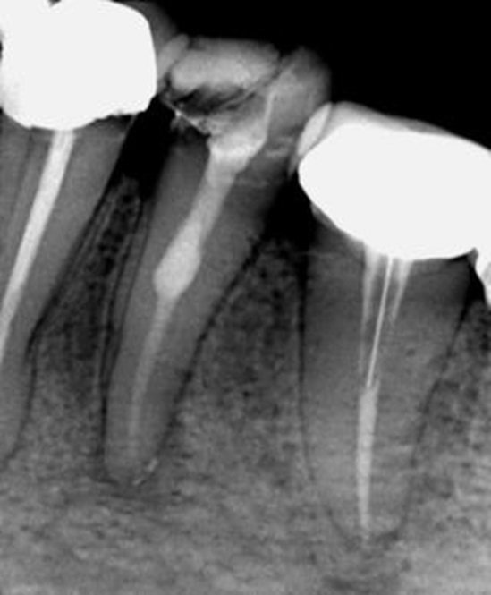
ICM PLACED
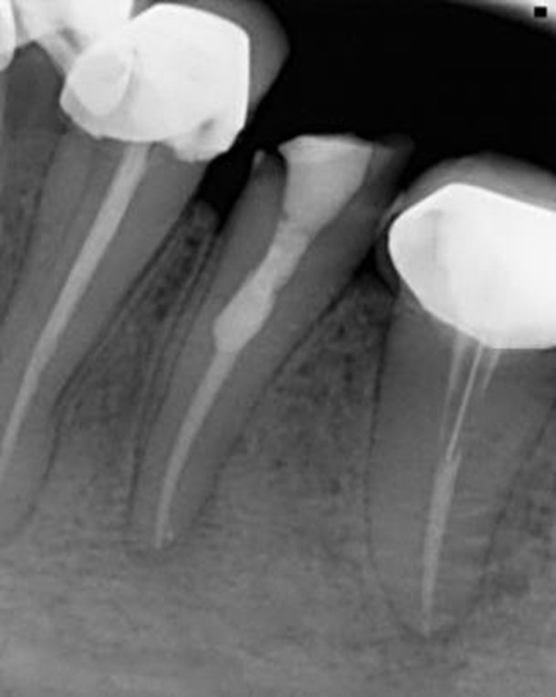
OBTURATION DONE

POST ENDO RESTORATION DONE

POST OPERATIVE RADIOGRAPH(6 MONTHS FOLLOW UP)
CASE-4
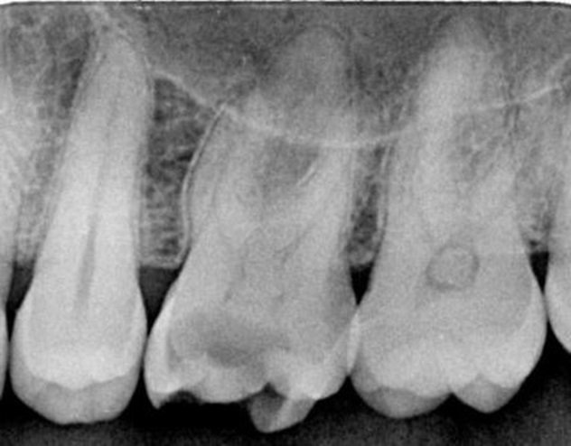
A 40-year-old male patient was referred to the clinic with severe pain to cold stimuli in his upper left maxillary arch. The medical history was noncontributory. All teeth in the area responded within normal limits to thermal cold tests except for the second left maxillary bicuspid. Probing depths were within 3 mm for all teeth of the region. Preoperative radiograph revealed a distal decay in the second left maxillary bicuspid and a double curve or s-shaped anatomy. After considering all findings, a diagnosis of irreversible pulpitis was made. Sizes. 10 K and .08 K files were initially used with the EDTA to try to reach working length. At the completion of the use of the large-sized files, 5.25% NaOCl was irrigated into the root canal preparation and a size .10 K file was taken to working length. 5.25% sodium hypochlorite was used to irrigate the root canal system between every instrument and patency was maintained with a .10 K file throughout the cleaning and shaping procedure and filled with gutta percha and obturated.6

PRE OP STRAIGHT VIEW

MESIAL VIEW
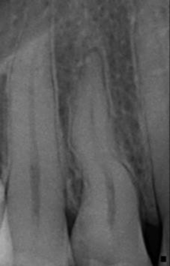
DISTAL VIEW
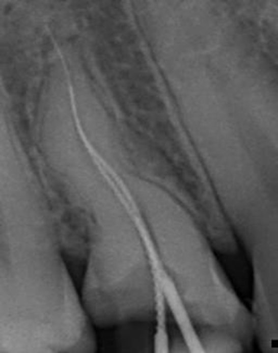
WORKING LENGTH
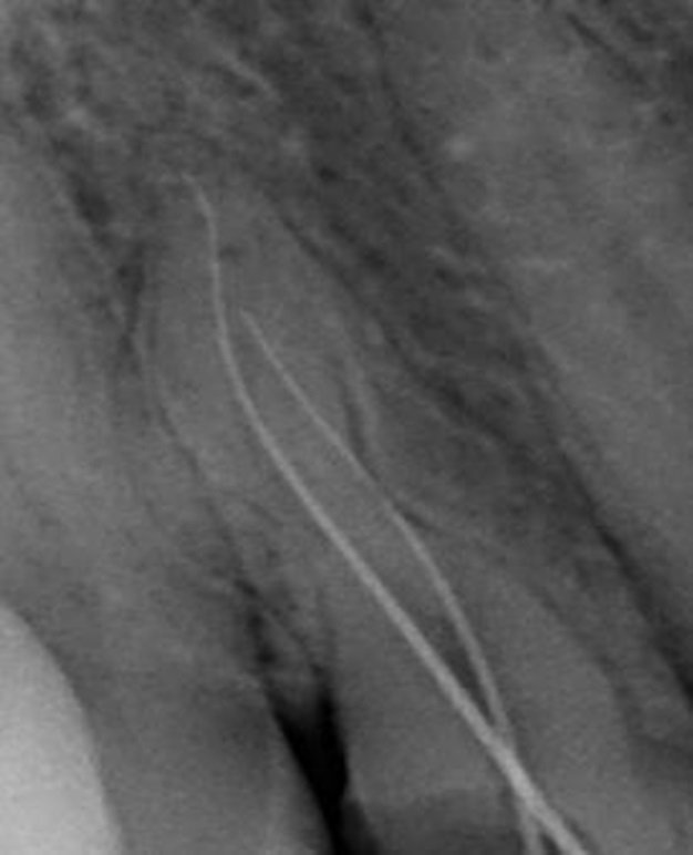
MESIAL VIEW
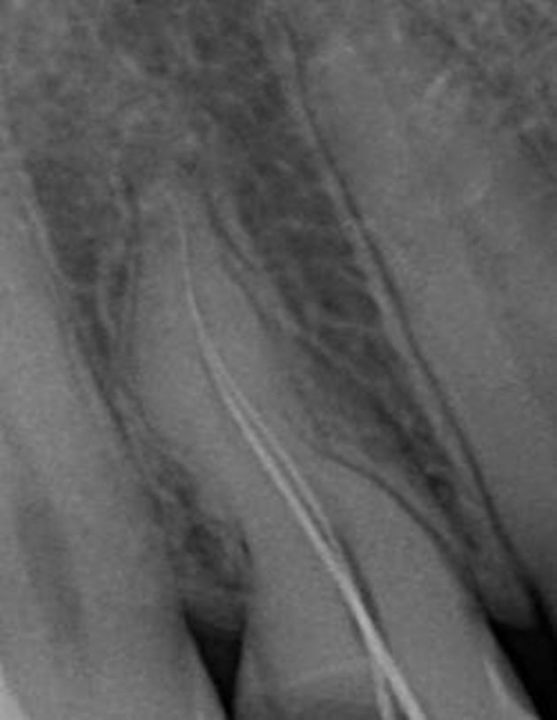
DISTAL VIEW
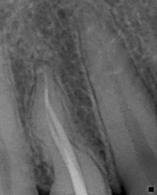
MASTER CONE (ST. VIEW)
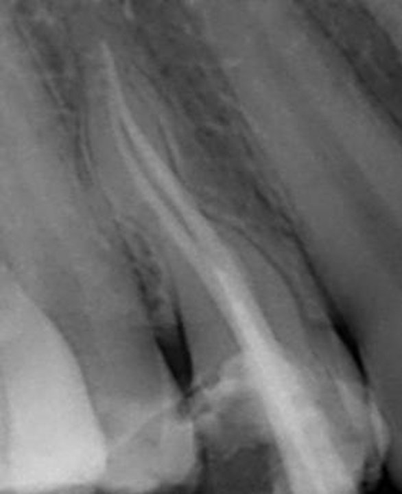
MESIAL VIEW

DISTAL VIEW
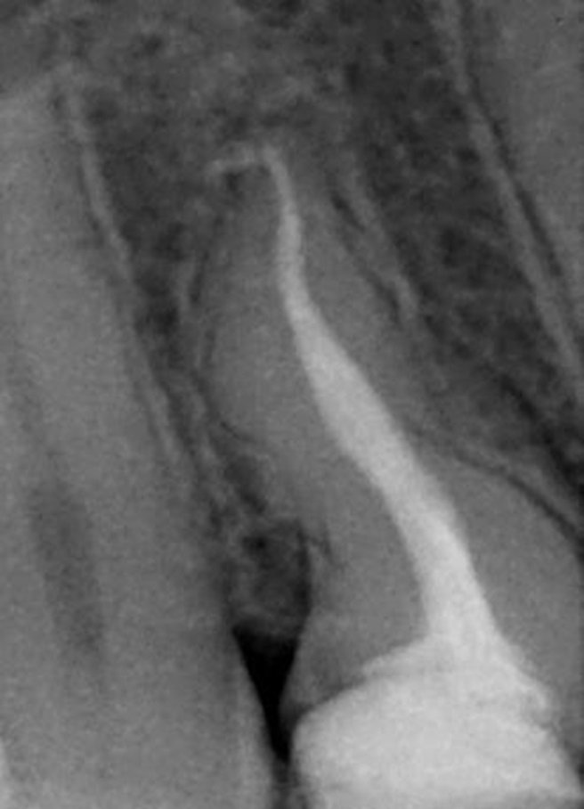
OBTURATION (ST VIEW)

MESIAL VIEW
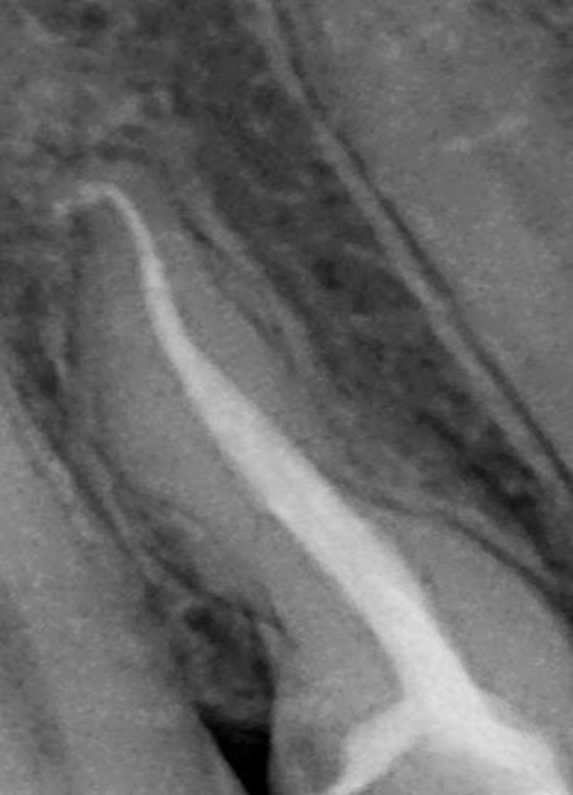
DISTAL VIEW
CASE-5
A 35-year-old female was referred for endodontic treatment of the mandibular right first molar. An initial opening of the pulp chamber had already been performed by the referring dentist to relieve acute throbbing pain (acute pulpitis). Radiographical examination showed no signs of apical periodontitis. No distinct distal root was visible, but tiny projection lines of the periodontal ligament indicated a strong curvature of (one of) the distal root(s) to the mesial . On adjusting the opening cavity, four distinct canal orifices were found and were coronally enlarged with Gates Glidden drills. Initial negotiation of the root canals was performed with a K-file ISO 15.
Although the coronal enlargement and relocation of the canal orifices allowed straight-line access in three canals, insertion of the file in the fourth, distolingual canal showed a more lingually oriented access inclination. Upon removal of the file, the tip was deformed with a strong curvature to the mesial. This, together with the different access inclinations between the two distal canals, indicated the presence of two separate distal roots. The lengths of these canals were measured electronically. The canals were cleaned with sodium hypochlorite solution (5.25%) and EDTA, and shaped with ProTaper instruments. A second distal canal was found and prepared. The two distal canals merged at the mid-root level. Ultrasonic tips) were used to remove the isthmus between the distal canals. The gutta cone fit, with radiographical exposure 30 degrees from the mesial, confirmed the presence of an RE .All canals were filled with gutta- percha and AH26 sealer and the opening cavity was sealed with c omposite.7

PRE OPERATIVE RADIOGRAPH
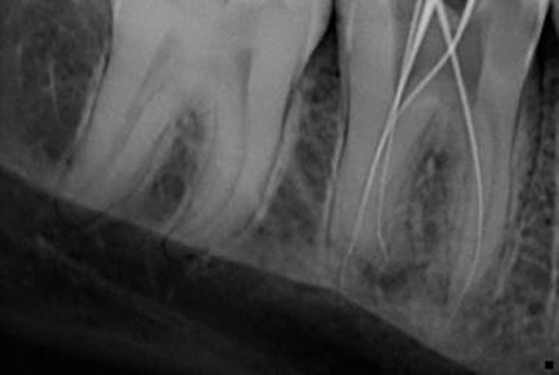
WORKING LENGTH

MASTER CONE
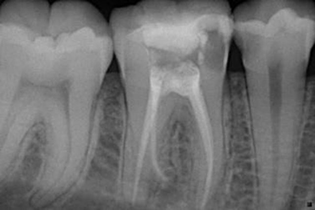
OBTURATION

POST OPERATIVE RADIOGRAPH
CONCLUSIONS
Knowledge of the phenomenon of the complexity of the root canal system, canal obliteration and configuration, and the potential for additional root canal systems and radix entomolaris will improve the medical practice of the general dental practitioner.2
REFERENCES
- DIFFICULT CASES IN ENDODONTICS – PROGNOSIS AND PROPHYLAXIS OF COMPLICATIONS Janet Kirilova, Snezhanka Topalova-Pirinska. Department of Conservative Dentistry, Faculty of Dental Medicine, Sofia, Bulgaria
- Mayhall JT. Taurodontism: a review of the condition and endodontic treatment challenges. Inter Endod J. 2008 May; 41(5):375-88. [PubMed] [CrossRef]
- R. M. Al Shalabi, O. E. Omer, J. Glennon, M. Jennings, and N. M. Claffey, “Root canal anatomy of maxillary first and second permanent molars,” International Endodontic Journal, vol. 33, no. 5, pp. 405–414, 2000.View at: Google Scholar
- M. Hulsmann and E. Schafer, “Problems in gaining access to the root canal system,” Problems in Endodontics: Etiology, Diagnosis and Treatment, vol. 1, pp. 145–172, 2009.
- Internal root resorption: An endodontic challenge”: A case series Sunandan Mittal, Tarun Kumar, Shifali Mittal, and Jyotika Sharma
- S-Shaped Canals: A Series of Cases Performed by Four Specialists around the World
- The Radix Entomolaris and Paramolaris: Clinical Approach in Endodontics Filip L. Calberson, DDS, MMS, Roeland J. De Moor, DDS, MMS, PhD, and Christophe A. Deroose, DDS, MMS.
Disclaimer from Guident
The views and opinions expressed in articles published in Guident are solely those of the authors and do not necessarily reflect the views of the editors, the editorial board or the publisher, Ivory India. Guident does not endorse any product, technique or treatment described. Clinical content is intended for qualified dental and medical professionals and is not a substitute for individual clinical judgement. Guident, its editors and the publisher accept no liability for any consequence arising from the use of information published in the journal.
How to cite this article
Dr. Rishika Lahuch, Dr. Sugandha Sati, Dr. Annil Dhingra. Endodontic Pecularities-a Case Series. Guident. 2021 Jun;14(7). Available from: https://guident.in/articles/endodontics/endodontic-pecularities-a-case-series.html