
Endodontic Perforation Repair in Maxillary Molar Using Glass Ionomer Cement- a Case Report
Department of Conservative Dentistry & Endodontics, K.D. Dental College & Hospital, Mathura
ABSTRACT
Endodontic perforation can pose serious complication that may even result in tooth loss. Various materials have been suggested & used to repair the perforation defect that include calcium hydroxide, mineral trioxide aggregate, biodentin, and Glass ionomer cement. This report describes the clinical management of a maxillary first molar with lateral perforation in mesiocervical region. Treatment involved sealing of perforation defect with glass ionomer cement and completion of root canal treatment.
INTRODUCTION
The main purpose of endodontic therapy is complete debridement of pulpal tissues, thorough cleaning and shaping and ultimately total obturation of the root canal system that will provide an effective barrier which prevent passage of microorganisms & their toxic byproducts into the surrounding periapical & periodontal tissues. In some clinical scenarios, complications can impede the treatment outcome and may even lead to a questionable prognosis.1 Root perforation, once regarded as one of the obnoxious complication of endodontic treatment.2 According to Ingle et al. it is the second most common reason for failure of endodontic therapy.3 Endodontic perforation is defined as an artificial passage created within a tooth or its root which produces a communication between the pulp cavity & the periodontal tissues. Perforation may occur either iatrogenically (e.g. root canal preparation, post space preparation) or pathologic (e.g. carious decay, resorption).4 The most severe complication is the potential for secondary periodontal inflammation & loss of bone attachment that leads to tooth loss ultimately.
Root perforations at the alveolar crest level & epithelial attachment i.e. “critical zone” has the worst prognosis because of microbial contamination & subsequent periodontal destruction. Perforations that occur more coronally can be repaired more safely without involving periodontium. Perforations that are apical to critical zone, if adequately treated endodontically with sufficient bacterial tight perforation repair also result in good prognosis.5
In order to repair the perforation adequately, sealing of the tract is necessary. The properties that a material should possess for perforation repair include excellent sealing ability, biocompatibility, should be easy to compound & is able to promote osteogenesis & cementogenesis.6
Glass ionomer cement, is an alternative for sealing of perforation, especially in the cervical third region as it has been recommended to use adhesive restorative material there. The properties that make glass ionomer eligible for repairing perforation include its biocompatibility, bonding to dentin and cementum, fluoride releasing ability & low coefficient of thermal expansion.7
This case report describes a clinical case in which a lateral perforation in the mesiocervical region of a maxillary molar was detected. The decision was made to perform endodontic treatment and to seal the perforation with glass ionomer cement as well.
CASE REPORT
A 38-year old male patient reported to our department of conservative dentistry & endodontics with chief complaint of moderate to severe pain in upper left back teeth region since 7-10 days. He stated that the endodontic treatment had been initiated. His medical history was unremarkable. Extra oral examination revealed no abnormalities. Intraorally, tooth 26 presented with improper access opening. Tooth was tender to vertical percussion. Vitality test was omitted as the tooth already had undergone initial endodontic treatment. Radiograph revealed area of radiolucency in mesiocervical aspect & periodontal ligament space widening in relation to palatal root (Figure 1). A diagnosis of symptomatic apical periodontitis with mesiocervical lateral perforation was made. Planned treatment was to repair the perforation defect with glass ionomer cement alongwith definitive endodontic and restorative treatment.

Figure 1 : Preoperative Radiograph
After modifying the access opening, an oval perforation defect measuring about 2mm Ñ… 3mm was detected mesiocervically. The perforation showed minor haemorrhage (Figure 2). NaOCl irrigation followed by pressure packing with sterile gauze was used to achieve haemostasis.
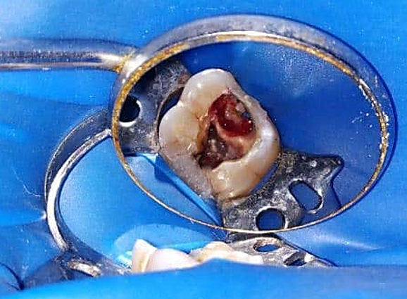
Figure 2: Preoperative clinical photograph
After rinsing with distilled water & drying, the perforation site was repaired using Glass ionomer cement. Care was taken not to occlude the orifices of the root canal (Figure 3). Subsequently, conventional endodontic treatment was performed. Working length was established using radiograph and apex locator, cleaning & shaping was done using rotary crown down technique (Hyflex CM, Coltene). Canal preparation was completed upto 30/0.04 in palatal canal & 25/0.04 in MB & DB canals. Irrigation was performed with 3% sodium hypochlorite solution, Normal saline, 17% EDTA and 2% chlorhexidine digluconate was used as the final irrigant. Canals were dried with paper point & Calcium hydroxide dressing was placed. In the follow up visit, as the patient become aymptomatic, obturation was completed using GP & zinc oxide eugenol as sealer with single cone technique. Tooth was then restored with composite and patient was asymptomatic during the follow-up period of 2 months.
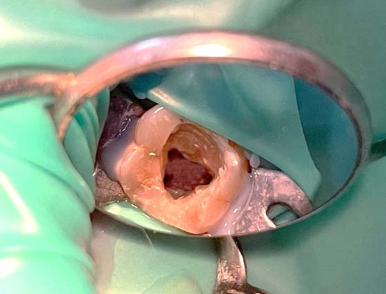
Figure 3: Perforation defect repaired with GiC

Figure 4 : Working Length Determination

Figure 5: Master cone radiograph

Figure 6: Postoperative radiograph
DISCUSSION
Endodontic therapy play a vital role in maintaining the integrity of the natural dentition which is necessary for function & esthetics. Occasionally, iatrogenic errors do occur i.e. perforating a wall, pulp chamber floor or root canal wall during carious removal, access cavity preparation, locating the canals & mechanical debridement which can affect the prognosis of the treatment.8 Perforations that are present in the furcation region or cervical third of the root have a bad impact on the prognosis of endodontic therapy, as they trigger an inflammatory reaction in periodontal region which may further lead to loss of attachment & occasionally loss of affected tooth. Once the alveolar bone gets destroyed, granulation tissue may form & invaginate into the tooth through the communicating tract.9
There is wide range of treatment strategies as well as materials available in order to conserve the affected tooth & make it to function normally.
In the present case, radiograph revealed a lateral perforation in the mesiocervical region of the left maxillary first molar, the prognosis was deemed favourable in aspect of the short time since the occurrence of perforation (less than 10 days). The prognosis of a perforation is highly dependent on the sealing ability of the chosen repair material and is related to the size and site of the perforation, the time since perforation, and the presence of microbial agents, as well as on the feasibility of sealing and the accessibility of the main canal.10 Proper sealing of the perforated region is very necessary because it prevents contamination from the microbes as well as restores th physiological condition of tooth and surrounding periodontal tissues. Measures should be taken in order to improve the treatment outcome of perforation repair, such as recognition, assessment of result, avoidance of file contact with bone & surrounding tissues during the cleaning & shaping of the canals.
Material used for repairing the perforation defect should be placed in intimate contact with the tooth structure as well as with the soft tissues of periodontium. Glass ionomer cement by the formation of chemical bonds between the cement & the calcium ions of dentin bonds very well to the tooth structure. It is a good material for repairing the perforation at the cervical region especially where use of restorative material is indicated.11 In the present case glass ionomer cement was chosen because of its physical properties & fluoride releasing property.
It is essential for a practitioner to incorporate preventive management strategies in order to avoid the iatrogenic perforations. A thorough analysis of the radiograph should be done in order to assess the pulp chamber and root canal anatomy, root direction & curvature in relation to adjacent teeth. Proper knowledge of both internal & external tooth morphology provide important guidance while performing access cavity preparation.
CONCLUSION
Proper diagnosis, thorough treatment planning, use of appropriate restorative material along with appropriate procedures provide good clinical outcome in case of teeth with perforation preventing the coronal damage that further might result in loss of tooth. Based on its biocompatibility glass ionomer material may be considered to be part of the clinician’s armamentarium for the treatment of endodontic perforations and it should, therefore, be of interest to many practitioners.
REFERENCES
- Dotto RF, Barbosa AN, Dotto SR, Hermes CR. Sealing of root perforation with glass ionomer cement: a case report. Stomatos 2014; 20(38): 29-46.
- Seltzer S, Bender IB, Smith J, Freedman I, Nazimov H. Endodontic failures- an analysis based on clinical, roentgenographic, and histologic findings. II. Oral Surg Oral Med Oral Pathol 1967; 23: 517-530.
- INGLE JI . Endodontics. 3rd Ed. Philadelphia: Lea & Febiger, 1985; 35-37.
- Jew RC, Weinw SW, Keene JJ, Smulson MH. A histologic evaluation of periodontal tissues adjacent to root perforations filled with cavit. Oral Surg Oral Med Oral Pathol 1982; 54: 124-135.
- Clauder T, Shin SJ. Repair of perforations with MTA: clinical applications and mechanism of action. Endod Topics 2009; 15:32-55.
- Menezes, R., da Silva Neto, U.X., Carneiro, E., Letra, A., Bramante, C.M., Bernadinelli, N. MTA repair of a supracrestal perforation: A case report. J Endod 2005; 31:212-214.
- Behnia A, Strassler HE, Campbell R. Repairing iatrogenic root perforation. J Am Dent Assoc. 2000;131:196-201.
- Breault, L.G., Fowler, E.B., Primack, C.B.D. Endodontic Perforation Repair with Resin-Ionomer: A Case Report. J Contemp Dent Pract 2000;1: 1-7.
- Juarez Broon, N., Bramante, C.M., de Assis, G.F., Bortoluzzi, E.A., Bernardineli, N., de Moraes, I.G. Healing of root perforations treated with mineral trioxide aggregate (MTA) andPortland cement. J Appl Oral Sci 2006;14: 305-11.
- Chevigny, C., Dao, T.T., Basrani, B.R., Marquis, V., Farzaneh, M., Abitbol, S., Friedman, S.Treatment outcome in endodontics: the Toronto study--phases 3 and 4: orthograde retreatment. J Endod 2008; 34:131-7.
- Gomes SC, Miranda LA, Soares I, Oppermann RV. Clinical and histologic evaluation of the periodontal response to restorative procedures in the dog. Int J Periodontics Restorative Dent. 2005; 25:39-47.
Disclaimer from Guident
The views and opinions expressed in articles published in Guident are solely those of the authors and do not necessarily reflect the views of the editors, the editorial board or the publisher, Ivory India. Guident does not endorse any product, technique or treatment described. Clinical content is intended for qualified dental and medical professionals and is not a substitute for individual clinical judgement. Guident, its editors and the publisher accept no liability for any consequence arising from the use of information published in the journal.
How to cite this article
Dr. Bhavya Varshney, Dr. Ajay Kumar Nagpal, Dr. Sunil Kumar. Endodontic Perforation Repair in Maxillary Molar Using Glass Ionomer Cement- a Case Report. Guident. 2021 Aug;14(9). Available from: https://guident.in/articles/endodontics/endodontic-perforation-repair-in-maxillary-molar-using-glass-ionomer-cement-a-case-report.html


