"A Comparative Evaluation of Surface Texture and Stain Absorption of Microfill and Nanofill Composite Resins Using Different Methods of Finishing and Polishing - in Vitro Study"
(Professor & H.O.D)
Abstract
Aim: To evaluate surface texture and stain absorption of microfill and nanofill composite resins using different methods of finishing and polishing before and after simulated tooth brush abrasion
Objectives: To evaluate surface texture before and after simulated tooth brush abrasion of microfill and nanofill composite resins using different methods of finishing and polishing. To evaluate Stain absorption before and after simulated tooth brush abrasion of microfill and nanofill composite resins using different methods of finishing and polishing. To comparatively evaluate the surface texture and stain absorption before and after simulated tooth brush abrasion of microfill and nanofill composite resins. Materials and methods: A. Master die, B. Materials for composite resin samples, C. Materials for finishing, polishing and application of glaze, D. Materials used for mounting and staining of samples, E. Materials used for abrasion. Method: In this study 66 composite resin disc samples were prepared and were divided into the following:- 1. Preparation of composite resin samples. 2. Evaluation of surface texture by scanning electron microscope. 3. Evaluation of stain absorption by spectrophotometer. Result: finished and polished surface of microfill and nanofill showed better surface texture and least amount of stain absorption. Conclusion: Application of glaze gave rough surface texture and maximum stain absorption.
Keywords: Microfill and Nanofill Composite resin, Biscover Glaze, stain absorption and surface texture .
INTRODUCTION
Esthetics has emerged as the most important aspect of dentistry. With development in science and research in dental materials a massive improvement has taken place in the quality of materials used to attain esthetics.
The esthetics and durability of composite resins is based on the particle size, its manipulation and the process of finishing and polishing.1 The luster of composite resins contributes to the overall esthetic appearance of the restoration. The potential staining of composite resin surface is directly related to surface texture.1
In the last decades composite resins have evolved to their highest with the hybrid and nanofill particles. Though the microfill composite resins were ideal for anterior restorations as they maintained their gloss but were weak and susceptible to fracture in high stress areas. The newer generation of hybrid composite resins – the nanofill have physical properties equivalent to the original hybrid composite resins with handling characteristics and higher polishability.2
None of the composite resins attained and retained polish for a long period of time, which further lead to absorption of stain and discoloration of composite resins.4 Though, the newer composite resins claimed to have better esthetics a study was planned to evaluate their colour stability and surface texture with varied finishing and polishing procedures and application of surface glaze and also to analyze the effect of tooth brushing on the surface texture and stain absorption of composite resins.
METHOD
The study was divided into the following:-
1. Preparation of composite resin samples.
2. Evaluation of surface texture by scanning electron microscope.
3. Evaluation of stain absorption by spectrophotometer.
1. PREPARATION OF COMPOSITE RESIN SAMPLES
Split brass mould was used for fabrication of the test samples. The mould was placed on the glass slab with mylar strip interposed between them. The test samples were prepared by placing the first layer of composite resin and light cured for 20 seconds (Fig.1). Second and final increment of composite resin was placed to fill the mould completely and cured for another 20 seconds. Finally the samples were removed from the mould (Fig.2) and light cured for an additional 40 seconds.2 Seventy two samples -thirty six of microfill composite resin and thirty six of nanofill composite resin were prepared in the similar manner to get standardized discs of 2mm thickness and 10 mm diameter.
For the study 36 samples each of microfill and nanofill were divided into three groups depending on the method of finishing and polishing –
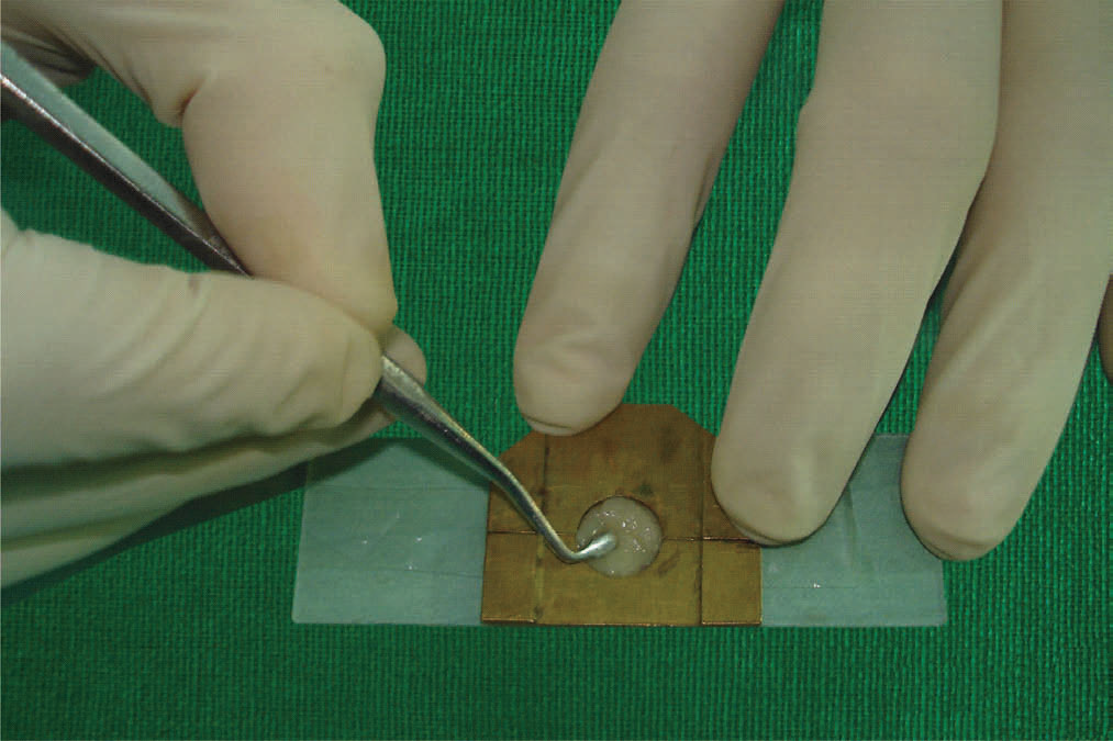
Fig 1: Placement of Composite Resin

Fig 2: Removal of Composite Resin Sample From Mould
Group I -- Finish and Polish
- Microfill composite resin – 12 samples
- Nanofill composite resin – 12 samples
Group II -- Finish, Polish and application of Glaze
- Microfill composite resin – 12 samples
- Nanofill composite resin -- 12 samples
Group III --Finish and application of Glaze
- Microfill composite resin – 12 samples
- Nanofill composite resin – 12 samples
GROUP I –Finish and Polish
This was done by reorienting the samples in the split cast mold. Gross irregularities on the surface were removed using a tapered finishing carbide bur.
A. Microfill composite resin:-
Finishing was done using astropol F, astropol P, astropol HP in a sequential order (Fig.3, 4). Each of them was used for 20 seconds with 15 strokes. After finishing, polishing was done with astropol polishing brush for 20 seconds following unidirectional strokes.2
B. Nanofill composite resin:-
Samples were reoriented in the split cast mould. Gross irregularities on the surface were removed using tapered finishing carbide bur.
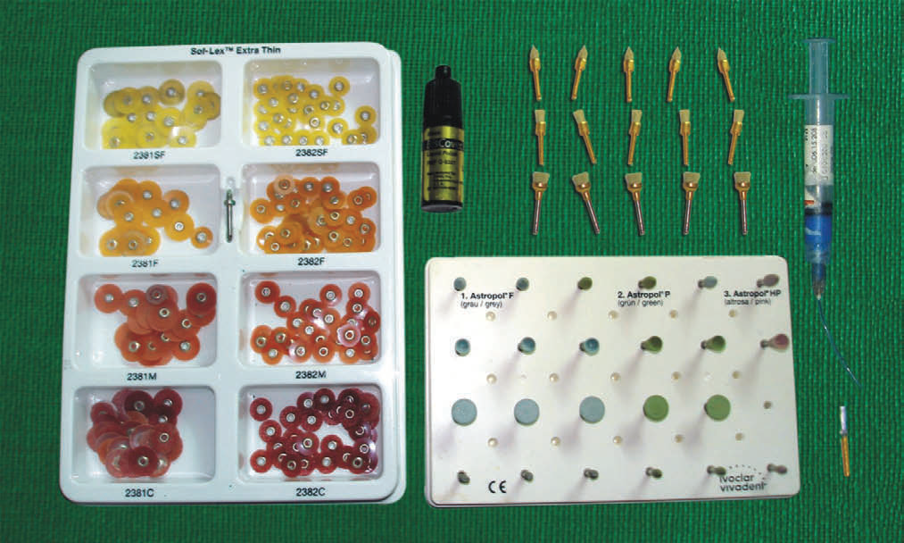
Fig 3: Finishing, Polishing And Glazing Kit

Fig 4: Finishing of Microfill Composite Resin Sample Using Astro

Fig 5: Finishing of Nanofill Composite Resin Samples Using Course
Finishing was done using coarse, medium and fine soflex disc in a sequential order (Fig.3, 5). Each soflex disc was used for 20 sec with 15 strokes. After finishing, polishing was done with extra fine soflex disc for 20 sec following unidirectional stroke.2 (Fig.6).
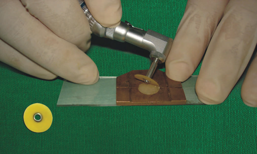
Fig 6: Polishing of Nanofill Composite Resin Samples Using Extra.
GROUP II – Finish, Polish and application of Glaze
Similar procedure of group I was followed for finishing and polishing of group II [A. Microfill & B.Nanofill] composite resin samples. On the polished surface of samples 37% of orthophosphoric acid was applied with a paint brush for 15 seconds (Fig.7).. Surface was
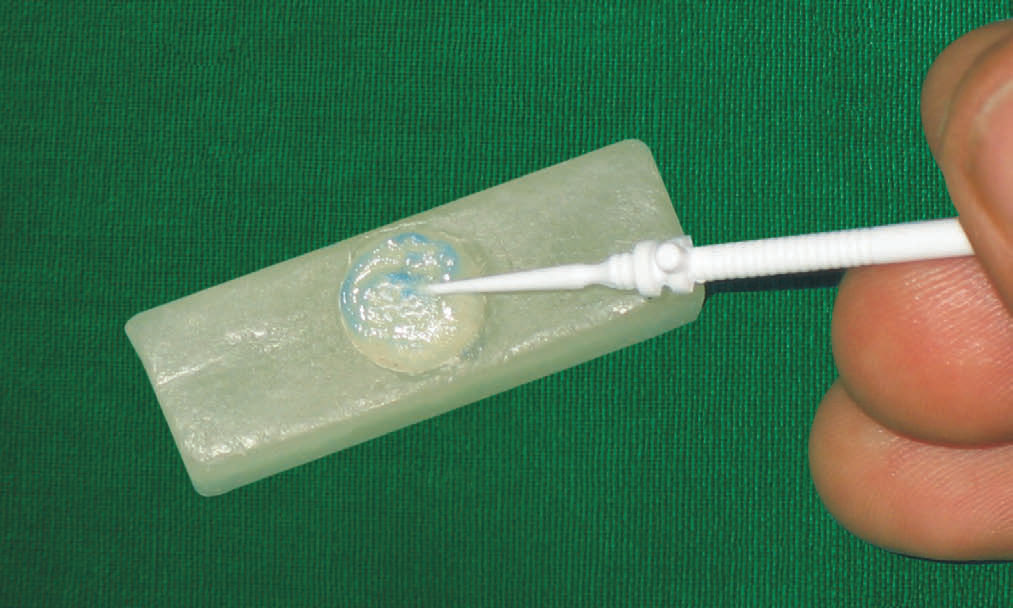
Fig 7: Application Of Etchant on Composite Resin Sample.
then rinsed with distilled water for 30 seconds and dried with compressed air. A single layer of glaze (biscover liquid) was applied with a paint brush. It was light cured for 15 seconds as per the manufacturer’s instructions (Fig.8).1
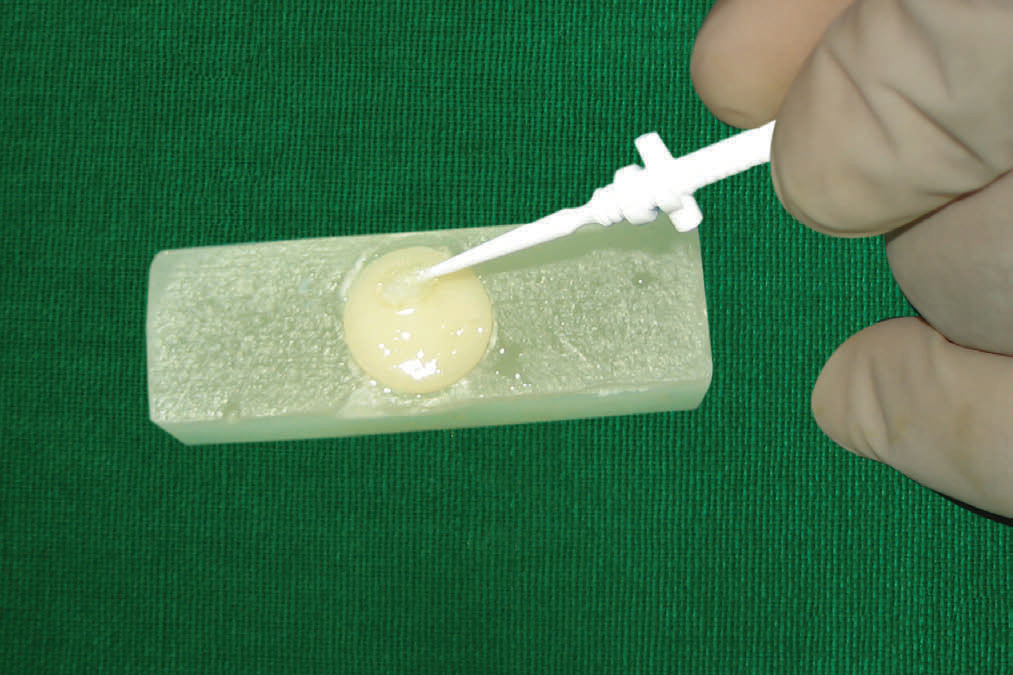
Fig 8: Application of Biscover Liquid on Composite Resin Sample.
GROUP III – Finish and application of Glaze
Similar procedure for finishing of group III [A. Microfill & B. Nanofill] was followed as done in group I, but no polishing was carried out. Glaze was applied on the finished surface as done in group II. All samples were stored in distilled water until further use.
2. EVALUATION OF SURFACE TEXTURE BY
SCANNING ELECTRON MICROSCOPE;-
Two samples from each of the three groups were randomly selected for scanning electron microscopic evaluation for the surface texture before and after simulated tooth brush abrasion.
Each sample was divided into two.
Sub group a. - Before tooth brushing
Sub group b. - After tooth brushing
1. Sub group a. ---- Evaluation of surface texture:-
Samples were gold sputtered and observed under scanning electron microscope at 130x magnification for evaluation of surface texture.
2. Sub group b. ---- Evaluation of surface texture:-
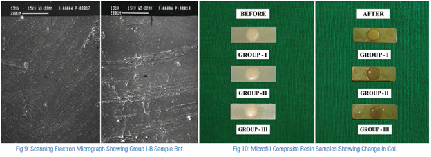
Samples were subjected to simulating tooth brushing in an assembly prepared for the abrasion using a powered tooth brush and Colgate total tooth paste. Brush was suspended in the lever arm of the assembly. Tooth paste was placed on the bristle of the tooth brush. A weight of 250 gms was placed on the head of the assembly to simulate the pressure exerted during brushing.5 Samples were placed below the bristles of the tooth brush at a distance that would allow the bristle to just be in contact with the surface. All samples were abraded for 2 minute each day for seven days, during this period samples were kept in distilled water. At the end of the seventh day surface texture evaluation was carried out under the scanning electron microscope as done for the earlier samples (Fig.9).
3. EVALUATION OF STAIN ABSORPTION BY SPECTROPHOTOMETER
Sixty samples of group I, II, III were embedded in a hard carving wax leaving only the processed surface uncovered. They were numbered on the under side [1 to 60] and stored in distilled water for 24 hours. At the end of the first day, samples were subjected to baseline spectrophotometric evaluation.
I. Preparation of tea solution:-
Tea solution was prepared by boiling 5gms of tea [Girnar GTC strong tea] in 100ml of distilled water for 5minutes and filtered through a strainer. Fresh solution was made each day for the study for seven days.2
II. Spectrophotometric evaluation of change in colour after placement in tea solution at the end of day one:-
Once the base line evaluation of each composite resin sample was done, they were immersed in a standard tea solution for 1 day at room temperature 370c in a covered petri dish. They were washed with distilled water and then subjected to spectrophotometeric evaluation.
III. Spectrophotometric evaluation of change in colour at the end of 7 days, after placement in tea solution & subjected to simulated tooth brushing:-
Samples were placed in tea solution each day for the next seven days at room temperature at 370C. They were subjected to simulated tooth brushing as described earlier each day for the next seven days and then subjected to spectrophotometric evaluation. Each sample was placed in front of the spectrophotometer and the reading of change in colour was noted (Fig.10, 11).
RESULTS
It was concluded from the study that the surface texture and stain absorption of microfill and nanofill composite resin, seven days after tooth brushing showed the smooth surface and least stain absorption of finishing and polishing followed by finishing, polishing and application of glaze and the roughest surface with maximum stain absorption was observed for the finishing with application of glaze, both at first day and seventh day after simulated tooth brushing. Nanofill composite resin showed smoother surface texture and less stain absorption compared to microfill composite resin (Fig.12).
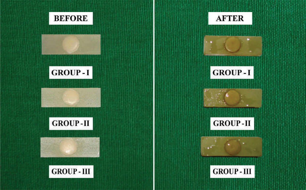
Fig 11: Nanofill Composite Resin Samples Showing Change In Colou.

Fig 12
DISCUSSION
Composite resins have become increasingly popular due to their esthetic qualities. However, it has been seen that long term esthetics of composite resin have not been very promising due to their property of stain absorption.1 The stain absorption could be reduced to a great extent by meticulous process of finishing and polishing. Application of unfilled resin over the composite resin restoration has also been recommended by some workers.4 Though the final product of restoration depends both on the size of the filler particle and method of finishing and polishing it is still to be seen which method of finishing polishing and application of unfilled resin would give better long term esthetics.1,4
The application of glaze has been recommended by manufacturer to be applied simply after finishing of the composite resin. Whether application of unfilled resin [biscover liquid] with only finishing gives less stain absorption or finishing and polishing is required to be done before application of unfilled resin was to be investigated.1
In the present study composite resin with microfill particles and nanofill particles were chosen for evaluation of their surface texture soon after finishing and polishing was done and later when surface of both the composite resin were subjected to tooth brushing for successive seven days.
The recommended method of finishing and polishing of composite resin has been laid down by the manufacturer. The microfill composite resin required Astropol finishing and Astrobrush polishing kit of IVOCLAR VIVADENT. While nanofill composite resin required Soflex finishing and polishing kit of 3M ESPE DENTAL.
The present study has shown a smooth surface texture initially of all samples finished polished, finished polish and application of glaze and finished and application of glaze but, after tooth brushing for seven days the surface did not maintain its surface smoothness. In those cases where glaze [Biscover liquid] was applied after finishing, the surface was most rough. The samples which were finished and polished with glaze showed less roughness as compared to the earlier method. But it was the samples which were finished and polished without application of glaze gave the smoothest surface after tooth brushing.
From the study it has been shown that less stain absorption were observed when the composite resin was only finished and polished. The glaze applied after finishing and polishing showed more stain absorption. Maximum amount of stain absorption was seen in finishing and application of glaze. However this was not in co-relation to the result by other workers who belived that glaze gave better finishing of the restoration.1,3,5,8
However, the best results were obtained when only finishing and polishing was carried out without the application of glaze .That is to say that least stain absorption was observed when a proper finishing and polishing was done. The composite resins were subjected to simulated tooth brushing for seven days. In case where only finishing and polishing was done smooth surface texture was observed at the end of first day and after seven days.
The glaze which is a layer of unfilled resin does not have any filler particles. It was obvious that the unfilled resin did give initially smooth glossy surface, but with passage of time for the first day to the seventh day absorption of stain took place. The absorption seen in the glaze applied samples was much more than with only finishing and polishing. Therefore glaze should not be applied as a routine, but the composite resin should be meticulously finished and polished to prevent absorption of stain.
The microfill composite resin had more absorption than nanofill composite resin which is probably due to the size of the filler particle. The nanofill composite resin as claimed have particles which are of the size 20 nm.2 Thus polishing of the composite resin gave a smoother surface which can be maintained for a longer time and also absorbs less amount of stain.
Within the limitation of the study we could conclude that a well polished composite resin is a longer lasting esthetic restoration with smooth surface and less stain absorption.
CONCLUSION
Following conclusions were drawn from the study:-
- The microfill and nanofill composite resin showed a smooth surface texture soon after finishing and polishing, finishing polishing and application of glaze and finishing and application of glaze.
- The surface texture of microfill and nanofill composite resin, seven days after tooth brushing showed the smooth surface of finishing and polishing followed by finishing, polishing and application of glaze and the roughest surface was observed for the finishing with application of glaze.
- Least stain absorption was observed in finishing and polishing followed by finishing polishing and application of glaze and maximum stain absorption was seen in finishing with application of glaze, both at first day and seventh day after simulated tooth brushing.
- Nanofill composite resin showed smoother surface texture and less stain absorption compared to microfill composite resin at the end of first day and seventh day after simulated tooth brushing.
REFERENCES
- Duygu Sarac, Sinasi Sarac, Safak Kulunk and Tolag Kulunk. The effect of polishing technique on the surface roughness and colour change of composite resins. J Prosthet Dent 2006; 57: 683-9.
- Huan LS. Effect of surface roughness on stain resistance of dental resin composite. J Esthet Restor Dent 17; 102-109,2005.
- Kazuo Itoh. Effectiveness of glazing composite resin restoration. J Prosthet Dent 1981; 45: 606-13.
- Erica C. N. Teixeria. In vitro tooth brush – dentifrice abrasion of two restorative composite resin. J Esthet Restor Dent 17; 172-182,2005.
- Marie – France Bertrand. Effect of surface penetrating sealant on surface texture and micro hardness of composite resin. J Prosthet Dent 2000; 53: 658-63.
- C. Mclndie, F.D.Murray. Comparison of methods used in finishing of composite resin – a scanning electron microscope study. J Prosthet Dent 1974; 31: 163-71.
Disclaimer from Guident
The views and opinions expressed in articles published in Guident are solely those of the authors and do not necessarily reflect the views of the editors, the editorial board or the publisher, Ivory India. Guident does not endorse any product, technique or treatment described. Clinical content is intended for qualified dental and medical professionals and is not a substitute for individual clinical judgement. Guident, its editors and the publisher accept no liability for any consequence arising from the use of information published in the journal.
How to cite this article
Dr. Kunal B. Dalal, Dr. Anvesha Gupta, Dr. Sabita M. Ram Dean. "A Comparative Evaluation of Surface Texture and Stain Absorption of Microfill and Nanofill Composite Resins Using Different Methods of Finishing and Polishing - in Vitro Study". Guident. 2014 Jan;7(2). Available from: https://guident.in/articles/research/a-comparative-evaluation-of-surface-texture-and-stain-absorption-of-microfill-and-nanofill-composite-resins.html